SEARCH

Evans J, Negoescu A. Early recognition pivotal in the management of spondyloarthritis. Practitioner Dec 2017;261(1810):11-15
Early recognition pivotal in the management of spondyloarthritis
20 Dec 2017
AUTHORS
Dr Jobie Evans MB BS MRCP Rheumatology SpR
Dr Andra Negoescu Consultant Rheumatologist Rheumatology Department, Addenbrooke’s Hospital, Cambridge, UK
Competing interests: None
Article
Abstract
The spondyloarthritis group is divided into two main subgroups: axial spondyloarthritis and peripheral spondyloarthritis. These may exist as separate entities or coexist in the same patient. Axial spondyloarthritis can be further subdivided into non-radiographic axial spondyloarthritis and ankylosing spondylitis (AS). Peripheral spondyloarthritis encompasses psoriatic arthritis, reactive arthritis and enteropathic arthritis. Axial spondyloarthritis tends to occur before the age of 45 years but can occur in older age groups, while peripheral spondyloarthritis tends to affect a wider age group. The association with the HLA-B27 gene is well established. The vast majority of patients with AS are male, while non-radiographic spondyloarthritis affects both genders equally. Genitourinary and gastrointestinal infections with specific pathogens (Chlamydia trachomatis and Shigella, Salmonella and Campylobacter, respectively) are recognised triggers for reactive arthritis. Classically, axial spondyloarthritis presents with insidious onset inflammatory lower back pain, which is typically worse in the morning and after rest, and improves with activity. Peripheral spondyloarthritis can present with peripheral joint pain and/or swelling, swelling of the digits (dactylitis), tendon and entheseal pain that is not secondary to a mechanical cause. Early referral of patients with suspected spondyloarthritis to specialist care is strongly recommended as this can improve long-term outcomes.
Article
Spondyloarthritis is a diverse group of inflammatory musculoskeletal diseases that can present as axial and/or peripheral disease. There is a well established association with human leukocyte antigen HLA-B27.
The spondyloarthritis group is divided into two main subgroups: axial spondyloarthritis and peripheral spondyloarthritis. These may exist as separate entities or may coexist in the same patient.
Axial spondyloarthritis can be further subdivided into non-radiographic axial spondyloarthritis1 and ankylosing spondylitis, where the first subgroup does not necessarily have a natural progression towards ankylosing spondylitis. Peripheral spondyloarthritis encompasses psoriatic arthritis, reactive arthritis and enteropathic arthritis.
The prevalence of spondyloarthritis has been estimated at 0.01-2.5%, with ankylosing spondylitis being the most prevalent.2 Extra-articular features include uveitis, psoriasis and inflammatory bowel disease.
The onset of axial spondyloarthritis tends to be before the age of 45 years but it can occur in older age groups, while peripheral spondyloarthritis tends to affect a wider age group.1
Risk factors
The association with the HLA-B27 gene is well established. The presence of this gene in the Caucasian population varies from 7.5% in the United States to around 15% in northern Scandinavia, with more than 90% of patients with ankylosing spondylitis possessing this gene. HLA-B27 prevalence in non-Caucasian populations is lower.
The vast majority of patients with ankylosing spondylitis are male, while non-radiographic spondyloarthritis affects both genders equally. Genitourinary (GU) and gastrointestinal (GI) infections with specific pathogens (Chlamydia trachomatis and Shigella, Salmonella and Campylobacter, respectively) are recognised triggers for reactive arthritis (see table 1).
Presentation in primary care
Because of the diverse nature of spondyloarthritis, patients can present with a variety of symptoms. Classically, axial spondyloarthritis presents with inflammatory lower back pain of insidious onset, which is typically worse in the morning and after rest, and improves with activity. In addition, patients can present with alternating buttock (sacroiliac) pain and neck pain and stiffness. The response to oral non-steroidal anti-inflammatory drugs (NSAIDs) is typically good.
Peripheral spondyloarthritis can present with peripheral joint pain and/or swelling, swelling of the digits (dactylitis), tendon and entheseal pain that is not secondary to a mechanical cause. In these cases, a past medical history or symptoms/signs suggestive of psoriasis, inflammatory bowel disease, uveitis or recent GI or GU infections are of great diagnostic value.
Practical tip 1: Always explore the possibility of inflammatory symptoms in younger patients (< 45 years) presenting with lower back pain and consider early referral if spondyloarthritis is suspected.
Referral
Early referral of patients with suspected spondyloarthritis to specialist care is strongly recommended as this can improve long-term outcomes.
The absence of serological markers, including rheumatoid factor (RhF), anti-cyclic citrullinated peptide (CCP) antibodies and anti-nuclear antibodies (ANA) raises the suspicion of spondyloarthritis in a patient with evidence of inflammatory joint disease.
In the UK, HLA-B27 is present in up to 90-95% of ankylosing spondylitis patients and approximately 50-60% of psoriatic arthritis patients but a negative result does not rule out a spondyloarthritis.3
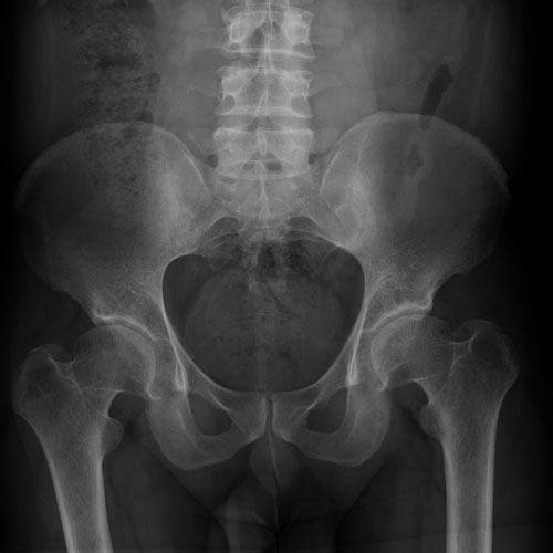
Inflammatory markers (ESR and CRP) can be elevated in spondyloarthritis but a normal ESR or CRP does not rule out the condition. Joint effusions can be aspirated and analysed to rule out a crystal arthritis. Plain X-rays can be used to assess for long-term joint damage such as erosive disease, syndesmophytes (calcification of the spinal ligaments) or sacroiliitis (see figure 1). However, X-rays can be normal in early disease and therefore MRI may be useful to detect suggestive inflammatory changes of the spine or sacroiliac joints. Ultrasound or MRI can also be used to assess for peripheral subclinical synovitis and enthesitis.
Ankylosing spondylitis has historically been diagnosed using the modified New York criteria (see table 2).4 New classification criteria were developed in 2011 by the American Society of Ankylosing Spondylitis (ASAS), see table 3.1
For psoriatic arthritis, CASPAR classification criteria may aid diagnosis, see table 4.5
There are no specific diagnostic guidelines for enteropathic arthritis but the diagnosis is normally based on the presence of an axial or peripheral spondyloarthritis in the context of a history of inflammatory bowel disease. Similarly, reactive arthritis diagnosis is usually based on the presence of a spondyloarthritis in the context of a recent GI or proven GU infection and in the absence of psoriasis or inflammatory bowel disease.
Practical tip 2: HLA-B27 testing and imaging with plain X-rays are extremely valuable in supporting the diagnosis of spondyloarthritis.
Treatment and monitoring
Axial spondyloarthritis
NSAIDs (COX-1 and COX-2 inhibitors) are the first-line treatment for axial spondyloarthritis and should be used at the lowest effective dose. The risks and benefits of using NSAIDs should be assessed and gastric protection should be considered.
Patients with intolerance or contraindications to NSAIDs, and patients who do not respond to at least two different NSAIDs at the maximum tolerated dose (2-4 weeks each), should be considered for biologic DMARDs. Biologic treatment response is assessed at 12 weeks and treatment is only continued if there is a reduction in the Bath Ankylosing Spondylitis
Disease Activity Index (BASDAI)6 of at least 50% or 2 points compared with pre-treatment and a reduction in the spinal visual analogue scale (VAS) by 2 cm or more.7 If there is an inadequate response to the first biologic therapy, then the patient should be switched to a second anti-TNF-alpha agent, or if they have ankylosing spondylitis/radiographic axial spondyloarthritis, they can be treated with the anti-IL-17 agent secukinumab8 (see table 5).
Psoriatic arthritis and other peripheral spondyloarthritis
For patients with psoriatic arthritis presenting with monoarthritis, local steroid injections may be beneficial, and delay the need for DMARD therapy. Standard DMARDs can be offered to patients presenting with oligoarthritis, polyarthritis or persistent monoarthritis. The type of DMARD used depends on the patient’s condition and circumstances. Methotrexate and leflunomide should be avoided in patients wishing to conceive, while sulfasalazine should be considered in patients with co-existing inflammatory bowel disease or uveitis. Methotrexate should be first choice in patients with co-existent psoriasis, because of its efficacy in treating both joint and skin disease. NSAIDs, oral, intra-articular or intra-muscular steroids can also be used as adjunct therapies.
Biologic (anti-TNF-alpha) therapy can be considered in patients who are unresponsive to two traditional DMARDs.9,10 In addition, ustekinumab (anti-IL-23/IL-12) or secukinumab can be considered for patients who have not responded to at least one anti-TNF-alpha agent11,12 (see table 5).
Monitoring of standard and biologic DMARDs requires regular follow-up in a specialist clinic to assess efficacy, and regular blood test monitoring to assess safety.
Non-pharmacological treatments may include physiotherapy, hydrotherapy and the use of other specialist therapies such as hand therapy and orthotics.13
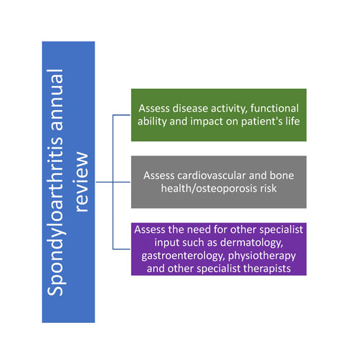
Stable spondyloarthritis patients can be reviewed annually to assess various aspects of the disease, see figure 2.
Practical tip 3: In the absence of contraindications, consider a trial of NSAIDs (COX-1 or COX-2 inhibitors) at the lowest effective dose in patients with suspected spondyloarthritis.
Management of flares
Flares can be managed with NSAIDs, or oral, intramuscular or intra-articular steroids depending on the nature of the flare. For patients with co-existent psoriasis, there is a small risk of psoriasis exacerbation with oral steroids. Advice from the specialist team may be obtained if needed.
Complications
Long-term complications of spondyloarthritis include chronic joint damage and associated chronic disability. Patients with spondyloarthritis are at greater risk of cardiovascular disease and additional risk factors for this should be addressed whenever possible. In addition, spondyloarthritis patients are at increased risk of osteoporosis and fractures and therefore bone health should be optimised with bone protective treatment if needed, including calcium and vitamin D supplements, with or without antiresorptive therapy (bisphosphonates). For the potential complications of treatment, see table 5.
Practical tip 4: During the assessment of patients with spondyloarthritis, consider reviewing other aspects of the condition including extra-articular manifestations, cardiovascular and bone health, mood and functionality/disability.
Conclusion
Spondyloarthritis encompasses a diverse range of conditions that can present with a variety of clinical features. The diagnosis of spondyloarthritis involves clinical assessment, HLA-B27 testing and the use of a variety of imaging modalities including X-ray and MRI. Early recognition and treatment with both pharmacological and non-pharmacological therapies can reduce joint damage and disability and improve long-term outcomes. Regular review is essential for monitoring disease activity, safety and efficacy of treatment, and for the assessment of potential complications.
REFERENCES
1 Rudwaleit M, van der Heijde D, Landewe R et al. The development of Assessment of SpondyloArthritis international Society classification criteria for axial spondyloarthritis (part II): validation and final selection. Ann Rheum Dis 2009;68(6):777-83
2 Stolwijk C, Boonen A, van Tubergen A et al. Epidemiology of spondyloarthritis. Rheum Dis Clin North Am 2012;38(3):441-76
3 Sheehan NJ. The ramifications of HLA-B27. J R Soc Med 2004;97(1):10-14
4 van der Linden S, Valkenburg HA, Cats A. Evaluation of diagnostic criteria for ankylosing spondylitis. A proposal for modification of the New York criteria. Arthritis Rheum 1984;27(4):361-68
5 Taylor W, Gladman D, Helliwell P et al. Classification criteria for psoriatic arthritis: development of new criteria from a large international study. Arthritis Rheum 2006;54(8):2665-73
6 Garrett S, Jenkinson T, Kennedy LG et al. A new approach to defining disease status in ankylosing spondylitis: the Bath Ankylosing Spondylitis Disease Activity Index. J Rheumatol 1994;21(12):2286-91
7 National Institute for Health and Care Excellence. (TA383). TNF-alpha inhibitors for ankylosing spondylitis and non-radiographic axial spondyloarthritis. NICE. London. 2016
8 National Institute for Health and Care Excellence. (TA407). Secukinumab for active ankylosing spondylitis after treatment with non-steroidal anti-inflammatory drugs or TNF-alpha inhibitors. NICE. London. 2016
9 National Institute for Health and Care Excellence (TA199). Etanercept, infliximab and adalimumab for the treatment of psoriatic arthritis. NICE. London. 2010
10 National Institute for Health and Care Excellence (TA220). Golimumab for the treatment of psoriatic arthritis. NICE. London. 2011
11 National Institute for Health and Care Excellence. (TA445). Certolizumab pegol and secukinumab for treating active psoriatic arthritis after inadequate response to DMARDs. NICE. London 2017
12 National Institute for Health and Care Excellence. (TA340). Ustekinumab for treating active psoriatic arthritis. NICE. London. 2015 (updated 2017)
13 National Institute for Health and Care Excellence. (NG65). Spondyloarthritis in over 16s: diagnosis and management. NICE. London. 2017
Further reading
1 Van der Heijde D, Ramiro S, Landewe R et al. 2016 update of the ASAS-EULAR management recommendations for axial spondyloarthritis. Ann Rheum Dis 2017;76(6):978-91
2 Sieper J, Rudwaleit M, Baraliakos X et al. The Assessment of SpondyloArthritis international Society (ASAS) handbook: a guide to assess spondyloarthritis.
Ann Rheum Dis 2009;68 Suppl 2:ii1-44
3 McKenna F. Spondyloarthritis. Arthritis Research UK. Reports on the Rheumatic Diseases. 2010
ARTICLE IN PDF
 Download this article in PDF format
EXTERNAL WEBLINKS
National Ankylosing Spondylitis Society
The Physio Matters Podcast, Session 26: Solving Spondyloarthropathy with Dr Karl Gaffney