SEARCH

Lewis G, Maxwell AP. Timely diagnosis and treatment essential in glomerulonephritis. Practitioner 2015;259 (1779)13-17
Timely diagnosis and treatment essential in glomerulonephritis
23 Feb 2015
AUTHORS
Dr Gareth Lewis MB MSc PhD MRCP,Specialist Registrar in Nephrology
Professor Alexander P. Maxwell MD PhD FRCP, Consultant Nephrologist, Regional Nephrology Unit, Belfast City Hospital, Belfast, UK
Article
Glomerulonephritis includes a variety of inflammatory and immune-mediated disorders that result in inflammation within the glomerulus and other structures within the kidney. It is an important cause of kidney disease and is also the most common diagnosis in patients receiving chronic dialysis or waiting for kidney transplantation in the UK.1,2
Reported global incidence rates of 0.2-2.5/100,000 per year3 are likely to underestimate the scale of glomerulonephritis within the community. In spite of the diversity of conditions and complex terminology associated with glomerulonephritis there are straightforward approaches to its recognition and investigation.
Presentation
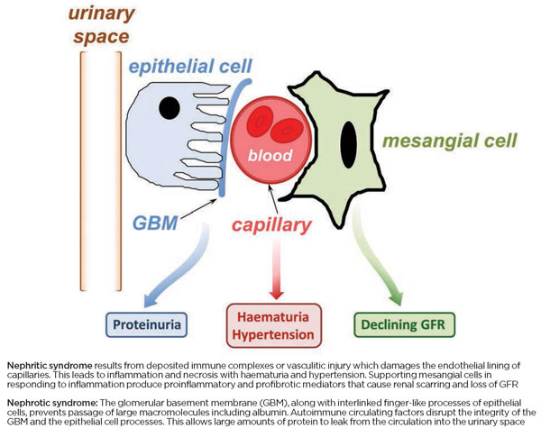
Glomerulonephritis has a wide range of clinical presentations, see table 1, but a key feature in all patients is the presence of urinary abnormalities (proteinuria ± haematuria). Inflammation and injury to cellular and molecular components of the glomerulus result in disruption of the normal glomerular filtration barrier between the blood and urinary space. Protein leakage into the urine (proteinuria) is a hallmark of glomerular injury. If the pathological damage to glomeruli is particularly severe then blood will also enter the urinary space (haematuria). Clinical descriptions of glomerulonephritis can be confusing particularly because nephrotic and nephritic are similar terms but describe different syndromes (see figure 1 and figure 2).
Nephrotic syndrome
Patients with nephrotic syndrome typically present with peripheral oedema, massive urinary protein loss (> 3.5 g per day) and associated low serum albumin levels. This nephrotic syndrome triad (oedema, proteinuria and hypoalbuminaemia) is caused by disruption of the glomerular filtration barrier.4 Blood pressure and renal function, as measured by estimated glomerular filtration rate (eGFR), are usually normal initially.
Minimal change disease (MCD), membranous nephropathy (the most common cause in adults in the UK), and focal and segmental glomerulosclerosis (FSGS) are the leading three causes of nephrotic syndrome.
Nephritic syndrome
Patients presenting with nephritic syndrome typically tend to be hypertensive with dipstick-positive or visible haematuria. There may be rapidly progressive renal dysfunction and fall in eGFR. The nephritic syndrome is caused by autoimmune or vasculitic injury that leads to mechanical disruption of the renal microvasculature and supporting mesangial cells. Despite extensive cellular proliferation and glomerular disruption (leading to histological crescents on renal biopsy) proteinuria may be relatively modest.
Anti-neutrophil cytoplasmic antibody (ANCA)-associated vasculitis and anti-glomerular basement membrane (anti-GBM) disease are serious conditions that can both cause rapidly progressive renal failure progressing to end-stage within weeks.
Risk factors
Glomerulonephritis may be primary (no associated disease elsewhere) or secondary to a systemic autoimmune disease, infection, drug or malignancy.
It is now appreciated that many patients will have a background genetic susceptibility to glomerulonephritis which may be triggered by environmental, infective or autoimmune factors.5 For example, IgA nephropathy, the most common cause of glomerulonephritis, may result from environmental factors, such as alcohol abuse, interacting with genetic and autoimmune mechanisms resulting in abnormal glycosylation of IgA molecules. A throat infection may then trigger clinically obvious nephritis with haematuria.
Autoimmunity, in combination with genetic factors, is responsible for a significant proportion of cases of glomerulonephritis. Systemic lupus erythematosus (SLE) can cause almost any type of glomerulonephritis histology in addition to its many systemic manifestations. Recently, an antibody responsible for the majority of primary membranous nephropathy has been characterised.6 MCD is more common in atopic individuals.
Some rare cases of familial nephrotic syndrome are caused by discrete genetic mutations. Other types of glomerulonephritis are more likely in those with combinations of single gene variants that have been characterised by genome-wide scans.7 A careful family history may reveal other relatives with, or at risk of, a primary glomerulonephritis.
Infective agents such as viruses can precipitate MCD. Hepatitis B and C are implicated in the development of polyarteritis nodosa and cryoglobulinaemic vasculitis respectively. HIV can cause a variant of FSGS. Bacterial infection with Staphylococcus aureus can cause relapse of previously treated ANCA vasculitis. Streptococcal throat infections can lead to a flare of IgA nephropathy (typically two to three days post infection) or a post-infectious glomerulonephritis (usually taking two to three weeks to develop).
Socioeconomic status and differential access to healthcare may be responsible for the lower rates of post-infectious glomerulonephritis in developed countries compared with Sub-Saharan Africa.8
NSAIDs, lithium, penicillamine and heroin can cause nephrotic syndrome. Cocaine adulterated with levamisole can result in an ANCA-like vasculitis.
An underlying solid organ malignancy may be responsible for the onset of membranous nephropathy in adults, while MCD may be the presenting feature of Hodgkin’s lymphoma.9
Diagnosis
Almost any symptom could conceivably be associated with an underlying glomerulonephritis, see table 1, above.
GPs are often the first contact for patients with oedema, visible urinary abnormalities (frothy urine or haematuria) and constitutional upset in addition to those with incidentally discovered hypertension and/or an eGFR < 60 ml/min/1.73m2.
Identifying those patients with a primary glomerular problem can be challenging. Recognition of glomerulonephritis is often delayed, especially in cases of multisystem disease where the natural history is not clearly known, with detectable renal involvement occurring only late on. For example, worsening ankle swelling may be caused by an underlying nephrotic syndrome but unless a dipstick urinalysis (for proteinuria) and a serum albumin (for hypoalbuminaemia) are performed then this diagnosis will be missed.
Another patient may have chronic ENT symptoms and a positive ANCA but normal eGFR. Despite the reassuring renal function result a dipstick urinalysis may show haematuria which, in this context, warrants urgent nephrology referral, as in this situation it may only be a matter of time before renal function declines sharply.
In order to confirm or refute a diagnosis of glomerulonephritis we suggest performing the investigations in table 2.
The following findings should prompt urgent discussion with a nephrologist:
• Dipstick urinary abnormalities (proteinuria ± haematuria) in the setting of acute kidney injury
• A urine ACR > 100
• Uncontrolled hypertension
• Positive antibody tests
The only definitive way to establish a diagnosis of glomerulonephritis is by kidney biopsy. Of all renal biopsies performed for any indication on adults in one UK centre, 49% had evidence of a primary glomerulonephritis with IgA nephropathy being the most common glomerulonephritis diagnosis.10
Biopsy confirmation of the subtype of glomerulonephritis helps inform best management.
Potential complications
Timely diagnosis and treatment of glomerulonephritis can help to minimise both the occurrence and severity of complications. Ongoing glomerular inflammation triggers the upregulation of kidney repair mechanisms that ultimately lead to fibrosis and distortion of normal nephron architecture. Worsening proteinuria and hypertension may follow with an irreversible decline in eGFR towards end-stage kidney failure and dialysis dependence along with all the attendant cardiovascular morbidity and risk of premature death. For example, those individuals with anti-GBM disease who require haemodialysis at presentation (and have 100% crescents on kidney biopsy) remain dialysis-dependent whereas those who begin treatment earlier may recover self-supporting kidney function.11 Vigilance and early telephone consultation with a nephrologist can improve both renal and patient survival.
Uncontrolled hypertension and prolonged acute kidney injury increases the risk of chronic kidney disease, cardiovascular disease and heart failure.12 Those with nephrotic syndrome are prone to acute kidney injury from accompanying intravascular volume depletion. Patients with nephrotic syndrome have a significantly increased risk of thromboembolic events because of the relatively greater loss of anticoagulant compared with procoagulant proteins in the urine. Urinary losses of immunoglobulin proteins increase susceptibility to infection.
Treatment
General management of patients with nephrotic syndrome includes loop diuretics along with a low-salt diet, fluid restriction and daily weight monitoring aiming for gradual reduction in oedema of < 1 kg/day.
Those who are proteinuric and hypertensive may benefit from careful introduction of an angiotensin converting enzyme inhibitor (ACEi) or angiotensin receptor blocker (ARB) and a statin for hypercholesterolaemia.
Blood pressure control will help retard underlying progression of renal disease. Many individuals with glomerulonephritis and persistent urinary abnormalities do not have progressive kidney failure or only develop chronic kidney disease after many years of follow-up.
For the minority of glomerulonephritis patients who present with a predominantly nephritic syndrome and rapidly declining kidney function the treatment is designed to induce remission and then provide a maintenance regimen.13
The best evidenced approach for induction of remission is the use of high-dose glucocorticoids, e.g. prednisolone 1 mg/kg, along with an immunosuppressive agent, usually cyclophosphamide 1-2 mg/kg.
Patients with severe renal failure, anti-GBM disease or pulmonary haemorrhage are usually also given multiple sessions of plasma exchange. Cyclophosphamide is then stopped after three to six months and the patient maintained on azathioprine or similar agent for at least 18 months. During this time the steroid dosage is tapered down and stopped, if possible.
Maintenance immunosuppressive agents can include azathioprine or newer biologics such as rituximab which show promise in controlled trials.14 Patients with a predominantly nephrotic picture may be prescribed drugs such as tacrolimus or ciclosporin if they are becoming steroid resistant.
Blood sugar should be checked regularly while patients are on high-dose steroids. Calcium, vitamin D3 and a bisphosphonate should be prescribed if the total steroid course is prolonged (> 3 months) along with a proton pump inhibitor or H2-receptor antagonist for gastric protection.
Co-trimoxazole is prescribed as prophylaxis against Pneumocystis jiroveci pneumonia and many routinely prescribe nystatin mouthwash and aciclovir orally for fungal and viral prophylaxis.
Immunosuppressed patients should be offered pneumococcal vaccination and annual influenza vaccine — they should not have live vaccines such as the oral polio, varicella zoster or BCG vaccine.
Monitoring and follow-up
It is not possible to predict the course of glomerulonephritis accurately in any individual. Some will achieve spontaneous remission without any specific therapy while others will need to be maintained on some form of long-term immunosuppression to avoid relapse.
Careful monitoring and nephrology follow-up aims to identify those patients in whom treatment can be safely tapered. During the active treatment phase patients will be reviewed initially weekly or fortnightly, with those on cyclophosphamide and azathioprine having weekly monitoring of blood counts, electrolyte profile and liver function tests for the first month. Cyclophosphamide or azathioprine should be stopped if the white cell count is < 4,000 and patients are encouraged to phone the nephrology unit if they develop a fever or become unwell as they are at risk of neutropenic sepsis. General advice should be given to avoid people with active infections.
Patients taking tacrolimus or ciclosporin may develop elevated drug levels, through cytochrome P450 enzyme inhibition, when co-prescribed clarithromycin or certain azole antifungals.
Women of child-bearing age should be advised to use contraception until at least six months after stable remission and/or the last dose of cyclophosphamide. Drugs that should be discontinued or substituted in pregnancy are mycophenolate, cyclophosphamide, ACEi and ARBs. Tacrolimus, ciclosporin, azathioprine, hydroxychloroquine and steroids are considered relatively safe in pregnancy.15
All patients with glomerulonephritis should be managed according to chronic kidney disease (CKD) guidelines with CKD stage-appropriate measurement of renal function, blood pressure, and proteinuria.16, 17
REFERENCES
1 Shaw C, Pitcher D, Pruthi R, Fogarty D. UK Renal Registry 16th Annual Report: Chapter 2. UK RRT Prevalence in 2012: National and centre-specific analyses. Nephron Clin Pract 2013;125:29-54
2 Pruthi R, Hilton R, Pankhurst L et al. UK Renal Registry 16th Annual Report: Chapter 4. Demography of patients waitlisted for renal transplantation in the UK: National and centre-specific analyses. Nephron Clin Pract 2013;125:81-98
3 McGrogan A, Franssen CFM, de Vries CS. The incidence of primary glomerulonephritis worldwide: a systematic review of the literature. Nephrol Dial Transplant 2011;26:414-430
4 Lewis G, Maxwell AP. Tracking down the cause of proteinuria in primary care. Practitioner 2013;257:19-22
5 Couser WG, Johnson RJ. The etiology of glomerulonephritis: roles of infection and autoimmunity. Kidney Int 2014;86:905-914
6 Beck LH, Salant DJ. Membranous nephropathy: from models to man. J Clin Invest 2014;124:2307-2314
7 Kiryluk K, Novak J. The genetics and immunobiology of IgA nephropathy. J Clin Invest 2014;124:2325-32
8 Jackson SJ, Steer AC, Campbell H. Systematic review: Estimation of global burden of non-suppurative sequelae of upper respiratory tract infection: rheumatic fever and post-streptococcal glomerulonephritis. Trop Med Int Health 2011;16:2-11
9 Audard V, Larousserie F, Grimbert P et al. Minimal change nephrotic syndrome and classical Hodgkin’s lymphoma: Report of 21 cases and review of the literature. Kidney Int 2006;69:2251-60
10 Hanko, JB, Mullan RN, O’Rourke DM et al. The changing pattern of adult primary glomerular disease. Nephrol Dial Transplant 2009;24:3050-3054
11 Moroni G, Ponticelli C. Rapidly progressive crescentic glomerulonephritis: Early treatment is a must. Autoimmun Rev 2014;13:723-729
12 Coca SG, Singanamala S, Parikh CR. Chronic kidney disease after acute kidney injury: a systematic review and meta-analysis. Kidney Int 2012;81:4442-44813
13 Galesic K, Ljubanovic D, Horvatic I. Treatment of renal manifestations of ANCA-associated vasculitis.
J Nephropathol 2013;2:6-19
14 Guillevin L, Pagnoux C, Karras A et al. Rituximab versus azathioprine for maintenance in ANCA-associated vasculitis. N Engl J Med 2014;371:1771-80
15 Pepper RJ, Salama AD. Practical immunosuppression guidelines for patients with glomerulonephritis. In: Harber M (Ed.) Practical Nephrology. p249-256. Springer-Verlag. London. 2014
16 McLaughlin MJ, Courtney AE. Early recognition of CKD can delay progression. Practitioner 2013;257(1758):13-17
17 Larmour KE, Maxwell AP, Courtney AE. Improving early detection of chronic kidney disease. Practitioner 2015;259(1779):19-23
ARTICLE IN PDF
 Download this article in PDF format
EXTERNAL WEBLINKS
Clear and well illustrated presentations for non-specialists which reinforce and expand on the points in this article
Guidance on chronic kidney disease
Guidelines on the management of glomerulonephritis. Useful summaries of treatment options are on pages 143-153