SEARCH

Keenan DB, O’Rourke DM, Courtney AE. Pyelonephritis can lead to life-threatening complications. Practitioner 2017;261(1801):17-20
Pyelonephritis can lead to life-threatening complications
22 Feb 2017
AUTHORS
Dr Declan B Keenan MB BCh BAO MRCP, Specialist Registrar in Nephrology
Dr Declan M O’Rourke MB BCh BAO FRCP FRCPath, Consultant Histopathologist
Dr Aisling E Courtney MPhil FRCP, Consultant Nephrologist, Regional Nephrology Unit, Belfast City Hospital, Belfast, UK
Article
Abstract
Acute pyelonephritis is suggested by the constellation of fever (temperature ≥ 38.5° C), flank pain (typically unilateral), nausea and vomiting, and costovertebral angle tenderness. Complaints typical of lower UTI are variably present. The severity of symptoms ranges from a mild pyrexial illness to life-threatening sepsis. The diagnosis of acute pyelonephritis should be suspected on the basis of the history and clinical examination. If the urine dipstick is negative for nitrites and leukocyte esterase this does not exclude the diagnosis, but it should prompt a re-evaluation of the clinical features and consideration of other potential diagnoses. Antibiotic therapy should be initiated without delay; this can be modified subsequently depending on the culture result. Antibiotics that are typically effective in lower urinary tract infections are frequently inadequate in acute pyelonephritis, and more prolonged therapy is necessary. Review of the clinical course and urine culture results is necessary to ensure that the patient is improving. Patients who have not improved within two days of commencing antimicrobial treatment should be referred to secondary care unless the infecting pathogen is not susceptible to the agent originally used, an alternative appropriate antibiotic is available, and the patient remains well enough for community care.
Pyelonephritis is derived from the Greek pyelitis meaning inflammation of the renal pelvis and nephritis meaning inflammation of the kidney.
As distinct from cystitis and lower urinary tract infection (UTI), which are much more common, pyelonephritis involves the upper tract with potentially more serious sequelae.
It is most commonly caused by bacterial infections, typically ascending from the lower urinary tract; haematological seeding from bacteraemia is less common.
Diagnosis
Acute pyelonephritis is suggested by the constellation of:
• Fever: temperature ≥ 38.5°C
• Flank pain: typically unilateral
• Nausea and vomiting
• Costovertebral angle tenderness
Complaints typical of lower UTI are variably present. The severity of symptoms ranges from a mild pyrexial illness to life-threatening sepsis.
The range of differential diagnoses is wide. Acute abdominal conditions must be considered, particularly if nausea and vomiting are prominent.
The character of the pain in renal colic can help in differentiation. Obstetric and gynaecological pathology may cause similar symptoms but typically tenderness is not in the costovertebral region.
If this is a conspicuous feature, then the aetiology may be musculoskeletal. Occasionally shingles may be the diagnosis.
In immunocompromised individuals and those at extremes of age typical features may be masked making the diagnosis of acute pyelonephritis more challenging. When the diagnosis is uncertain, the most important clinical decision is whether the patient requires hospitalisation or not. If not but there is ongoing concern, then clinical review within 24-48 hours is essential. Typically the evolving clinical picture provides diagnostic certainty.
A number of pathogens have been identified as causative agents in pyelonephritis. Most commonly identified is E. coli which accounts for half of all cases in some reports. Enterococci and Enterobacteriaceae such as Proteus mirabilis or Klebsiella species are the next most frequently identified. Table 1, lists pathogens which have been identified in pyelonephritis and their frequency of identification.1
The diagnosis of acute pyelonephritis should be suspected on the basis of the history and clinical examination.
The most important investigations are urine dipstick testing and culture.
Typically, leukocyte esterase and nitrite are positive on urinalysis.
The former has good sensitivity but not specificity and the latter good specificity but not sensitivity.
A negative result does not exclude the diagnosis, however it should prompt a re-evaluation of the clinical features and other potential diagnoses need to be considered.
Urine culture results, typically cultured using CLED (cysteine-, lactose-, and electrolyte-deficient) agar, can be confirmatory.
In those with indwelling urinary catheters or who perform intermittent self-catheterisation, urinalysis is frequently positive as the urine is commonly colonised and a significant growth of organisms on culture may not be of any clinical significance. Conversely, particularly in patients who have already been treated with antibiotics or who are on prophylactic antibiotic treatment, the laboratory may report that culture is negative. However, lower colony counts can be significant and in the setting of clinical features and urinalysis that support the diagnosis of acute pyelonephritis, antibiotic therapy should be continued.
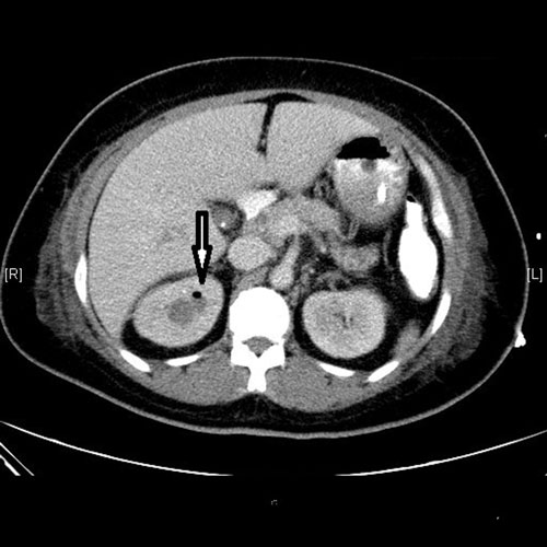
Imaging studies are not performed to confirm the diagnosis of acute pyelonephritis. When other features suggest an alternative diagnosis and contrast-enhanced CT is performed, an infected kidney is usually enlarged with decreased opacification of the affected parenchyma, typically in a wedge-shaped or linear pattern.
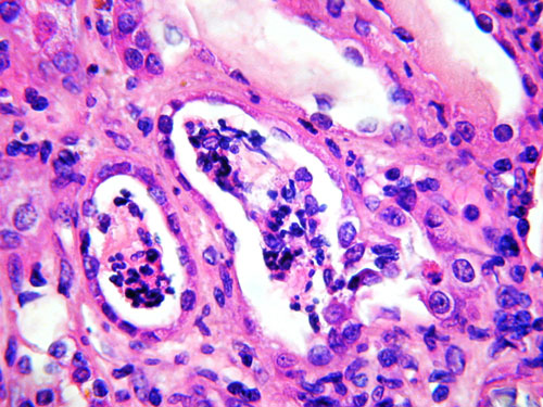
Renal biopsy should never be performed to confirm a diagnosis of pyelonephritis, but may be necessary, particularly in a kidney transplant, to exclude other causes of pain, fever and graft dysfunction (such as acute rejection). Typical pathological features are a focal inflammatory reaction with neutrophil and monocyte infiltrates, tubular damage and interstitial oedema.
Potential complications
With prompt and appropriate antimicrobial treatment acute pyelonephritis can be self-limiting with complete resolution. However, there can be serious sequelae.
Early complications
• Life-threatening septicaemia and multi-organ failure
• Abscess formation (cortical, medullary, perinephric)
• Papillary necrosis
• Emphysematous pyelonephritis
(if gas-forming Enterobacteriaceae are involved, see figure 1)
The following groups of patients are at increased risk of clinical deterioration and warrant closer attention:2,3
• Immunocompromised patients
o transplant recipients, HIV-positive patients
o those treated with chemotherapy or some biologic therapies
• Anatomical urinary tract variation
o polycystic kidneys
o urinary diversion (ileal conduit,
neo-bladder)
o vesicoureteric reflux
• Functional urinary tract problems
o neurogenic bladder
• Instrumentation or foreign material within the urinary tract
o urethral catheterisation (permanent or intermittent self-catheterisation)
o ureteric stent
o nephrostomy
• Pregnancy
• Diabetes mellitus
Children less than three months of age should be referred immediately to secondary care; those aged three months or over should be referred where there are features of concern (in accordance with the NICE guideline on urinary tract infection in children4), otherwise antibiotic treatment should be initiated in the community and referred if there is no clinical improvement after 24-48 hours.
Late complications
• Cortical scarring which can act as a potent stimulant for systemic hypertension
• Chronic kidney disease and progression to end stage renal disease
• Xanthogranulomatous pyelonephritis (a poorly understood condition in which the renal parenchyma is replaced with cellular infiltrate of lipid-laden macrophages)
Chronic pyelonephritis
These complications more typically characterise recurrent episodes of acute pyelonephritis. In the UK, 5.9% of people starting renal replacement therapy do so because of chronic pyelonephritis.5
The significant scarring in chronic pyelonephritis typically occurs as a result of recurrent episodes of acute pyelonephritis in infancy or childhood, although self-supporting renal function can be maintained until adulthood.
Frequently there is an incidental finding of CKD when laboratory tests are performed for an unrelated presentation, with bilaterally scarred kidneys on ultrasound examination being diagnostic. There may be a childhood history of stomach pains or ‘a chill in the kidneys’ sufficient to require time off school. With reduced nephron numbers and subsequent hyperfiltration in remaining glomeruli progression to end stage renal disease can occur at any age (depending on the severity of the initial insult, and presence or absence of additional causes of hyperfiltration such as obesity).
Rate of decline in renal function can be reduced by good blood pressure control, minimisation of proteinuria (blockade of renin-angiotensin axis), and avoidance of, or reduction in, obesity.
If the scarring is unilateral only, then lifelong self-supporting renal function can be anticipated, provided there is care to avoid further renal insults e.g. with hypertension, obesity etc.
Acute pyelonephritis
An algorithm to guide management is shown in figure 2. After a clinical diagnosis of acute pyelonephritis is made, ideally (but not invariably) supported by positive findings on urinalysis, the first step is to decide if community or hospital treatment is most appropriate.
Patients warranting hospital admission are those already showing signs of sepsis,6 and those at increased risk of developing complications (detailed above). Pregnant women7 and patients who are immunocompromised are particularly at risk. If not referred to secondary care they require early review and a very low threshold for hospitalisation if not improving.
In primary care management of acute pyelonephritis, urinalysis and submitting urine for culture at the time of presentation are essential. Antibiotic therapy should be initiated without delay; this can be modified subsequently depending on the culture result.
The Health Protection Agency (HPA) and the British Infection Association (BIA) joint 2013 guideline8 recommends empirical treatment for women who are not pregnant and men (including those with indwelling catheters):
• Ciprofloxacin 500 mg, twice daily for 7 to 10 days
or
• Co-amoxiclav 500/125 mg three times daily for 14 days
The HPA and BIA guideline is based on the need to cover the most common causative pathogens associated with pyelonephritis and have good renal tissue penetration. Notably antibiotics that are typically effective in lower UTIs are frequently inadequate in acute pyelonephritis, and more prolonged therapy is necessary. If there have been previous episodes then review of past urinary samples is useful to ascertain whether previous pathogens have been identified; for example patients who are known to have a beta-lactamase-inhibiting organism (ESBL) should receive an aminoglycoside or carbapenem antimicrobial.
Pregnant women may warrant hospital admission for observation because of the increased risk of premature labour. However, if hospital admission is not required, then treatment must be with an antibacterial agent which has a safe profile in pregnancy, cephalexin 500 mg twice daily for 10-14 days is advised.8
Cyst infections can be difficult to manage in patients with polycystic kidneys. A causative organism may not be cultured from urine samples because the cysts usually do not communicate with the renal tract. Antibiotics may not penetrate the larger cysts, lipid-soluble antibiotics such as ciprofloxacin are preferred as they can diffuse into cyst fluid more readily than penicillins and prolonged treatment is usually necessary to eradicate infection.9,10
Additional advice to patients is to take regular paracetamol to manage symptoms and ensure adequate hydration until resolution. Potential nephrotoxic agents (ACE inhibitors, angiotensin receptor blockers, non-steroidal medications) should be discontinued until symptoms resolve and the patient is eating and drinking normally.
For those being managed in primary care it is essential that culture results are reviewed after 48 hours to confirm the causative pathogen is sensitive to the prescribed antibiotic. Particularly in patients with risk factors for complications it is prudent to confirm there is no clinical deterioration that would merit hospitalisation 24-48 hours after commencing treatment.
Referral
In the acute setting, patients who have not improved within two days of commencing antimicrobial treatment should be referred to secondary care. An exception is those cases in which the infecting pathogen is not susceptible to the agent originally used, an alternative appropriate antibiotic is available, and the patient clinically remains well enough for community care.3
Patients who fully recover from a single episode of acute pyelonephritis do not need referral to secondary care. Women with recurrent pyelonephritis and men with a single episode without obvious cause should be referred to urology for a specialist opinion and investigation of possible urological abnormalities.
REFERENCES
1 Nicolle LE. A practical guide to the management of complicated urinary tract infection. Drugs 1997;53:583-92
2 Efstathiou SP, Pefanis AV, Tsioulos DI et al. Acute pyelonephritis in adults: prediction of mortality and failure of treatment. Arch Intern Med 2003;163(10):1206-12
3 Grabe M, Bartoletti R, Bjerklund-Johansen TE et al. Guidelines on urological infections. European Association of Urology 2015. www.uroweb.org/wp-content/uploads/ 19-Urological-infections_LR2.pdf Accessed 06.02.2017
4 National Institute for Health and Care Excellence. CG54. UTI in children. Urinary tract infection in children: diagnosis, treatment and long-term management. NICE. London. 2007. www.nice.org.uk/guidance/cg54 Accessed 06.02.2017
5 UK Renal Registry. The 18th Annual report. Nephron 2016;132 (suppl 1):9-40 www.renalreg.org/wp-content/ uploads/2015/01/01-Chap-01.pdf Accessed 06.02.2017
6 Neumann I, Moore P. Pyelonephritis (acute) in non-pregnant women. BMJ Clin Evid 2011:2011;0807
7 Ramakrishnan K, Scheid DC. Diagnosis and management of acute pyelonephritis in adults. Am Fam Physician 2005; 71(5):933-42
8 HPA and British Infection Association. Management of infection guidance for primary care for consultation and local adaption. pp39-40. Health Protection Agency. 2013
https://www.gov.uk/government/uploads/system/uploads/attachment_data/file/586766/managing_common_infections.pdf Accessed 06.02.2017
9 Schwab SJ, Bander SJ, Klahr S. Renal infection in autosomal dominant polycystic kidney disease. Am J Med 1987;82:714-18
10 Sklar AH, Caruana RJ, Lammers JE, Strauser GD. Renal infections in autosomal dominant polycystic kidney disease. Am J Kidney Dis 1987;10:81-88
ARTICLE IN PDF
 Download this article in PDF format
PRESET SEARCHES
PubMed
NICE evidence search
Immediate CPD prompts
These handy templates*, associated with all articles, prompt drafting of personal learning, reflection and planning.
Save your record straight into your personal development folder on your device or cloud storage system.
*Adobe fillable PDFs. Adobe reader needed for iPad
Comment or question
If you, or your peer group, would like to comment on an article or have a question for the authors, write to:
Editor
Case report submissions
Do you have an interesting case that you would like to share with other readers?
Your article would need to be around 1,500 words long, include a brief review of the literature to put the case in context, and be fully referenced in the Vancouver style.
Submissions for consideration for publication should be sent to: [email protected]
EXTERNAL WEBLINKS
NICE
European Association of Urology