SEARCH

Lewis G, Maxwell AP. Tracking down the cause of proteinuria in primary care. Practitioner 2013;257 (1758):19-22
Tracking down the cause of proteinuria in primary care
21 Feb 2013
AUTHORS
Dr Gareth Lewis MB BCh MRCP PhD Specialist Registrar in Nephrology
Professor Alexander P. Maxwell MD PhD FRCP Consultant Nephrologist
Regional Nephrology Unit, Belfast City Hospital, Belfast, UK
Article
The finding of proteinuria on dipstick urinalysis poses a number of diagnostic questions.
Proteinuria is relatively common in the general population with reported point prevalence of up to 8% 1 but the prevalence falls to around 2% on repeated testing. 2,3
Common causes
Proteinuria originates from the kidney and occurs as a result of injury to either the glomerulus or the renal tubule or both. The molecular mechanisms causing proteinuria will vary depending on whether primarily glomerular or tubular pathology is present.4
Chronic glomerular injury resulting in proteinuria may be secondary to prolonged duration of diabetes or hypertension. Autoimmune disorders, such as glomerulonephritis, can result in proteinuric states through subtle derangements in the molecular signalling pathways and structural scaffolding of glomerular epithelial (podocyte) cells and their adjacent glomerular basement membrane that provide the final barrier to glomerular filtration.5
In contrast, a tubular origin of proteinuria may be associated with inflammation of renal tubules triggered by prescribed drugs or ingested toxins. There is substantial variation in both the natural history and the response to therapies in the different types of proteinuric kidney disease.
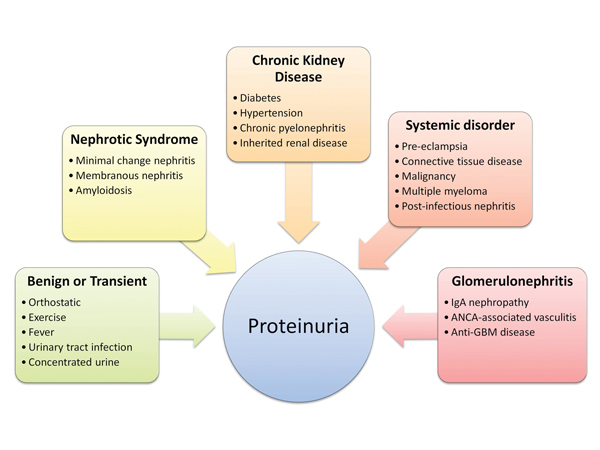
Proteinuria may be a clinical feature in a variety of conditions encountered in general practice (see figure 1). NICE guidelines recommend screening for proteinuria in individuals at higher risk for chronic kidney disease (CKD).6 These include patients with diabetes, hypertension, cardiovascular disease, connective tissue disorders, a family history of renal disease and those prescribed potentially nephrotoxic drugs. Screening for CKD should also include tests of renal function such as serum creatinine and estimated glomerular filtration rate (eGFR).
Proteinuria is usually asymptomatic unless a multisystem disorder, e.g. vasculitis or systemic lupus erythematosus, is present or there are very significant urinary protein losses causing frothy urine and associated peripheral oedema. In the absence of obvious clues to the cause of persistent proteinuria on history or clinical examination it is worthwhile reviewing the patient’s prescribed drugs to identify any potentially nephrotoxic agents e.g. NSAIDs.
Annual screening for the development of microalbuminuria in patients with diabetes is now well established.7 Microalbuminuria, the earliest clinically detectable sign of diabetic nephropathy, represents low level albuminuria and is not detected by dipstick urinalysis but requires measurement by a sensitive quantitative assay, the urinary albumin:creatinine ratio (ACR).
Screening for urinary protein excretion is also routine practice in pregnancy; the onset of proteinuria is a feature of pre-eclampsia.8
Persistent proteinuria indicates there is underlying renal pathology but it may in itself be injurious to the kidney, with several mechanisms proposed to account for this.9
Patients with high, nephrotic-range, proteinuria (>3.5 g/day) have additional risks of hypercoagulability, thromboembolism, infection, malnutrition and acute kidney injury.
In CKD the amount of proteinuria tends to be directly correlated with a faster rate of decline of kidney function (more rapid fall in eGFR) as well as an associated substantially increased cardiovascular disease risk.10 These observations supply the evidence-based rationale for controlling levels of proteinuria.11
Investigations
Essential initial investigations are summarised in table 1, above. Patients with urine dipstick-positive proteinuria should have the remainder of their urinalysis checked for the presence of blood, or evidence of urinary tract infection (UTI). Menstruation and UTI can lead to transiently positive tests for protein and blood on dipstick urinalysis.12 Orthostatic proteinuria is present if early morning dipsticks are negative for protein but the dipsticks become positive for protein later in the day.
Dipstick urinalysis with a high specific gravity (>1.030) indicating a concentrated urine may lead to a false-positive protein result. Conversely, very dilute urine with a low specific gravity (a false-negative test for proteinuria.
The most useful investigation to quantify any proteinuria accurately is the urinary albumin:creatinine ratio (ACR) or a protein:creatinine ratio (PCR).13 Normal values are outlined in table 2, with a comparison between different methods of estimating proteinuria. Urinary ACR or PCR can be conveniently estimated on a single urine sample.14 ACRs are commonly used to screen for, and monitor the progress of, microalbuminuria in patients with diabetes.15
Blood pressure measurement and kidney function estimation are both important because of the association of proteinuria with hypertension and CKD. Blood glucose should be assayed to exclude diabetes. Patients with sudden onset of lower limb oedema and associated proteinuria should have a serum albumin level measured and if hypoalbuminaemia is present this indicates the patient has a nephrotic syndrome. Serum cholesterol levels are often substantially raised in those with nephrotic syndrome. Conversely, severe hypercholesterolaemia with even a trace of peripheral oedema should prompt the practitioner to perform dipstick urinalysis and send a urine sample for ACR.
A history of frank haematuria, joint pain, rash, ocular symptoms as well as weight loss, haemoptysis or sinusitis may suggest an underlying systemic disorder such as vasculitis. A drug history may reveal the use of NSAIDs, recent antibiotics or herbal and over the counter medications that can be associated with an interstitial nephritis. Clinical examination for evidence of rash, red eyes, xanthelasma, chest signs of fluid overload, pyrexia, anaemia or peripheral oedema may suggest a nephrotic or nephritic syndrome triggering an urgent nephrology referral. The presence of blood in the urine with onset or worsening of hypertension defines the nephritic syndrome which has many causes.
Renal tract ultrasound will measure kidney size, and detect scarring associated with chronic pyelonephritis or prior renal stone disease which can cause proteinuria.
Referral
Indications for urgent referral of patients with proteinuria are listed in table 3. All other referrals can be made routinely.
Patients with gross fluid overload from a nephrotic syndrome or in whom vasculitis or a serious underlying disorder is suspected should be discussed by telephone with a nephrologist to arrange early hospital assessment.
For all referrals it is helpful to have any documented serial urine dipstick or ACR measurements available in addition to the trend in serum creatinine levels. This can be of assistance in reassuring patients with a reduced likelihood of progressive CKD if a stable pattern of low level proteinuria has been demonstrated over a long period of time.
Management
For patients with persistent proteinuric renal disease the main therapeutic option is treatment with an ACE inhibitor or angiotensin receptor blocker (ARB). These agents also have an important role in the management of hypertension and cardiovascular disease, underlining the interconnection of these disorders.16 Thus, addressing components of overall cardiovascular risk is vital in the management of all proteinuric renal conditions. The two general areas in which GPs can take an active role are in managing proteinuria associated with CKD, including diabetes, and in the long-term follow-up of those with primary renal disorders resulting in nephrotic syndrome.
In the management of CKD, the aim is to up-titrate ACE inhibitor or ARB therapy to the maximum tolerated dose in order to reduce proteinuria to a urinary ACR> 70. Patients should be warned that they should stop these drugs temporarily in the presence of intercurrent illness leading to volume depletion, e.g. vomiting and diarrhoea, and to seek medical attention for blood pressure and renal function and electrolyte checks if not improving after 48 hours.
Slow up-titration of ACE inhibitor or ARB dosage every 2-4 weeks, checking blood pressure and renal function one week after any increase in dose, and aiming to have serum potassium concentration to no more than 25% above baseline is recommended by the Renal Association guidelines (see Useful information).
If an ACE inhibitor or ARB is being prescribed care should be taken with other medications such as NSAIDs, potassium-sparing diuretics and trimethoprim which can contribute to serious hyperkalaemia if not monitored carefully.
Control of blood pressure to recommended targets (70) and reduction of proteinuria have beneficial effects on both renal and cardiovascular disease progression.17,18
Many GPs will have patients with diagnosed nephrotic syndrome who are also being followed up in nephrology clinics. After a renal biopsy has demonstrated the specific cause of the nephrotic syndrome, many patients have initial fortnightly or monthly outpatient appointments with nephrologists to manage diuretic therapy and fluid overload.
Treatment with ACE inhibitor and ARB therapy as described above may be prescribed, while others may be given steroids, immunosuppressive treatment with agents such as tacrolimus or ciclosporin, or cytotoxic drugs such as cyclophosphamide. Statin therapy for hypercholesterolaemia, as well as gastric and bone protection with regular monitoring for hyperglycaemia for those on steroids is also required. Patients are encouraged to weigh themselves and adjust diuretic dosing to manage oedema.
All this requires coordinated management between primary and secondary care.
Macrolide antibiotics and some antifungal drugs can increase tacrolimus and ciclosporin levels so interactions with any newly prescribed medications should be checked. Those on cyclophosphamide require regular monitoring of white cell counts and may experience hair loss, nausea, altered taste and infertility. For all immunosuppressive and cytotoxic agents, patients are at relatively increased risk of infection, neutropenic sepsis, and malignancy with prolonged treatment. Thus higher doses and prolonged courses of antibiotics may be required for common infections, with immediate referral to the emergency department if neutropenia is confirmed or even suspected in the context of sepsis.
Patients should also be encouraged to use sun protection and participate in age-specific cancer screening programmes.
Conclusion
Proteinuria and the underlying pathologies causing it often present in primary care.
A subset of patients will have a primary renal diagnosis for which specific treatment is available. Accurate quantification of proteinuria by sending a spot sample of urine for ACR or PCR measurement is the most important test in addition to blood pressure and serum creatinine measurement.
Detecting the presence of a nephrotic or nephritic syndrome can be made using a small number of investigations and can direct timely referral to a nephrologist, while partnership with those managing chronic proteinuric conditions is paramount in reducing the risk of progressive cardiovascular and renal disease.
REFERENCES
1 de Lima AO, Kesrouani S, Gomes RA et al. Population screening for chronic kidney disease: a survey involving 38,721 Brazilians. Nephrol Dial Transplant 2012;27 Suppl 3:iii135-8
2 Garg AX, Kiberd BA, Clark WF et al. Albuminuria and renal insufficiency prevalence guides population screening: results from the NHANES III. Kidney Int 2002;61(6):2165-75
3 Atkins RC, Briganti EM, Zimmet PZ, Chadban SJ. Association between albuminuria and proteinuria in the general population: the AusDiab Study. Nephrol Dial Transplant 2003;18(10):2170-4
4 Garg P, Rabelink T. Glomerular proteinuria: A complex interplay between unique players. Adv Chronic Kidney Dis 2011;18(4):233-242
5 Mathieson PW. The podocyte cytoskeleton in health and disease. Clin Kidney J 2012;5:498-501
6 National Institute for Health and Clinical Excellence. CG73. Chronic kidney disease: Early identification and management of chronic kidney disease in adults in primary and secondary care. NICE. London. 2008
7 Karalliedde J, Viberti G. Proteinuria in diabetes: bystander or pathway to cardiorenal disease? J Am Soc Nephrol 2010;21:2020-27
8 Morris RK, Riley RD, Doug M et al. Diagnostic accuracy of spot urinary protein and albumin to creatinine ratios for detection of significant proteinuria or adverse pregnancy outcome in patients with suspected pre-eclampsia: systematic review and meta-analysis. BMJ 2012;345:e4342
9 Abbate M, Zoja C, Remuzzi G. How does proteinuria cause progressive renal damage? J Am Soc Nephrol 2006;17:2974-84
10 Rosansky SJ. Renal function trajectory is more important than chronic kidney disease stage for managing patients with chronic kidney disease. Am J Nephrol 2012;36:1-10
11 Perkovic V, Verdon C, Ninomiya T et al. The relationship between proteinuria and coronary risk: A systematic review and meta-analysis. PLoS Med 2008;5:1486-1495
12 Carter JL, Tomson CRV, Stevens PE, Lamb EJ. Does urinary tract infection cause proteinuria or microalbuminuria? A systematic review. Nephrol Dial Transplant 2006;21:3031-3037
13 Johnson DW, Jones GRD, Mathew TH et al. Chronic kidney disease and measurement of albuminuria or proteinuria: a position statement. Med J Aus 2012;197(4):1-6
14 Price CP, Newall RG, Boyd JC. Use of Protein: Creatinine ratio measurements on random urine samples for prediction of significant proteinuria: A systematic review. Clin Chem 2005;51(9):1577-1586
15 Johnson DW. Global proteinuria guidelines: are we nearly there yet? Clin Biochem Rev 2011;32:89-95
16 Linas SL. Are two better than one? Angiotensin-converting enzyme inhibitors plus angiotensin receptor blockers for reducing blood pressure and proteinuria in kidney disease. Clin J Am Soc Nephrol 2008;3:S17-S23
17 Jafar TH, Stark PC, Schmid CH et al. Progression of chronic kidney disease: The role of blood pressure control, proteinuria, and angiotensin-converting enzyme inhibition. Ann Int Med 2003;139:244-252
18 Bakris GL. Slowing nephropathy progression: Focus on proteinuria reduction. Clin J Am Soc Nephrol 2008;3:S3-S10
ARTICLE IN PDF
 Download this article in PDF format
PRESET SEARCHES
Immediate CPD prompts
These handy templates* prompt immediate drafting of personal learning, reflection and planning. Save your record straight into your personal development folder on your device or Cloud storage system.
*Adobe reader needed for iPad
EXTERNAL WEBLINKS
The Renal Association
Compilation of guidelines from NICE, SIGN and the Renal Association.
Links to proteinuria, hypertension, and referral indicators are accessible from the Pages of the eGuide box.
There are also links to GP and patient information leaflets on CKD.
www.renal.org/whatwedo/InformationResources/CKDeGUIDE.aspx
Edinburgh Renal Unit
Practical information for GPs
For patients
www.edren.org/pages/edreninfo/proteinuria.php
NICE Guidance on CKD