SEARCH

Todd P, Hill C. Suspected nephrotic syndrome warrants urgent referral. Practitioner January 2022;266(1854):17-20
Suspected nephrotic syndrome warrants urgent referral
24 Jan 2022
AUTHORS
Dr Peter Todd MBChB MSc MRCP, Specialist Registrar in Nephrology
Dr Christopher Hill MD FRCP Consultant Nephrologist, Regional Nephrology Unit, Belfast, UK
Competing interests: None
Article
Nephrotic syndrome results from dysfunctional glomerular filtration and consists of three clinical features: severe proteinuria (> 3.5 grams/24 hours); hypoalbuminaemia (serum albumin < 30 g/L); and peripheral oedema. It may occur in people of all ages. Patients with nephrotic syndrome may present with a normal eGFR. Others may have chronic renal impairment at presentation. If the nephrotic syndrome remains uncontrolled even those with normal kidney function at presentation can develop progressive chronic kidney disease. Acute kidney injury may occasionally be seen in association with nephrotic syndrome. Serum concentrations of cholesterol are raised in many patients with nephrotic syndrome. Nephrotic syndrome increases the risk of venous thromboembolism which can sometimes be the presenting symptom. Patients with nephrotic syndrome are at increased risk of bacterial infections because of urinary loss of immunoglobulins. Primary causes of nephrotic syndrome include membranous nephropathy, minimal change disease and focal segmental glomerulosclerosis. In children, minimal change disease is the most common cause. Secondary causes relate to glomerular dysfunction secondary to a systemic disease such as diabetes mellitus, systemic lupus erythematosus or amyloidosis. Initial investigations focus on confirming diagnosis and finding an underlying cause. A renal biopsy is indicated in almost all cases. This will confirm the underlying diagnosis, guide management and provide prognostic information. All patients with suspected nephrotic syndrome should be referred urgently to nephrology.
Nephrotic syndrome is a constellation of clinical features that result from dysfunctional glomerular filtration. It is not a specific disease itself but is the mode of presentation for several diseases that affect the glomerulus.
Nephrotic syndrome may occur in people of all ages, with an overall annual incidence of approximately 2-3 per 100,000.1,2
It is a rare presentation in primary care, but one which may lead to significant morbidity if untreated.
Diagnostic features
The three features required for diagnosis of nephrotic syndrome are:
• Severe proteinuria: > 3.5 grams/24 hours and
• Hypoalbuminaemia: serum albumin < 30 g/L and
• Peripheral oedema
Proteinuria
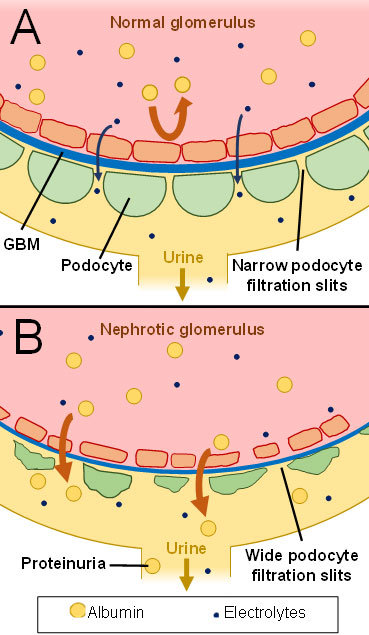
Severe proteinuria is the primary mechanism by which nephrotic syndrome arises. The glomerular filtration barrier, which includes the glomerular basement membrane (GBM) and epithelial podocytes, becomes dysfunctional.
This leads to pathological filtration of significant amounts of albumin, which is then excreted in the urine (see figure 1). Many patients will report noticing that their urine appears cloudy or frothy.
A urine dipstick to investigate for proteinuria should be performed on any patient with new onset peripheral oedema. Proteinuria should then be confirmed with a laboratory measurement; either an albumin: creatinine ratio (ACR) or a protein: creatinine ratio (PCR), depending on local protocols, see table 1.
In most cases, 24-hour urine protein collections are not required for the diagnosis of nephrotic syndrome, however table 1 gives approximate equivalent values for spot urine samples.
Hypoalbuminaemia
Hypoalbuminaemia occurs through profound loss of albumin in the urine. Although hepatic albumin synthesis is often upregulated, this does not fully compensate for urinary albumin loss in nephrotic syndrome. Other protein synthesis, including that of cholesterol binding proteins, may also be upregulated.
Peripheral oedema
The underlying mechanism behind peripheral oedema in nephrotic syndrome is the subject of ongoing debate.
One hypothesis is that development of hypoalbuminaemia reduces plasma oncotic pressure. This leads to net movement of fluid from the intravascular space to the interstitial fluid causing oedema and reducing intravascular volume. This volume reduction subsequently leads to increased tubular reabsorption of sodium and water in the kidneys, compounding the development of oedema.5
Another hypothesis suggests that high levels of protein in the urine are the primary cause of sodium and water reabsorption in the kidneys. This leads to increased plasma volume which increases intravascular hydrostatic pressure, ultimately causing fluid movement into the interstitial compartment, creating oedema.6
The distribution of oedema in nephrotic syndrome may differ from that typically seen in heart failure. Periorbital, facial or genital oedema are commonly reported. Pleural effusions and ascites can occur but pulmonary oedema is less common.7
Oedema will progressively worsen while the proteinuria remains uncontrolled.
Associated features
Hyperlipidaemia
Serum concentrations of cholesterol are raised in many patients with nephrotic syndrome.8
Hypercoagulability
Nephrotic syndrome raises the risk of venous thromboembolism (VTE).9
VTE can sometimes be the presenting symptom. The mechanism is believed to involve abnormal urinary loss of antithrombotic factors.
Susceptibility to infection
Patients with nephrotic syndrome are at increased risk of bacterial infections because of urinary loss of immunoglobulins. There is no evidence to support use of prophylactic antibiotics, but infections should be treated aggressively.10
Renal impairment
Some patients with nephrotic syndrome may present with a normal eGFR. Others may have chronic renal impairment at presentation. If nephrotic syndrome remains uncontrolled even those with normal kidney function at presentation can develop progressive chronic kidney disease. Acute kidney injury (AKI) may occasionally be seen in association with nephrotic syndrome.
Causes
Primary causes
Glomerular diseases that often have immune-mediated underlying mechanisms are a primary cause of nephrotic syndrome. Some of the most common glomerular diseases in adults are membranous nephropathy, minimal change disease and focal segmental glomerulosclerosis (FSGS). In children, minimal change disease is the most common cause of nephrotic syndrome.
Secondary causes
In some cases of nephrotic syndrome glomerular dysfunction is secondary to a systemic disease such as diabetes mellitus, systemic lupus erythematosus (SLE) or amyloidosis.
Investigations
In nephrotic syndrome, initial investigations focus on confirming diagnosis and finding an underlying cause, see table 2.
All patients with suspected nephrotic syndrome should be referred urgently to nephrology.
A renal biopsy is indicated in almost all cases of nephrotic syndrome. This will confirm the underlying diagnosis, guide management and give prognostic information.
Management
Management of nephrotic syndrome involves general management principles and specific management of the underlying disease.
General management
Diuretics
Early initiation of loop diuretics is vital in the management of oedema and may prevent development of life-threatening complications, such as skin breakdown and pleural effusions.
Regular monitoring of renal function is advisable when commencing or titrating up diuretics to monitor for electrolyte disturbances or AKI.
Patients with nephrotic syndrome may be relatively resistant to diuretics, so response to treatment should be closely monitored and the dosage increased if required.
Patients should check their weight on a daily basis aiming for a target weight loss of 0.5 to 1 kg per day until their oedema has resolved.11
Lack of improvement may necessitate hospital admission for intravenous diuretics.
Dietary salt and fluid restriction
Dietary sodium intake should be restricted to < 2 g sodium (< 5 g salt) per day. Fluid restriction may also be necessary if patients have very severe fluid overload.
Lipid-lowering treatment
Although, high quality trials involving lipid-lowering treatment in nephrotic syndrome are lacking, guidance suggests considering statin therapy for patients with additional cardiovascular risk factors.11
ACE inhibitors/angiotensin receptor blockers
ACE inhibitors (ACEi) or angiotensin receptor blockers (ARB) may reduce the degree of proteinuria and manage associated hypertension. If a patient is already taking these agents, nephrotic syndrome is not an indication to stop them if their eGFR is stable. Discussion with the nephrology service is advised before initiating treatment with ACEi or ARB as there may be a slightly increased risk of AKI, particularly in the setting of high diuretic requirements. For patients established on ACEi or ARB therapy in nephrotic syndrome, sick day rules apply as in other settings.
Anticoagulation
The risk of thromboembolic disease is increased in nephrotic syndrome. Any patients presenting with symptoms suggestive of VTE should be assessed urgently. Some patients with very low serum albumin and heavy proteinuria at presentation may be prophylactically anticoagulated but this will be decided by the nephrology team.11
Warfarin and low molecular weight heparin are the recommended anticoagulant agents in nephrotic syndrome. Direct oral anticoagulants (DOACs) are not currently recommended as there is no clinical trial evidence yet to support their use. DOACs are partially albumin-bound and the impact of urinary excretion on their efficacy in nephrotic syndrome is unclear.
Disease-specific management
Management of primary glomerular diseases often involves immunosuppressive agents, such as corticosteroids, mycophenolate mofetil, calcineurin inhibitors (tacrolimus or cyclosporin), cyclophosphamide or rituximab. These therapies will be initiated and monitored by the nephrology team, usually following a renal biopsy result.
In cases where nephrotic syndrome is secondary to a systemic disease, such as diabetes mellitus, treatment involves a combination of the general principles outlined above and disease-specific management.
Follow-up
Patients with nephrotic syndrome will be followed up closely by the nephrology service. Patients on long-term immunosuppression for a primary cause of nephrotic syndrome should also remain under regular review by nephrology. However, GPs may be asked to assist in blood monitoring or medication adjustments.
Patients with resolved or inactive disease may be discharged from nephrology follow-up, but many will remain under long-term review if they continue to be at risk of relapse or progressive CKD.
Conclusion
Nephrotic syndrome should be considered in all patients with new oedema and a dipstick urinalysis should be performed. It is characterised by severe proteinuria, hypoalbuminaemia and oedema.
Suspected cases of nephrotic syndrome should be referred urgently to the nephrology service.
REFERENCES
1 Hull RP, Goldsmith DJ. Nephrotic syndrome in adults. BMJ 2008;336(7654):11858-59
2 Banh TH, Hussain-Shamsy N, Patel V et al. Ethnic differences in incidence and outcomes of childhood nephrotic syndrome. Clin J Am Soc Nephrol 2016;11(10):1760-68
3 National Institute for Health and Care Excellence. NG203. Chronic kidney disease: assessment and management. NICE. London. 2021[Last accessed 22 Dec 2021]
4 UK Kidney Association. Proteinuria. [Last accessed 22 Dec 2021]
5 Gupta S, Pepper RJ, Ashman N, Walsh SB. Nephrotic syndrome: oedema formation and its treatment with diuretics. Front Physiol 2019;9:1868
6 Siddall EC, Radhakrishnan J. The pathophysiology of edema formation in the nephrotic syndrome. Kidney Int 2012;82(6):635-42
7 Koomans HA. Pathophysiology of oedema in idiopathic nephrotic syndrome. Nephrol Dial Transplant 2003;18(Supp6):vi30-vi32
8 Agrawal S, Zaritsky JJ, Fornoni A, Smoyer WE. Dyslipidaemia in nephrotic syndrome: mechanisms and treatment. Nat Rev Nephrol 2018;14(1):57-70
9 Kerlin BA, Ayoob R, Smoyer WE. Epidemiology and pathophysiology of nephrotic syndrome-associated thromboembolic disease. Clin J Am Soc Nephrol 2012;7(3):513-20
10 Wu HM, Tang JL, Cao L et al. Interventions for preventing infection in nephrotic syndrome. Cochrane Database Syst Rev 2012;2012(4):CD003964
11 Kidney Disease: Improving Global Outcomes (KDIGO) Glomerular Diseases Work Group. KDIGO 2021 clinical practice guideline for the management of glomerular diseases. Kidney Int 2021;100(4S):S1–S276
ARTICLE IN PDF
 Download this article in PDF format
EXTERNAL WEBLINKS
For patients
For patients/healthcare professionals