SEARCH

Fergie RJC, McCaughan JA. Prompt recognition key in glomerulonephritis. Practitioner Feb 2020;264(1834):17-20
Prompt recognition key in glomerulonephritis
24 Feb 2020
AUTHORS
Dr Ruth CJ Fergie MBChB MRCP, Specialty Registrar in Nephrology
Dr Jennifer A McCaughan PhD MRCP FRCPath, Consultant Nephrologist
Regional Nephrology Unit, Belfast City Hospital, Belfast, UK
Competing interests: None
Article
Abstract
Glomerulonephritis is the second most common cause of end stage renal disease (ESRD) in the UK and the most common cause in those under 35. Some forms can respond well to treatment if diagnosed early. However, if glomerulonephritis goes unrecognised, it can lead to irreversible loss of renal function. The most common type in the UK is IgA nephropathy, which may be associated with chronic liver disease. Other types of glomerulonephritis are familial and most are more common in older patients. Some forms, particularly membranous glomerulonephritis, can occur secondary to an underlying malignancy. Glomerulonephritis can present in a wide variety of ways, ranging from asymptomatic urinary abnormalities to fulminant disease with life-threatening acute kidney injury and extra-renal manifestations. Asymptomatic invisible haematuria is common. In the absence of infection or a urological cause, glomerulonephritis should be considered. Proteinuria is almost always glomerular in origin and is a strong predictor of progressive kidney disease. Significant proteinuria (> 1 g/day) in the absence of an identifiable cause warrants referral. Nephrotic syndrome is defined by: oedema, nephrotic range proteinuria (> 3.5 g/24 hr) and hypoalbuminaemia (< 30 g/L). Urgent referral to a nephrologist is indicated.
Registry data indicate that glomerulonephritis is the second most common cause of end stage renal disease (ESRD) in the UK.1 Around 14% of patients with ESRD have glomerulonephritis.1 In those under 35, it is the most common cause of ESRD, accounting for 27% of cases.1
Some forms of glomerulonephritis can respond well to treatment if diagnosed early. However, if it goes unrecognised glomerulonephritis can lead to irreversible loss of renal function.
Glomerulonephritis can present in a wide variety of ways, ranging from asymptomatic urinary abnormalities to fulminant disease with life-threatening acute kidney injury and extra-renal manifestations.
Asymptomatic urinary abnormalities
Invisible haematuria
Haematuria and proteinuria on urine dipstick testing is the classical finding in glomerulonephritis, but invisible (microscopic) haematuria may be the first urinary sign. This may be picked up incidentally at a routine medical visit, e.g. an early pregnancy assessment. Invisible haematuria is defined as more than + blood on more than one urine sample.
Asymptomatic invisible haematuria is common. Urinary tract infection and other urological conditions should be considered before evaluation for glomerular disease.
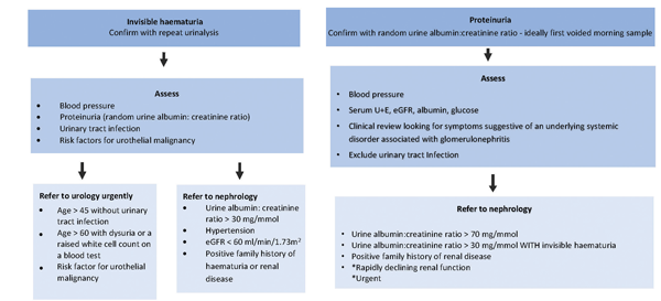
Urgent urology referral is recommended for patients aged ≥ 60 with unexplained invisible haematuria and dysuria or an elevated white cell count, via a suspected cancer referral pathway.2
In the absence of infection or a urological cause, glomerulonephritis should be considered, see figure 1. Invisible haematuria is common in patients with IgA nephropathy.
Proteinuria
The hallmark of glomerular disease is the excretion of protein into the urine through failure of the glomerular ‘filter’. Proteinuria is almost always glomerular in origin and is a powerful predictor of progressive kidney disease.
Significant proteinuria (> 1 g/day,urine albumin: creatinine ratio [ACR] >70 mg/mmol) in the absence of an identifiable cause, or an ACR > 30 mg/mmol in the presence of invisible haematuria should be further investigated, see figure 1.
Visible haematuria
Visible haematuria is usually caused by non-glomerular pathology e.g. urothelial malignancy or renal calculi. However, it can be glomerular in nature. Episodic painless visible haematuria associated with glomerular disease is often brown or ‘smoky’, and clots are unusual. It is mainly seen in children and young adults and may represent a post-infectious glomerulonephritis.
Patients with IgA nephropathy may develop visible haematuria in the context of intercurrent illness.
Nephrotic syndrome
Nephrotic syndrome is defined by a triad of features: oedema, nephrotic range proteinuria (> 3.5 g/24 hr) and hypoalbuminaemia (< 30 g/L). It is pathognomonic of glomerular disease. Urgent referral to a nephrologist is indicated. In severe cases, patients may develop significant fluid overload with pleural effusions and ascites. Nephrotic syndrome is also associated with a plethora of metabolic complications which require specific management, see table 1.
Extra-renal manifestations
Glomerulonephritis may form part of a multisystem disease where blood vessels in many organs are inflamed (vasculitis). In these cases, there may be extra-renal manifestations, see table 2. If there is suspicion of a multisystem vasculitis, the presence of blood and protein on urinalysis can confirm renal involvement.
Renal failure
Presentation with renal failure occurs in two situations:
• Individuals who have had progressive glomerulonephritis over many years and have never sought medical attention.
This is most commonly observed in young people with IgA nephropathy. These patients may present with established hypertension, proteinuria and renal impairment.
• Very aggressive and rapidly progressive glomerulonephritis.
These patients present with severe hypertension and uraemic features.
Causes and risk factors
There is a wide spectrum of both causes and risk factors for glomerulonephritis, see table 3. The most common type of glomerulonephritis in the UK is IgA nephropathy, which may be associated with chronic liver disease.
Other types of glomerulonephritis are familial and most are more common in older patients. Some types of glomerulonephritis, particularly membranous glomerulonephritis, can occur secondary to an underlying malignancy.
The definitive investigation for glomerulonephritis is kidney biopsy. History, physical examination, urinalysis and autoantibody testing are also helpful in the decision making.
Early referral to nephrology for evaluation is important, as prompt diagnosis and timely treatment may slow, or even prevent, development of chronic kidney disease (CKD).
Management
Treatment of glomerulonephritis can be classified into treatment to prevent progression and disease specific therapies.
Preventing CKD progression
Optimisation of blood pressure and reduction in proteinuria are vital in the management of glomerulonephritis. Primary care has a key role in this.
Hypertension is universal as chronic glomerulonephritis progresses and is the key modifiable risk factor in preserving renal function. The most recent KDIGO guideline recommends a BP target below 130/80 mmHg in proteinuric patients (ACR > 3 mg/mmol), but the SPRINT trial and other studies suggest that a systolic blood pressure < 120 mmHg may be more effective at reducing disease progression.3,4
ACE inhibitors and angiotensin receptor blockers (ARBs) should be the first-choice therapy because of their effect on both blood pressure and proteinuria. It is vital that these drugs are uptitrated as far as tolerated.
Lifestyle modification (salt restriction, weight normalisation, regular exercise and smoking cessation) should be an integral part of management.
Proteinuria is the second key modifiable risk factor to preserve glomerular filtration rate in patients with glomerulonephritis. In patients who are normotensive, ACE inhibitors or ARBs should still be initiated as their renoprotective effects are not confined to blood pressure control. They should be increased slowly in order to minimise symptomatic hypotension.
Common side effects of ACE inhibitors and ARBs include hyperkalaemia and a rise in creatinine. It is often possible to manage the hyperkalaemia with diet or the addition of a loop diuretic.
Guidelines suggest that a non-progressive rise in creatinine of up to 30% is acceptable after starting or increasing the dose of an ACE inhibitor.5 GPs should monitor urea and electrolytes and ensure uptitration of these drugs.
At times of high risk for acute kidney injury, e.g. acute volume depletion from vomiting and diarrhoea, ACE inhibitors and ARBs should be witheld until symptoms have resolved. It is important that these agents are resumed when the patient is well. Patients should be educated regarding these ‘sick day rules’ when initiating therapy, see box 1.
Cardiovascular risk factors should be addressed in patients with glomerulonephritis and managed according to national guidelines.
These individuals are at significant risk of major cardiovascular events because of their renal impairment and hypertension.
Disease specific therapies
A variety of immunosuppressive regimens are used for the treatment of glomerulonephritis, particularly in those who present with primary glomerular pathology causing nephrotic syndrome, or a multisystem disease, see table 4, below. These are initiated by a nephrologist or rheumatologist. There is usually high intensity induction therapy which is delivered in hospital followed by long-term maintenance therapy.
Shared care guidelines enabling prescribing and any associated monitoring to be shared between the specialist and primary care are invaluable. Primary care has an important role in prescribing as specified by the specialist, detecting and reporting adverse drug reactions to the initiating specialist and the usual bodies (e.g. MHRA) and identifying and reporting any suspected non-compliance to the specialist.5
Disease specific therapies are often immunosuppressant therefore it is important to have a low threshold of suspicion for infection if a patient presents acutely.
Monitoring and follow-up
Long-term follow-up for patients is essential. In many patients with asymptomatic urinary abnormalities, this can take place in a primary care setting. Patients should, at a minimum, have a yearly check of blood pressure, urine protein excretion (i.e. ACR) and eGFR.6
For patients receiving disease specific therapy, monitoring occurs in secondary care or as agreed in shared care guidelines. GPs should be vigilant for relapse of disease after immunomodulatory agents are stopped.
The presence of CKD and its complications requires more regular follow-up, often in secondary care.
As CKD progresses to ESRD, the emphasis of management moves to planning for renal replacement therapy, ideally pre-emptive renal transplantation.
REFERENCES
1 Byrne C, Caskey F, Castledine C et al. UK Renal Registry 20th Annual Report. UK Renal Registry. Bristol. 2018
2 National Institute for Health and Care Excellence. Urological cancers - recognition and referral NICE. London. 2015 [Last accessed 22 Jan 2020]. cks.nice.org.uk/urological-cancers-recognition-and-referral
3 KDIGO Blood Pressure Work Group. KDIGO clinical practice guideline for the management of blood pressure in chronic kidney disease. Kidney Int 2012;2:337-414
4 The SPRINT research group. A randomized trial of intensive versus standard blood-pressure control. N Engl J Med 2015;373:2103-16
5 Health and Social Care Northern Ireland. About Shared Care Guidelines – Interface Pharmacist Network Specialist Medicines Ipnsm.hscni.net. 2020 http://www.ipnsm.hscni.net/shared-care-guidelines/about-shared-care-guidelines [Last accessed 30 Jan 2020]
6 The Renal Association. Proteinuria. 2020. https://renal.org/information-resources/the-uk-eckd-guide/proteinuria/ [Last accessed 30 Jan 2020]
ARTICLE IN PDF
 Download this article in PDF format
PRESET SEARCHES
PubMed
glomerulonephritis in primary care
NICE evidence search