SEARCH

Sharkey NJ, Maxwell AP. Management and prevention of renal and ureteric stones. Practitioner Feb 2019 263(1823):11-14
Management and prevention of renal and ureteric stones
22 Feb 2019
AUTHORS
Dr Noel J Sharkey BSc Pharm MPharm MBChB, Academic Foundation Doctor
Professor Alexander P. Maxwell MD PhD FRCP, Consultant Nephrologist
Regional Nephrology Unit, Belfast City Hospital, Belfast, UK
Competing interests: None
Article
Abstract
Kidney stones are common with as many as 1 in 10 people developing a symptomatic stone. Up to 50% of stone formers will have at least one further symptomatic stone in their lifetime. Ureteric colic is unaffected by posture with patients often writhing in seemingly futile attempts to relieve pain. Immediate management comprises effective analgesia while urinalysis is the first step in confirming the diagnosis. One of the challenges facing the GP is to differentiate patients who warrant immediate hospital referral from those who can be seen less acutely. Symptoms indicative of ureteric colic along with confirmed haematuria on urine dipstick has a reported sensitivity of 84% and specificity of 99%. All patients with suspected stone disease should have imaging performed promptly because of the risk of renal failure developing. NICE suggests that this should be done within 24 hours of presentation. Non-contrast CT has replaced intravenous urography as the modality of choice. Risk of recurrence can be reduced by up to 60% with lifestyle advice alone. General measures to prevent stone recurrence include maintaining an adequate fluid intake (> 2 litres per day), weight reduction, avoiding sugary drinks and reducing dietary sodium and animal protein intake.
Kidney stones are common with as many as 1 in 10 people developing a symptomatic stone. Up to 50% of stone formers will have at least one further symptomatic stone in their lifetime.1,2 Stone disease most commonly presents between 30 and 60 years of age. Men are more likely to have kidney stones but the gender gap is narrowing due to a recent and disproportionate rise in female prevalence (7.1% versus 10.6% for men) linked to dietary and lifestyle changes and the increasing rate of obesity.3 An increased incidence in children is reported and referral to a paediatrician with an interest in stone disease is recommended.4
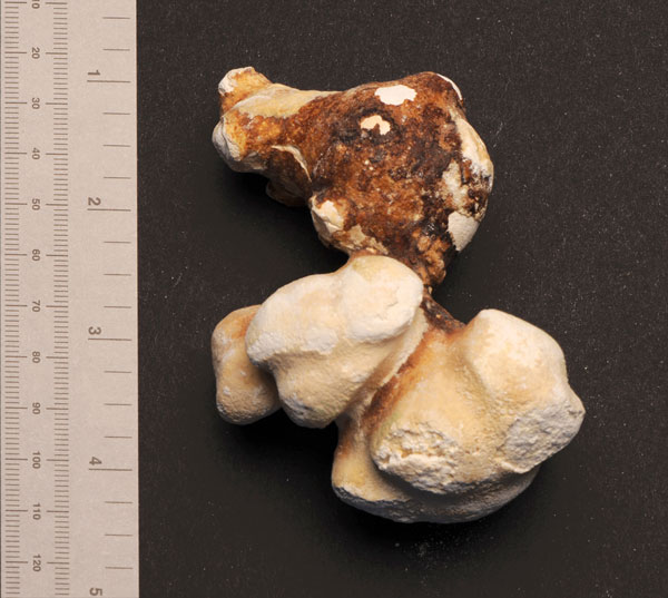
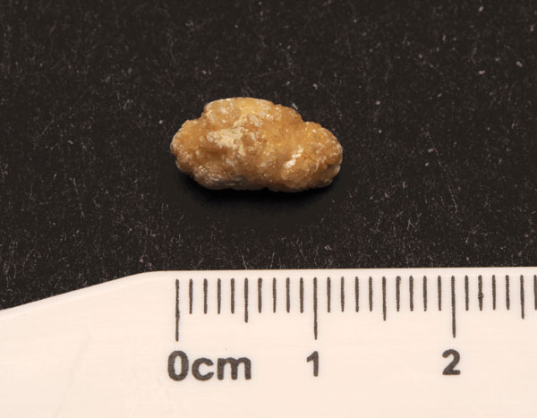
Increased ambulatory management in the UK has reduced the number of bed days used by stone patients by up to 15%, but waiting times are increasing.5 The number of hospital episodes for kidney stones continues to rise with the biggest increase observed in those aged 75 and over.6 GPs are often the first point of contact for patients presenting with kidney stones. Once imaging is completed and complications excluded, most are managed in primary care with lifestyle advice and pharmacological agents. Additional biochemical analysis of stones (see table 1, figure 1, and figure 2) can help inform personalised treatment plans to reduce recurrence and improve quality of life.
Presentation in primary care
Kidney stones may present with ureteric colic, urinary tract infection (UTI) or as an incidental finding on abdominal imaging. The incidence of asymptomatic kidney stones has risen with greater use of imaging. A third of these stones will become symptomatic with 50% requiring clinical intervention.7
Ureteric colic is caused by the passage of a calculus through the urinary tract. It typically presents as rapid onset, severe unilateral flank pain of short duration (colicky pain) radiating to the lower abdomen and/or ipsilateral groin and occasionally to the genitalia.
Gastrointestinal symptoms are usually absent, apart from nausea and vomiting, with appetite largely unaffected.8 The characteristics of the pain described can provide clues as to the anatomical position of the stone, with distal ureteric stones giving rise to symptoms of bladder irritability (see table 2). Ureteric colic is unaffected by posture with patients often writhing in seemingly futile attempts to relieve pain.9
History taking should aim to identify potential underlying conditions which might increase the risk of kidney stone formation, such as primary hyperparathyroidism, chronic malabsorption and diarrhoeal illnesses (e.g. ulcerative colitis), gout, and insulin resistance (type 2 diabetes, polycystic ovaries). Obesity is also a risk factor. Rarely, drugs such as topiramate, indinavir, furosemide, aspirin and antacids can predispose to stone formation. A personal or family history of kidney stone disease renders the diagnosis more likely. The patient’s history might also reveal dietary or occupational factors that increase the risk of kidney stones.8
Clinical examination of the abdomen and groin aims to exclude conditions that may mimic ureteric colic, such as scrotal pathology in men or gynaecological disease, including ectopic pregnancy, in women.
A compromised abdominal aortic aneurysm should be considered in older patients with known vascular disease and no previous history of nephrolithiasis. Assessment of vital signs is essential as a fever suggests an associated infection and should prompt urgent hospital referral. Immediate management comprises effective analgesia while urinalysis is the first step in confirming the diagnosis.
One of the challenges facing the GP is to differentiate patients who warrant immediate hospital referral from those who can be seen less acutely. Consistent with the new NICE guideline published in January 2019,5 GPs may increasingly find themselves prescribing drugs to expedite stone passage while in the longer term providing management advice to prevent recurrence.
Confirming diagnosis
Although stones may occur in the absence of haematuria, symptoms indicative of ureteric colic along with confirmed haematuria on urine dipstick has a reported sensitivity of 84% and specificity of 99%.10
All patients with suspected stone disease should have imaging performed promptly because of the risk of renal failure developing. The NICE guideline suggests that this should be done within 24 hours of presentation.5 Non-contrast CT has replaced intravenous urography as the modality of choice. It is more cost effective than plain abdominal X-ray, ultrasound and MRI, in part because it negates the need for a second scan in cases of diagnostic uncertainty. Although some have advocated prediction tools to reduce imaging in ureteric colic, CT frequently uncovers other intra-abdominal pathology after exclusion of stone disease.
Ultrasound is not as sensitive but is advised for pregnant women and children to minimise unnecessary radiation exposure. Plain radiographs can be useful for surveillance purposes. Controlled release drug preparations can sometimes mimic the appearance of calculi on plain radiographs and confound the diagnosis of stone disease.
After imaging, many stones are managed with watchful waiting following an open discussion with patients about the risks and benefits of such an approach. Useful investigations for patients with renal colic are summarised in table 3.
Management
Analgesia
Immediate pain relief is the first priority in the management of ureteric colic, although choice of analgesia and route of administration can vary. NSAIDs, such as diclofenac, are first-line analgesics and have demonstrated a reduced need for rescue medication when compared with intravenous paracetamol, opioids and antispasmodics. Prostaglandins induce ureteric smooth muscle spasm. NSAIDs inhibit prostaglandin release, thereby suppressing the ureteric contractions that cause colicky pain. Much of the evidence for NSAIDs in ureteric colic comes from data on intravenous and intramuscular administration, but they are more commonly administered orally or rectally.
If NSAIDs are contraindicated, for example in patients with a history of gastrointestinal bleeding or renal failure, or have not achieved adequate pain control, intravenous paracetamol is recommended rather than opioids. Although opioids are effective in suppressing pain via central nervous system pathways, they do not address the underlying muscular cause of pain in ureteric colic. Despite this, opioids are often used as first-line analgesia in emergency departments because of the severity of pain experienced by patients. If an opioid is used it is best to avoid pethidine, which is associated with increased incidence of vomiting.
Antispasmodic agents such as hyoscine butylbromide do not offer any benefits over NSAIDs.5 It is important to note that effective pain control should not delay timely intervention as complete obstruction, which may become more tolerable with high doses of analgesia, can lead to irreparable renal damage.
Medical expulsive therapy
The probability of spontaneous stone expulsion is inversely proportional to the size of the calculus with spontaneous passage more likely to occur in children than in adults. Delayed passage of a stone can cause significant morbidity. Patients who are still symptomatic at four weeks have a 20% increased risk of developing potentially life-threatening complications such as ureteric stricture or sepsis. Patients should maintain adequate hydration by increasing their oral fluid intake. There is limited evidence that intravenous fluids or diuretics aid stone expulsion.
The contractility of ureteric smooth muscle, mediated by alpha-adrenoceptor activity, is also an important factor in stone passage and has led to the use of alpha-blockers for this purpose. This is termed medical expulsive therapy (MET). Tamsulosin (0.4 mg once daily) is the most commonly prescribed alpha-blocker for stone elimination although there is probably a class effect for alpha-blockers. Nifedipine has also been studied for this purpose. MET is a relatively inexpensive treatment and its costs are likely to be offset by a reduction in subsequent interventions and improved quality of life.
The SUSPEND trial, the largest randomised placebo controlled trial addressing MET, recruited 1,136 patients in the UK.11 The authors concluded that neither tamsulosin nor nifedipine offered benefits for stone elimination, contradicting the findings of two previous systematic reviews on MET. However, NICE 5 suggests that alpha-blockers help the passage of distal ureteric stones < 10 mm, citing more recent evidence than the SUSPEND trial as the basis for their recommendation. Alpha-blockers have demonstrated superiority over nifedipine and this is reflected in both the NICE and European Association of Urology guidelines. MET is not a licensed indication for alpha-blockers but NICE has also recommended it for children with distal stones < 10 mm diameter, off label. Patients should be counselled that dizziness is the most common side effect, triggered by the hypotensive effects of MET.
Other treatment approaches
Further treatment options include extracorporeal shockwave lithotripsy (ESWL), ureteroscopy and percutaneous nephrolithotomy. The choice of treatment is determined not only by factors relating to calculi, but also patient and clinician preference as well as local availability of equipment and expertise.
A study of stone episodes requiring hospital admission in England, showed that over the five-year study period 2009/10 to 2014/15 the use of ESWL remained at a stable rate whereas the rate of ureteroscopy increased significantly over the same period.5 Although the gap between the number of ESWL and ureteroscopy cases continues to narrow, the NICE guideline advocates the use of ESWL.5 It is predicted that ESWL will confer cost savings over the longer term through an increase in outpatient interventions and a reduction in hospital admissions.
There is a growing body of evidence to support the use of MET as an adjunctive therapy. Steinstrasse (meaning stone street) is the term used to describe the accumulation of residual stone fragments that occurs following ESWL, in 4-7% of cases. One randomised controlled trial and several meta-analyses have shown tamsulosin to be effective in helping the clearance of stones after ESWL, reducing the incidence of steinstrasse as well as the need for analgesia. NICE recommends the use of MET after ESWL and has made a research recommendation for use of MET after ureteroscopy.5
Preventing recurrence
The underlying pathological process causing stone formation is the main predictor of recurrence risk. Approximately half of recurrent stone formers have just one stone recurrence (usually within five years) with increasing recurrence in patients left untreated.
Data from stone clinics suggests that the risk of recurrence can be reduced by up to 60% with lifestyle advice alone, with some UK centres launching virtual stone clinics to address this goal.
General measures to prevent stone recurrence include maintaining an adequate fluid intake (> 2 litres per day), weight reduction, avoiding sugary drinks and reducing dietary sodium and animal protein intake. Evidence supports the maintenance of a normal daily calcium intake of 700-1,200 mg for adults rather than dietary calcium restriction. These modifiable risk factors account for more than 50% of incident kidney stones. Increased fluid intake has been demonstrated by trial data to halve the risk of stone recurrence and is generally well tolerated.
Interventions to prevent recurrence aim to address urinary abnormalities that give rise to stone formation, by lowering urinary calcium, increasing urinary citrate excretion and/or altering urinary pH, see table 4. Potassium citrate is recommended to alkalinise urine in those with recurrent stones comprised of mainly calcium oxalate. NICE also advocates drinking water with added lemon juice (high in citrate). Thiazide diuretics are recommended for recurrent stone formers with hypercalciuria. Allopurinol may reduce stone formation in individuals with hyperuricosuria. Low-dose prophylactic antibiotics may be used for patients forming struvite stones associated with urinary infection. In the presence of hypercalcaemia, further investigation is advisable to identify a metabolic disorder contributing to nephrolithiasis e.g. hyperparathyroidism or renal tubular acidosis.
Conclusion
Renal and ureteric calculi are common, frequently symptomatic and have a high risk of recurrence. A detailed history, examination and simple investigations, such as urinalysis, urea & electrolytes and serum calcium, are helpful in determining predisposing risk factors, possible location of the stone and the potential cause of nephrolithiasis. Recurrence of stone formation is preventable in many individuals by lifestyle changes, adequate fluid intake and dietary manipulation. Medical and surgical management can be very successful in reducing the burden of symptomatic stone disease and epidemiological stone data can inform local service planning and population health initiatives.
REFERENCES
1 Morgan MS, Pearle MS. Medical management of renal stones. BMJ 2016;352:i52
2 Phillips R, Hanchanale VS, Myatt A et al. Citrate salts for preventing and treating calcium containing kidney stones in adults. Cochrane Database Syst Rev 2015 Oct 6;(10):CD010057
3 Scales CD Jr, Smith AC, Hanley JM et al. Prevalence of kidney stones in the United States. Eur Urol 2012;62(1):160-65
4 Issler N, Dufek S, Kleta R et al. Epidemiology of paediatric renal stone disease: a 22-year single centre experience in the UK. BMC Nephrol 2017;18(1):136
5 National Institute for Health and Care Excellence. NG118. Renal and ureteric stones: assessment and management. NICE. London. 2019 www.nice.org.uk/guidance/ng118 (Last accessed 11 Jan 2019)
6 Rukin NJ, Siddiqui ZA, Chedgy EC, Somani BK. Trends in upper tract stone disease in England: evidence from the hospital episodes statistics database. Urol Int 2017;98(4):391-96
7 Kittanamongkolchai W, Vaughan LE, Enders FT et al. The changing incidence and presentation of urinary stones over 3 decades. Mayo Clin Proc 2018;93(3):291-99
8 Bultitude M, Rees J. Management of renal colic. BMJ 2012;345:e5499
9 Portis AJ, Sundaram CP. Diagnosis and initial management of kidney stones. Am Fam Physician 2001;63(7):1329-38
10 Eskelinen M, Ikonen J, Lipponen P. Usefulness of history-taking, physical examination and diagnostic scoring in acute renal colic. Eur Urol 1998;34(6):467-73
11 Pickard R, Starr K, MacLennan G et al. Medical expulsive therapy in adults with ureteric colic: a multicentre, randomised, placebo-controlled trial. Lancet 2015;386(9991):341-49
 Download this article in PDF format