SEARCH

Shaikh M, Östör AJK. Evaluating the patient with low back pain. Practitioner 2015;259 (1788):21-24
Evaluating the patient with low back pain
22 Dec 2015
AUTHORS
Dr Maliha Shaikh MRCP, Clinical Research Fellow, Rheumatology Clinical Research Unit, Addenbrooke’s Hospital, Cambridge, UK
Dr Andrew J K Östör MB BS FRACP, Consultant Rheumatologist and Associate Lecturer, School of Clinical Medicine, University of Cambridge, Director, Rheumatology Clinical Research Unit, Cambridge, UK
Article
Low back pain is a major cause of morbidity worldwide.1 In the UK, it is the most common cause of disability in young adults. The main cause of working days lost in 2013 was musculoskeletal conditions, such as back and neck pain, accounting for 31 million days lost.2
Every year, 6-9% of adults in the UK consult their GP about back pain.3 While in most cases the symptoms are secondary to mechanical factors and self-limiting, GPs need to be aware of potentially sinister causes, or underlying systemic diseases. These include infection, malignancy, rheumatological and neurological disorders.
In order to help with classification, acute low back pain is usually defined as an episode lasting less than six weeks, subacute low back pain as lasting 6-12 weeks, and chronic low back pain as persisting for more than 12 weeks.
History and examination
In most patients with acute low back pain, a thorough history and examination is usually sufficient to make a diagnosis although the differential is wide, see table 1, p22. The main purpose is to exclude an alternative diagnosis, such as pain arising from the hip or trochanteric bursa and to categorise patients as having either: serious spinal pathology, nerve root/radicular pain or non-specific back pain.
The history should evaluate the duration of symptoms, previous episodes and the nature and severity of the current episode.
Risk factors for spinal infections such as diabetes, immunosuppression and intravenous drug use should be assessed. In patients with previous malignancy, the possibility of bony metastases should always be considered. Non-mechanical pain does not improve with bed rest and may be a marker for possible malignancy or inflammatory pain.
Red flag symptoms, see table 2, should raise concerns regarding a possible sinister cause such as malignancy or myeloma and more than one red flag mandates urgent further investigation. However, individual red flags have a low positive predictive value in primary care when used alone. Additionally, only a few are supported by a clear evidence base, for example a history of malignancy is associated with a 7% increase in spinal malignant disease.4
The red flags with the strongest association with fracture are older age, prolonged use of corticosteroid drugs, severe trauma and presence of a contusion or abrasion. The probability of spinal fracture is higher if more than one red flag is present. The red flag with the highest probability of spinal malignancy is a previous history of malignancy.
Psychosocial yellow flags increase the likelihood of chronicity and should also be explored, see table 3.5
The examination should be focused on elucidating a cause of the back pain and excluding any potentially sinister causes. Midline tenderness, for example, is often present in septic discitis. In mechanical lumbar back pain, one may observe tenderness and spasm of the paraspinal muscles, but this can also occur in patients with a psoas abscess. The musculoskeletal and neurological systems need to be assessed, looking specifically for any neural compromise. However, significant pathology can exist in the absence of hard clinical signs due to innervation from several levels.
Inflammatory back pain
Inflammatory back pain is often missed, particularly in the early stages. The primary features are pain arising in patients under the age of 40 years, thoracolumbar or sacroiliac pain and alternating buttock pain.
Early morning stiffness and stiffness after rest (the gelling phenomenon) is a hallmark of inflammatory back pain.6 There may also be peripheral joint involvement with evidence of inflammatory arthritis as well as extra-articular manifestations such as iritis, psoriasis and colitis.
Any history of preceding bowel infection or sexually transmitted infection as part of a reactive spondyloarthropathy should be explored. There may be a family history of ankylosing spondylitis or psoriasis. Objectively, a reduction in movement of the lumbar spine may be seen but is a late feature of disease.
Schober’s test is positive if there is less than a 5 cm increase between two points 5 cm below and 10 cm above the dimples of Venus at the L5 level on lumbar spine flexion. Sacroiliitis may be picked up by sacroiliac joint stress tests but these are non-specific.7
It is worth noting that in the early stages of inflammatory back pain examination may be normal.
Mechanical back pain
Mechanical back pain is extremely common and typically worse at the end of the day or after a period of movement. There is no associated morning stiffness. The pain arises from facet joint osteoarthritis, lumbar paraspinal muscle spasm and degenerative disc disease. There is often a history of previous similar episodes.
Vertebral fractures
Spinal fractures cause severe localised pain of sudden onset and can be associated with minor trauma in the case of osteoporosis, such as twisting movements of the back or stepping off a kerb. Major trauma such as a fall from a height or road traffic accident in younger patients may also lead to vertebral fracture.
Lumbar disc herniation
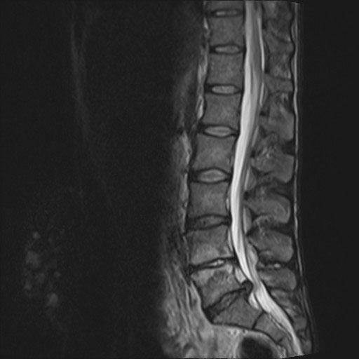
Acute disc prolapse is often preceded by a history of trauma such as heavy lifting. The most common discs to prolapse are L4/5 and L5/S1 (see figure 1) and result in sciatica. There is usually a history of low back pain of acute onset with concurrent severe pain radiating to the leg.7 Passive straight leg raise helps identify increased spinal root tension.
Spinal cord compression and cauda equina syndrome
Sphincter disturbance leading to loss of bladder or bowel control should always be explored as it is a sign of spinal cord compression (or cauda equina syndrome with lesions lower than the L1-2 level). There may also be saddle anaesthesia in the sacrum and reduced anal tone on rectal examination.8
Both spinal cord compression and cauda equina syndrome are neurosurgical emergencies and need urgent referral for further investigation and possible intervention.
Investigations
Investigations should be reserved for patients in whom a systemic or serious cause of low back pain is suspected. Blood tests may include full blood count, ESR, C-reactive protein, serum protein electrophoresis, urine for Bence-Jones protein, corrected calcium and prostate specific antigen.
In patients with suspected inflammatory back pain, an HLA B27 test can be helpful in stratifying risk, especially in young men with prolonged early morning stiffness but should not be performed routinely.
In primary care HLA B27 should be checked in patients aged under 45 with fewer than two of the following symptoms suggesting inflammatory back pain: bilateral buttock pain, pain which improves on movement and psoriasis. Suspected inflammatory back pain should be referred without HLA testing in patients if at least two of these symptoms are present.9
Plain radiography
This is the most common imaging requested as it is widely available and cheap compared with other imaging methods. However, in younger patients with mechanical or non-specific low back pain, routine radiographs do not provide any further benefit over clinical evaluation and are not cost effective.8 While information regarding soft tissue structures is limited, in older patients plain radiographs are more helpful.
Anteroposterior and lateral views provide information on alignment, vertebral body and intervening disc height. Plain X-rays may demonstrate compression fractures of the spine, scoliosis, spondylolisthesis and evidence of osteoarthritis (including facet joints) as well as providing a crude estimation of bone density.10
MRI
MRI is currently recommended for patients with red flags and for evaluation of possible radiculopathy.11 It has advantages in terms of better visualisation of the spinal canal, ligaments, discs and other soft tissues and provides a more complete evaluation for all potential sources of back pain.
It is also useful for identifying spinal infections such as discitis or osteomyelitis as well as infections such as paraspinal or psoas abscess. It is the investigation of choice in patients with suspected inflammatory back pain and in these patients an MRI of the spine and sacroiliac joints should be performed to detect bone marrow oedema.
Other imaging modalities
In some cases, CT scanning may be performed but its use is mainly limited to visualising the bones and nerve roots, and may be used to guide nerve root injections.12
Nuclear medicine bone scanning may be used in a secondary care setting to detect occult fractures, investigate whether a fracture is new or old and to detect bone metastases and differentiate these from degenerative changes.13
Referral
The majority of patients with low back pain can be managed in primary care as the pain will usually be self-limiting. However, it may take three months or longer to resolve.
Patients with suspected inflammatory back pain should be referred to rheumatology as soon as possible in order to institute early management and prevent long-term deformity and disability.
Patients with more than one red flag symptom should be referred urgently for further investigation.
Management
Management of back pain depends on the underlying cause. In the primary care setting, treatment of acute low back pain should aim to:
• Provide adequate information and reassurance that low back pain is usually not a serious disease and most patients recover, although this may take weeks to months
• Provide adequate symptomatic relief
• Encourage patients to stay active and return to normal activities as soon as possible. Complete bed rest should be avoided as this can be a factor in promoting chronicity.
The NICE clinical knowledge summaries on the management of acute and chronic low back pain were updated in April 2015.11 In chronic low back pain the main aim of intervention is to modify the patient’s underlying fears and beliefs regarding pain and improve their coping mechanisms and physical ability.14
A wide range of therapies exist that can be utilised by patients suffering from low back pain, see table 4.
Pharmacological therapies are commonly used for symptomatic relief. These include paracetamol, NSAIDs and opioids. In chronic low back pain, low doses of tricyclic antidepressants are often helpful and are recommended in the NICE guidelines. However, there is no clear evidence that antidepressants are more effective than placebo and they are not recommended by SIGN.15 It is of course important to recognise and treat depression and optimise antidepressant treatment in patients with moderate depression.15
Patients with radicular pain can benefit from treatments used for neuropathic pain such as gabapentin or pregabalin.
Muscle relaxants and benzodiazepines may also be used for a short defined period to enable early return to activity. Secondary prevention with alendronate or risedronate should be instituted in patients with osteoporotic vertebral fractures.
Non-pharmacological treatment is also a critical element of back pain management. Complete bed rest should be discouraged and instead, patients advised to continue normal activities as this leads to faster symptomatic recovery.
Back pain is often best managed using a multidisciplinary approach. This may incorporate medical treatment with psychotherapy, physiotherapy, relaxation techniques, patient education or vocational therapies to provide a holistic approach and has been shown to have better long-term outcomes.15 Physical therapy is useful especially back strengthening exercises.16 In chronic low back pain, this may be incorporated into a multidisciplinary programme. Some secondary care units offer back pain programmes to help patients deal with their pain. CBT focuses on patients’ reactions to pain and coping strategies and can be helpful in long-term management.
Conclusion
Low back pain in primary care needs careful and thorough evaluation to distinguish patients with serious causes, such as systemic disease or malignancy, from the vast majority of back pain which is self-limiting.
Red flag symptoms will alert GPs to patients who require further investigation and onward referral.
Long-term management is aimed at symptomatic relief, patient education and treatment of any underlying causes.
REFERENCES
1 Global Burden of Disease Study 2013 Collaborators. Global, regional, and national incidence, prevalence, and years lived with disability for 301 acute and chronic diseases and injuries in 188 countries, 1990–2013: a systematic analysis for the Global Burden of Disease Study 2013. Lancet 2015 DOI: 10.1016/S0140-6736(15)60692-4
2 Office for National Statistics. Sickness absence in the labour market 2014. www.ons.gov.uk/ons/rel/lmac/sickness-absence-in-the-labour-market/2014/sty-sickness-absence.html
3 Dunn KM, Croft PR. Classification of low back pain in primary care: using “bothersomeness” to identify the most severe patients. Spine 2005;130:1887–92
4 Downie A, Williams CM, Henschke N et al. Red flags to screen for malignancy and fracture in patients with low back pain: systematic review. BMJ 2013;347: f7095
5 Kendall NAS, Linton SJ, Main CJ. Guide to assessing psychosocial yellow flags in acute low back pain: risk factors for long-term disability and work loss. New Zealand: Accident Rehabilitation & Compensation Insurance Corporation of New Zealand the National Health Committee; 1997. http://kendallburton.com/Library/Resources/Psychosocial_Yellow_Flags.pdf
6 Braun A, Saracbasi E, Grifka J et al. Identifying patients with axial spondyloarthritis in primary care: how useful are items indicative of inflammatory back pain? Ann Rheum Dis 2011;70:1782-87 doi:10.1136/ard.2011.151167
7 Kataria RK, Brent LH Spondyloarthropathies. Am Fam Physician 2004;69(12):2853-60
8 Deyo RA, Rainville J, Kent DL. What can the history and examination tell us about low back pain? JAMA 1992;268:760-65
9 Braun A, Gnann H, Saracbasi E et al. Optimizing the identification of patients with axial spondyloarthritis in primary care—the case for a two-step strategy combining the most relevant clinical items with HLA B27. Rheumatology 2013 doi:10.1093/rheumatology/ket115
10 Kerry S, Hilton S, Patel S et al. Routine referral for radiography of patients presenting with low back pain: is patients’ outcome influenced by GPs’ referral for plain radiography? Health Technol Assess 2000;4(20):i-iv,1-119
11 Wilmink JT. CT morphology of intrathecal lumbosacral nerve-root compression. Am J Neuroradiol 1989;10:233-48
12 Han LJ, Au-Tong TK, Ton WC et al. Comparison of bone single-photon emission tomography and planar imaging in the detection of vertebral metastases in patients with back pain. Eur J Nucl Med 1998;25:635-8
13 National Institute for Health and Care Excellence. Clinical knowledge summaries. Back pain (low) without radiculopathy. Clinical knowledge summary. http://cks.nice.org.uk/back-pain-low-without-radiculopathy
14 Vlaeyen JW, Linton SJ. Fear-avoidance and its consequences in chronic musculoskeletal pain: a state of the art. Pain 2000;85(3):317-32
15 Scottish Intercollegiate Guidelines Network. SIGN 136. Management of chronic pain. SIGN. Edinburgh. 2013 www.sign.ac.uk/pdf/SIGN136.pdf
16 Hayden JA, van Tulder MW, Tomlinson G. Systematic review: strategies for using exercise therapy to improve outcomes in chronic low back pain. Ann Intern Med 2005;9:776-85
ARTICLE IN PDF
 Download this article in PDF format
PRESET SEARCHES
PubMed
Low back pain treatment review
NICE Evidence search
Immediate CPD prompts
These handy templates* prompt immediate drafting of personal learning, reflection and planning. Save your record straight into your personal development folder on your device or Cloud storage system.
*Adobe reader needed for iPad
EXTERNAL WEBLINKS
Arthritis Research UK
Provides patient information leaflets on back pain which include simple exercises that patients can do at home
LETTERS TO THE EDITOR
If you would like to comment on this article or have a question for the authors, write to: [email protected]