SEARCH

Practitioner 2011; 255 (1746):21-24
Early diagnosis crucial in ankylosing spondylitis
16 Dec 2011
AUTHORS
Dr Anshuman P Malaviya MBBS MD MRCP
Rheumatology Clinical Research Fellow
Dr Andrew JK Ostor MA MBBS FRACP
Director, Rheumatology Research Unit and Consultant Rheumatologist
Rheumatology Clinical Research Unit, Addenbrooke’s Hospital, Cambridge
Article
Back pain is common with up to 9% of the population consulting their GP with this problem each year.1
As the vast majority of these patients have non-specific back pain, it is often difficult to identify those with inflammatory disease such as ankylosing spondylitis (AS). It is critical to make this distinction as AS, if untreated, may cause significant morbidity with a substantial socio-economic impact both on the individual and society. Early diagnosis is particularly important as newer therapies are able to contain this condition and even induce remission in some cases.
AS is an inflammatory autoimmune disorder that predominantly affects the spine. It is one of a group of conditions called inflammatory spondyloarthropathies, see table 1, p22. AS affects about 0.2-0.5% of the population.2
It is at least twice as common in men than women and most often manifests in the third to fifth decades. It is estimated that up to 5% of patients with chronic lower back pain in primary care have inflammatory disease.3 Diagnosis is usually delayed at least partly through lack of awareness of this condition.4
Pathogenesis
The association between AS and HLA-B27 is the strongest known association between a disease and a major histocompatibility complex antigen. The prevalence of HLA-B27 varies vastly between different ethnicities and is estimated to be 8% in the general population in the UK.5 Although only 1% of patients with HLA-B27 develop AS, 90-95% of patients with AS are positive for HLA-B27. Various hypotheses have been proposed to explain the pathogenetic role of HLA-B27 although its exact role remains unclear. Immune dysfunction is the hallmark of this condition and it may be triggered by infection.5
The primary site of inflammation in AS is the entheses, the sites of insertion of tendons and ligaments into bone. Tumour necrosis factor (TNF) and other cytokines play a role in perpetuating the inflammatory process. If the inflammation remains untreated, there is resultant fibrosis and ultimately ossification at the entheseal sites, so-called bamboo spine which is now infrequently seen.
Diagnosis
AS should be suspected in patients who report back pain and stiffness with rest, especially in the morning, which improves with exercise. Although the condition affects both the sacroiliac joints, a proportion of patients report pain radiating into the buttocks which may be unilateral or alternate, particularly in the early stages.
In addition to the spine, large joint synovitis may develop as well as features of entheseal involvement (such as tennis/golfer’s elbow, Achilles tendinitis and plantar fasciitis).
Symptoms are usually associated with a modest elevation of inflammatory markers (ESR/CRP). Pelvic radiographs may reveal sacroiliitis, although MRI is a more sensitive tool, particularly in early disease.
The 1984 modified New York criteria formed the mainstay for the diagnosis of AS for several decades and required evidence of sacroiliitis on plain radiographs and at least one of the following three criteria to be met:
• low back pain and stiffness for more than three months which improves with exercise but is not relieved by rest
• restriction in lumbar movements in both plains
• limitation of chest expansion
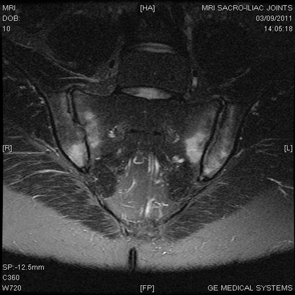
These diagnostic criteria are somewhat obsolete and have been replaced by new classification criteria that take into account early sacroiliitis evident on MRI scan, see table 2, opposite. Figure 1 on p21 shows the appearance of sacroiliitis on MRI scan.
Management
A simplified algorithm for the treatment of AS is shown in figure 2, opposite.
A proportion of patients respond well to NSAIDs coupled with a structured physiotherapy and exercise programme. However, about half these patients need escalation to biologic therapy.6
We recommend that patients with a suspected diagnosis be referred to secondary care in order to confirm the diagnosis and commence treatment.
NSAIDs and physiotherapy
NSAIDs form the mainstay of treatment of patients with AS. There is often a dramatic improvement in symptoms; however, it would appear that these drugs do not alter the natural course of the disease. In addition to gastrointestinal side effects, there is evidence to support adverse cardiovascular risk with long-term use. It is therefore recommended that patients take the lowest dose of an NSAID with the best safety profile to control symptoms. COX-2 inhibitors have been found to be effective in AS and are often used in patients as alternatives; albeit sparingly.
A recent Cochrane systematic review suggested that physiotherapy and exercise therapy was beneficial in AS.7
It concluded that home-based exercise therapy was better than no therapy; that group physiotherapy was superior to home-based therapy and that inpatient physiotherapy followed by group therapy was the best. It is, however, not clear which patients are most likely to benefit from these interventions.
DMARDs and corticosteroids
Conventional DMARDS such as methotrexate and sulfasalazine have no role in the treatment of axial spondyloarthropathy and enthesopathy. There is some evidence of efficacy in peripheral arthritis; and they should be reserved for this indication.
Corticosteroids are also less efficacious in AS than in rheumatoid arthritis and are seldom used to treat acute flares.
Biologic therapy
Biologic therapy has revolutionised the management of AS with four drugs currently licensed for treatment: infliximab, etanercept, adalimumab and golimumab. These drugs bind to, and eliminate, TNF and are highly efficacious in AS. Numerous large randomised controlled trials have demonstrated their efficacy and safety in AS.8, 9 NICE has approved the use of these drugs for patients with active disease who have failed a trial of two NSAIDS.10, 11 There is evidence to suggest that this cohort of patients is most likely to progress if left untreated.
Although there is significant improvement in clinical outcomes and quality of life indices, the evidence to support radiological non-progression is limited.12 Furthermore, up to 20-40% of patients do not respond to anti-TNFs.12 As a result there continues to be an unmet need and a wide range of novel biologics therefore are being trialled in AS to try to fill the void.
Conclusion
AS with its striking clinical features is likely to be encountered by primary care physicians. The role of the GP in identifying AS is critical. This requires
skill in sifting inflammatory disease from the sea of patients who present with back pain. Early diagnosis and therapy may reduce the impact of this condition on the individual and also help save healthcare costs in the long term.
We would strongly recommend referral of suspected cases to the local rheumatology service. MRI helps identify patients early in the course of the disease and current management strategies, including anti-TNFs, are very effective at reducing symptoms and improving quality of life. However, despite all the recent advances, anti-inflammatory drugs, patient education and physiotherapy remain the cornerstone of therapy.
REFERENCES
1 World Health Organization. The burden of musculoskeletal conditions at the start of the new millennium. World Health Organ Tech Rep Ser 2003; 919:1–218
2 Reveille JD. Epidemiology of spondyloarthritis in North America. Am J Med Sci 2011;341(4):284-6
3 O’Shea FD, Boyle E, Salonen DC et al. Inflammatory and degenerative sacroiliac joint disease in a primary back pain cohort. Arthritis Care Res (Hoboken) 2010;62:447-54
4 Hamilton L, Gilbert A, Skerrett J et al. Services for people with ankylosing spondylitis in the UK - a survey of rheumatologists and patients. Rheumatology 2011;50(11):1991-8
5 Sheehan NJ. The ramifications of HLA-B27. J R Soc Med 2004;97(1):10-14
6 Sieper J, Rudwaleit M, Baraliakos X et al. The Assessment of SpondyloArthritis international Society (ASAS) handbook: a guide to assess spondyloarthritis (Supplement). Ann Rheum Dis 2009;68:ii1-ii44
7 Dagfinrud H, Kvien TK, Hagen KB. Physiotherapy interventions for ankylosing spondylitis. Cochrane Database Syst Rev 2008;1:CD002822
8 Baraliakos X, Listing J, Fritz C et al. Persistent clinical efficacy and safety of infliximab in ankylosing spondylitis after 8 years- early clinical response predicts long-term outcome. Rheumatology 2011;50(9):1690-9
9 Braun J, van der Horst-Bruinsma IE, Huang F et al. Clinical efficacy and safety of etanercept versus sulfasalazine in patients with ankylosing spondylitis: a randomized, double-blind trial. Arthritis Rheum 2011;63(6):1543-51
10 National Institute for Health and Clinical Excellence. TA143. Adalimumab, etanercept and infliximab for ankylosing spondylitis. NICE. London. 2008
11 National Institute for Health and Clinical Excellence. TA233. Golimumab for the treatment of ankylosing spondylitis. NICE. London. 2011
12 Dougados M, Baeten D. Spondyloarthritis. Lancet 2011;377(9783): 2127-37
USEFUL INFORMATION
National Ankylosing Spondylitis Society (NASS)
NASS is an independent charity that provides information for patients, their families and healthcare professionals.
ARTICLE IN PDF
 Download this article in PDF format
EXTERNAL WEBLINKS
NICE Evidence
PubMed
Osteoarthritis general practice