SEARCH

The Practitioner 2009 – 253 (1724): 19-24
GPs have key role in shared care of patients with SLE
15 Dec 2009
AUTHORS
Dr Maame-Boatemaa Amissah-Arthur MBChB MRCP, Clinical Research Fellow, SWBH NHS Trust, Birmingham
Professor Caroline Gordon MA MD FRCP, Centre for Immune Regulation, Division of Immunity and Infection, University of Birmingham
Article
SLE (Systemic lupus erythematosus) is a complex, heterogeneous autoimmune disease involving inflammatory processes in multiple organ systems. Classically, patients have an exacerbating and remitting course with increased morbidity and higher mortality compared with the general population.
Twenty to thirty years ago, the mortality rate was as high as 50% at five years. However, survival rates have improved considerably as a result of earlier recognition, introduction of corticosteroids and immunosuppressive therapy and the availability of effective treatment for co-morbid conditions. The current 20-year survival rate is 80%.1,2
GPs play a key role in early referral to specialist centres and subsequent shared care pathways for the management of these patients.
SLE mainly affects adults and is common in women between the ages of 20 and 40 years, with a female to male ratio of 9:1. First onset of SLE before puberty is uncommon, with paediatric lupus accounting for 8-15% of all cases.3 Patients who are diagnosed postmenopause (late onset lupus) tend to have a milder form of the disease with an insidious onset, lower degree of disease activity, less multi-organ involvement but a significant risk of complications including accelerated atherosclerosis and premature death.
The prevalence in the UK is 28 per 100,000 suggesting that approximately 1 person in 3,500 will have lupus, but this is subject to ethnic variation.4,5 The disease is most common in people of Caribbean or Asian descent.
The pathogenesis of SLE is multifactorial and encompasses multiple immunological, vascular and inflammatory processes,6 which are triggered by a combination of environment (sunlight), genetics, infection and hormonal milieu. The hallmark of the disease is the formation of autoantibodies resulting in immune complex formation and complement activation.
Clinical features
Diagnosing SLE can be challenging because of the myriad of clinical features and substantial variability between patients.
GPs will commonly encounter constitutional symptoms, as well as skin and musculoskeletal manifestations, as these are the most common features. Non-specific symptoms such as overwhelming fatigue despite a good night's sleep, fever, weight loss and lymphadenopathy may be present. In such cases it is important to exclude infection, lymphoma, and solid organ tumours.
Major organ involvement may also occur (see table 1) and includes renal, neuropsychiatric, cardiopulmonary, gastrointestinal, haematological and ocular features.
Skin
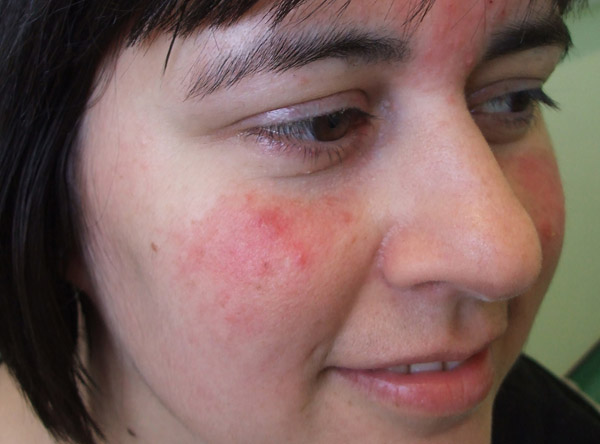
Cutaneous involvement is present in about 60% of cases7,8,9 and typically manifests as a malar or butterfly rash that is often sun-induced. This is a macular or papular rash on the face that may spread to become a confluent symmetrical erythema over the bridge of the nose and malar bones, sparing the nasolabial folds (see figure 1).
Active systemic disease is also associated with a generalised lupus rash, which can occur anywhere, and this may or may not be photosensitive. It can range from a maculopapular rash to annular, violaceous or urticarial lesions that are sometimes pruritic.
Discoid lupus is a cutaneous form of lupus characterised by a hyperkeratotic lesion with central atrophy, scarring and hypopigmentation.
Disfigurement caused by scarring, particularly on exposed areas such as the face, can pose cosmetic and psychological problems for patients.
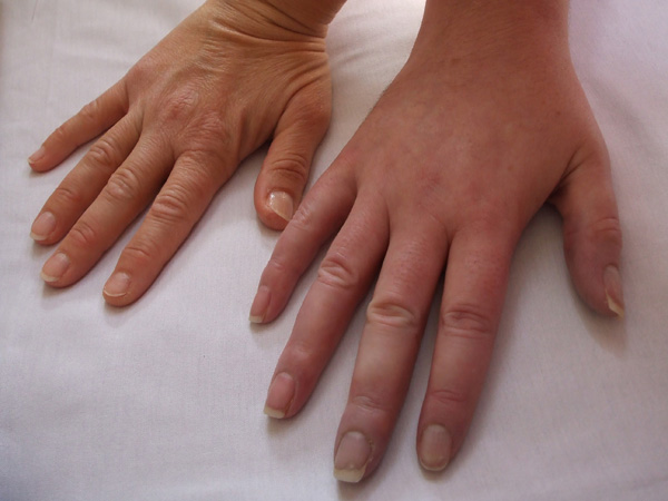
Raynaud's phenomenon may also occur in SLE (see figure 2)
Joints
Joint involvement is inflammatory in nature with arthralgia, arthritis and/or tendinitis and occurs in about 90% of patients with SLE.1 The knees, wrists and hands are the most frequent areas involved. Characteristically, it is symmetrical, episodic and accompanied by early morning stiffness. Joint damage is uncommon, non-erosive and associated with reducible deformities resulting from ligamentous and capsule laxity. This pattern is referred to as Jaccoud's syndrome. Early in the disease it can mimic changes in rheumatoid arthritis.
Mucosal ulceration
Mucosal ulceration is another common feature, occurring in up to half of all patients with SLE. Typically, the ulcers are superficial and painless and mainly found on the hard palate, buccal cavity and vermiform border, but may occur in the nose.3 Lupus-induced mucosal ulceration may need to be distinguished from that caused by drugs such as methotrexate which can also cause oral mucositis as well as ulceration.
Alopecia
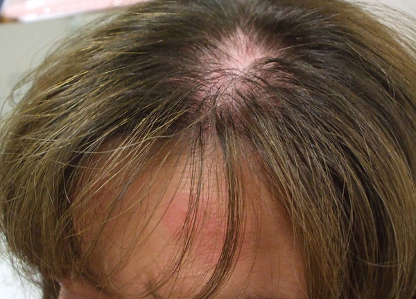
Patients may develop diffuse alopecia when the disease is active but this is usually reversible. Hair regrowth normally occurs once disease control is achieved. Patchy alopecia (see figure 3) may lead to scarring, particularly if it is associated with discoid rash and this can be permanent and very distressing for patients.
Cardiorespiratory symptoms
Cardiorespiratory symptoms are common. Patients may present with chest pain on inspiration caused by lupus-induced pleurisy or pericarditis, which may be associated with effusions. This needs to be distinguished from pleurisy secondary to infection and pulmonary embolus, both of which can occur in lupus patients. Chest pain on exertion may occur because of premature or accelerated coronary artery disease. Breathlessness on exertion may be caused by ischaemic heart disease or less commonly acute pneumonitis, chronic lung fibrosis or pulmonary hypertension.
Renal complications
Lupus glomerulonephritis is one of the most important systemic complications, occurring in about 30% of patients with SLE in the UK.10 Other manifestations of renal involvement include microvascular thrombosis presenting with a slow increase in proteinuria, haematuria, worsening renal function, and uncontrolled hypertension, or rarely vasculitis.
Careful screening tests for renal disease need to be undertaken as it is asymptomatic. Urinalysis, looking for haematuria and proteinuria, is the most important and effective method to detect and monitor lupus nephritis. However, it is essential to exclude other conditions that can present with these features such as infection, menstrual blood loss and renal stones.
It is important for patients with confirmed proteinuria and/or haematuria to be referred promptly to hospital for further assessment. This will involve quantifying protein loss by measurement of 24-hour urine protein excretion, urine protein:creatinine ratio or albumin:creatinine ratio on spot urine samples. Renal function should be assessed by serum creatinine and estimated GFR.
Beware of new onset or worsening hypertension as this correlates strongly with proliferative nephritis and seek expert advice if this happens.
Renal disease in lupus is classified using the International Society of Nephrology/Renal Pathology Society (ISN/RPS) 2003 classification of lupus nephritis, which is based on biopsy findings as was its predecessor the WHO classification.11 This has important prognostic implications and is used to guide therapeutic decisions, see table 2, right.12
Neuropsychiatric manifestations
Neuropsychiatric manifestations of lupus are well recognised but uncommon and include:
- epileptic seizures
- psychosis
- aseptic meningitis
- stroke
- transverse myelitis
Stroke may be caused by premature atherosclerosis, thrombosis, emboli and occasionally vasculitis.
Other symptoms
Ophthalmic manifestations are uncommon but potentially serious and need prompt specialist assessment.13 Gastrointestinal manifestations are also possible but these are uncommon and rarely recognised (see table 1, p21). Anorexia and abdominal pain with guarding because of serositis are the most common manifestations.
Diagnosis
The diagnosis of SLE is traditionally based on a combination of clinical features and laboratory findings. Classification criteria have been established by the American College of Rheumatology (ACR), 14 see table 1). These stipulate that a minimum of four out of a possible 11 ACR criteria should be present in order for a patient to be classified as having SLE for the purposes of clinical trials and other studies.
Some patients may never achieve four criteria or may fall within the spectrum of overlap syndromes or mixed connective tissue disease. In such cases, patients with three ACR classification criteria, of which at least one must be clinical and one must be serological (an autoantibody), may be diagnosed by an expert (rheumatologist or nephrologist) as having lupus if other diagnoses are excluded and they have relevant features. These include biopsy-proven glomerulonephritis or other clinical manifestations that are not in the ACR criteria but suggestive of lupus, for example alopecia, skin vasculitis, Raynaud's phenomenon (see figure 2) and lung fibrosis.
Investigations
Any patient with suspected clinical features of lupus should be investigated for the presence of autoantibodies including anti-nuclear antibody (ANA), anti-double stranded DNA (ds-DNA) or antibodies to extractable nuclear antigens (ENA), as well as low complement (C3 and C4) levels. Early clues often come from abnormal haematological indices (low haemoglobin because of haemolysis, low platelet count, low white cell count most often lymphopaenia) and raised ESR without raised CRP. Inflammatory markers showing raised ESR and normal CRP levels are characteristic of SLE and related conditions such as primary Sjögren's syndrome.
Drug-induced lupus
It is important to establish what medications the patient is taking to rule out drug-induced lupus (see box 1). Newer agents, such as anti-TNFα drugs, have been associated with a lupus-like illness.
Management
Treatment is varied and dependent on the clinical manifestations. It is best planned by an appropriate specialist depending on disease severity and organ involvement. GPs should refer patients initially to a rheumatologist and, if available to them, one with a specialist interest in SLE, who can oversee/co-ordinate the overall care of the patient. However, other specialist care may be enlisted, for example a nephrologist in the case of diagnosing and managing lupus nephritis. Other system-specific disease may also require relevant specialist input. Treatment will often include the use of corticosteroids, by various routes, at different points in disease management.
In addition, some experts advocate the use of hydroxychloroquine, an antimalarial, as a principal drug in all SLE patients. It is beneficial in the management of mucocutaneous, musculoskeletal, serosal and constitutional symptoms. Recently, it has been shown in several studies to have cardioprotective and survival enhancing properties, by reducing total cholesterol, low density lipoprotein (LDL) cholesterol and triglycerides (TG).15,16,17 Patients require annual visual acuity monitoring because of the risk of hydroxychloroquine-related maculopathy but this is rare if the dose does not exceed 6.5mg/kg/day.18
Other agents used in addition to reduce the risk of cardiovascular disease in these patients include aspirin, anti-hypertensive agents especially ACE inhibitors, and cholesterol-lowering drugs. Patients should also be given advice on lifestyle to influence modifiable risk factors such as smoking, obesity and exercise.
As UV light is a major factor known to initiate or exacerbate cutaneous disease, patients must be fully informed about the importance of sun avoidance and/or protection with clothing and high factor sunscreen, usually SPF 25 and above. Local treatment is with topical or intralesional steroids, but long-term use must be avoided. In severe and/or refractory cases, systemic steroids are used. Topical tacrolimus, a calcineurin inhibitor, may also be used as adjunctive therapy.19
Joint symptoms can be managed with analgesics and NSAIDs. The latter need to be used with caution to avoid worsening the disease-associated risk of premature atherosclerosis. Control of active disease may require immunosuppressive treatment with corticosteroids, methotrexate, azathioprine or ciclosporin. These disease modifying anti-rheumatic drugs (DMARDS) need to be regularly monitored. Only hydroxychloroquine, corticosteroids and azathioprine are licensed for use in lupus.
Severe organ disease is managed initially with pulsed intravenous corticosteroids and cyclophosphamide therapy and once remission is achieved, patients are put on maintenance therapy usually with azathioprine. Mycophenolate mofetil (MMF) is increasingly being used for inducing and maintaining remission but is not yet licensed for lupus. Newer biological drug therapies, such as rituximab, a monoclonal anti-CD20 chimeric antibody that targets and depletes B cells but not plasma cells, are being investigated for use in lupus. Further guidance may be sought by consulting the EULAR recommendations on the management of SLE.9
As most women who develop SLE are of childbearing age, pregnancy and the issues surrounding it are of major importance (see box 2).
Monitoring
Monitoring disease activity, response to therapy and accumulation of long-term damage can be performed using validated objective scoring systems. Common methods include the BILAG (British Isles Lupus Assessment Group) Index and SLEDAI (Systemic Lupus Erythematosus Disease Activity Index) for measuring disease activity and SLICC/ACR (Systemic Lupus International Collaborating Clinics) damage index for recording multi-organ damage.20,21
Prognosis
SLE has a bimodal pattern of mortality, demonstrating an early peak, less than five years from diagnosis and a later peak, more than five years after diagnosis, increasingly due to cardiovascular complications.2 Early death in lupus is usually due to active disease, lupus nephritis and/or sepsis. As longevity of patients with lupus increases they are at risk of increasing co-morbidities, either due to the disease and/or its treatment. These include:
- premature atherosclerosis
- hypertension
- dyslipidaemias
- diabetes
- osteoporosis
- avascular necrosis
- infection
- malignancies (especially non-Hodgkin's lymphoma and lung cancer in smokers)
Lupus patients develop cardiovascular disease earlier than the general population and overall women have a 5-6 fold increased risk of myocardial infarction, stroke and congestive heart failure.22,23,24
GPs can assist specialists in addressing risk factors, as well as the prompt evaluation and management of these co-morbidities.
REFERENCES
1 Isenberg DA, Horsfall A. Systemic lupus erythematosus in adults. In: Oxford Textbook of Rheumatology. pp1145-19. Oxford Medical Publications. Oxford. 1998
2 Urowitz M, Bookman A, Koehler B et al. The bimodal mortality pattern of systemic lupus erythematosus. Am J Med 1976; 60: 221-5
3 Tsokos GC, Gordon C, Smolen JS. Systemic Lupus Erythematosus: A Companion to Rheumatology. First Edition. Mosby Elsevier. Philadelphia. 2007
4 Jonsson H, Nived O, Sturfelt G et al. Estimating the incidence of systemic lupus erythematosus in a defined population using multiple sources of retrieval. Br J Rheumatolol 1990; 29(3): 185-8
5 Johnson A, Gordon C, Palmer R et al. The prevalence and incidence of systemic lupus erythematosus in Birmingham, UK, related to ethnicity and country of birth. Arthritis Rheum 1995; 38: 551-8
6 Mok CC, Lau CS. Pathogenesis of systemic lupus erythematosus. Br Med J 2003; 56(7): 481
7 Patel P, Werth V. Cutaneous lupus erythematosus: a review. Dermatol Clin 2002 ; 203: 73-385
8 Wysenbeek A, Guedji D, Amit M et al. Rash in systemic lupus erythematosus: prevalence and relation to cutaneous and non-cutaneous disease manifestations. Ann Rheum Dis 1992; 517: 17-19
9 Mosca M, Tani C, Aringer M, Bombardieri S et al. EULAR Recommendations for the management of Systemic Lupus Erythematosus (SLE) Report for the Task Force of the European Standing Committee for International Clinical Studies including Therapeutics (ESCISIT). Ann Rheum Dis 2008; 67: 195-205
10 MacGowan JR, Ellis S, Griffiths M et al. Retrospective analysis of outcome in a cohort of patients with lupus nephritis treated between 1977 and 1999. Rheumatology 2002; 41(9): 981-987
11 Weening JJ. The classification of glomerulonephritis in systemic lupus erythematosus revisited. J Am Soc Nephrol 2004; 15(2): 241-250
12 Illei GG, Balow JE. Kidney involvement in systemic lupus erythematosus. In: Systemic Lupus Erythematosus: A Companion to Rheumatology. pp 336-350. Mosby Elsevier. Philadelphia. 2007
13 Sivaraj RR, Durrani OM, Denniston AK et al. Ocular manifestations of systemic lupus erythematosus. Rheumatology 2007; 46(12): 1757
14 Hochberg M. Updating the American College of Rheumatology revised criteria for the classification of systemic lupus erythematosus. Arthritis and Rheum 1997; 40(9): 1725-34
15 Wallace D, Metzger A, Stecher V et al. Cholesterol-lowering effect of hydroxychloroquine in patients with rheumatic disease: reversal of deleterious effects of steroids on lipids. Am J Med 1990; 89: 322-6
16 Petri M, Lakatta C, Magder L et al. Effect of prednisolone and hydroxychloroquine on coronary artery disease risk factors in systemic lupus erythematosus: a longitudinal data analysis. Am J Med. 1994; 96: 254-9
17 Rahman P, Gladman D, Urowitz M et al. The cholesterol lowering effect of antimalarial drugs is enhanced in patients with lupus taking corticosteroid drugs. J Rheumatol 1999; 26: 325-330
18 Royal College of Ophthalmologists, The British Society of Rheumatology and British Association of Dermatologists. Hydroxychloroquine and Ocular Toxicity: Recommendations on Screening (Replacing the Royal College of Ophthalmologists Guidelines for Screening 2004 and 1998). London. 200919 Bohm M, Gaubitz M, Luger T et al. Topical tacrolimus as a therapeutic adjunct in patients with cutaneous lupus erythematosus. Dermatology 2003; 207: 381-5
20 Hay E, Gordon C, Emery P. Assessment of lupus: where are we now? Ann Rheum Dis 1993; 52: 169-172
21 Griffiths B, Mosca M, Gordon C. Assessment of patients with systemic lupus erythematosus and the use of lupus disease activity indices. Best Practice & Research. Clinical Rheumatol 2005; 19(5): 685-708
22 Manzi S, Meilahn EN, Rairie JE et al. Age-specific incidence rates of myocardial infarction and angina in women with systemic lupus erythematosus: comparison with the Framingham Study. Am J Epidemiol 1997; 145(5): 408-415
23 Esdaile JM, Abrahamowicz M, Grodzicky T et al. Traditional Framingham risk factors fail to fully account for accelerated atherosclerosis in systemic lupus erythematosus. Arthritis Rheum 2001; 44(10): 2331-2337
24 Bessant R, Hingorani A, Patel L et al. Risk of coronary heart disease and stroke in a large British cohort of patients with systemic lupus erythematosus. Rheumatology 2004; 43(7): 924-929
25 Lima F, Buchanan N, MA Khamashta. Obstetric outcome in systemic lupus erythematosus. Semin Arthritis Rheum 1995; 25: 184-192
26 Clowse M, Witter F, Madger L. The impact of increased lupus activity on obsterical outcomes. Arthritis Rheum 2005; 52: 514-521
USEFUL INFORMATION
Lupus UK
http://www.lupusuk.org.uk/
European League Against Rheumatism (EULAR)
http://www.eular.org/
Arthritis Research Campaign UK
http://www.arc.org.uk/
ARTICLE IN PDF
 Download this article in PDF format
EXTERNAL WEBLINKS
NICE Evidence
PubMed