SEARCH

Practitioner 2011; 255 (1736): 19-22
Preventing osteoporotic fractures in older people
24 Jan 2011
AUTHORS
Professor Eugene McCloskey MD MRCP FRCPI
Professor of Adult Bone Disease, Academic Unit of Bone Metabolism, University of Sheffield
Article
While fractures at the spine, wrist and hip are regarded as classical osteoporotic fractures, all fragility fractures in the elderly should be considered as osteoporotic once pathological fracture (e.g. metastatic disease) has been excluded. The working definition of a fragility fracture is a fracture occurring during a fall from standing height or less or equivalent injury.
Until recently, the management of osteoporosis was centred around the use of diagnostic thresholds based on bone mineral density (BMD) measured at the spine or hip by dual x-ray absorptiometry (DXA). This was reflected in UK guidance for the identification of individuals at high fracture risk from the Royal College of Physicians (RCP).1
The guidance was based on an opportunistic case-finding strategy where physicians were alerted to the possibility of osteoporosis and high fracture risk by the presence of clinical risk factors associated with fracture. This provided a trigger for the measurement of BMD, and treatment was considered in those with a BMD value that lay in the range of osteoporosis as defined by the WHO.
Treatment was, however, also recommended for women with a prior fragility fracture without necessarily measuring BMD.
Since the development of these guidelines, it has become apparent that the presence of several of the risk factors used to trigger a BMD test is associated with a fracture risk greater than can be accounted for by BMD alone.2,3
Thus, the assessment of fracture risk should also take account of specific risk factors in addition to BMD, since this increases the detection rate of individuals at high risk.
Modern osteoporosis management therefore comprises:
- assessment of future fracture risk to determine the need for intervention
- diagnosis or exclusion of underlying causes of osteoporosis
- institution of appropriate long-term therapy
Identifying the high-risk patient
Clinical risk factors
Several risk factors for fracture have been well established. Many of these risk factors impact on BMD but also contribute independently to future fracture risk. Clinicians frequently take account of these other risk factors in deciding whether treatment is required and a number of algorithms have been developed to improve the prediction of fracture risk.
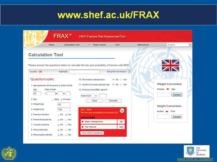
The WHO has produced FRAX, a well validated tool that estimates the probability of a major osteoporotic fracture (clinical vertebral, hip, wrist or proximal humerus) or a hip fracture in the next 10 years.4
The algorithm, specifically designed for use in primary care, incorporates BMD as an additional, measurable risk factor with information gleaned from clinical risk factors (see figure 1, p19) that adds value to the prediction of risk.
A history of prior low trauma fracture in adult life is an important risk factor to identify and can usually be obtained by a good clinical history. The only means of identifying prior vertebral fractures, including those deemed to be asymptomatic, is by spinal imaging.
Spinal radiographs
More than two-thirds of vertebral fractures are relatively asymptomatic but may be suspected from height loss and the development of kyphosis. The latter features may also result from degenerative spinal disease, however, and radiographs of the thoracic and lumbar spine are important to differentiate fractures from degenerative changes. The importance of vertebral fractures for future fracture risk cannot be overstated and many current strategies involve the assessment of patients by low radiation DXA scans to identify prevalent fractures.5,6,7
Bone densitometry
After age and prior fragility fracture, BMD is the next major determinant of a person's risk of fracture. If access to bone densitometry is limited, it is appropriate to consider treatment of elderly individuals who have had previous low trauma fractures or who have other strong risk factors for fracture such as high-dose corticosteroid therapy.1,8 If BMD is available, rather than scanning all individuals with a risk factor, measurements should be targeted to individuals whose probability of fracture lies close to the intervention threshold, where knowledge of BMD will influence management.9
This is the approach encapsulated within the guideline from the National Osteoporosis Guideline Group, published in 2008, and integrates BMD with clinical risk factors.10,11
The clinical risk factors are not totally independent of BMD and when clinical risk factors alone are used in women aged 70 years or more, BMD is approximately 1 SD lower in the high-risk group compared with the low-risk group.9 The higher the probability of fracture estimated from FRAX, but without inputting BMD, the greater the response to intervention in elderly women recruited from the general population.12
underlying causes of osteoporosis
Individuals with a low trauma vertebral fracture or low BMD for age should be investigated for underlying causes of osteoporosis (secondary osteoporosis). Secondary causes account for up to 40% of cases of osteoporosis in women and 60% of cases in men
(see table 1, above).
In addition to a good clinical history, a small number of investigations can exclude the most common secondary causes of osteoporosis (see table 2, above). Treating the underlying cause often leads to at least partial recovery of bone mass.
Reducing fracture risk
The ultimate goal of osteoporosis management is to reduce the future risk of fracture and involves educating the patient about the nature of the disease, their fracture risk, lifestyle modification and, if necessary, the different types of therapy available.
Lifestyle modification includes measures to reduce falls risk and bone loss including exercise (regular and weight bearing such as walking, aerobics or tai chi), adequate dietary calcium intake and avoidance of smoking and excessive alcohol consumption.
In some patients, particularly those with recent vertebral fractures, additional approaches should aim to reduce pain and improve mobility. Good pain relief is frequently achieved with analgesics, but physical measures such as hydrotherapy or transcutaneous nerve stimulation may be useful.
While acute pain usually resolves within a few weeks or months, patients with vertebral fractures may need to be given long-term analgesia because of secondary degenerative disease. Techniques such as vertebroplasty and kyphoplasty are now available for use in selected patients with recent vertebral fractures and persistent or severe pain.
Preventing falls
As the majority of non-vertebral fragility fractures are caused by falls, it is important that all patients with an osteoporotic fracture and those deemed to be at high risk of fracture are assessed for falls risk. Predisposing factors, such as postural hypotension or drowsiness caused by drugs, should be eliminated where possible.
International guidelines recommend asking older people annually about falls and carrying out a simple assessment of gait/balance to direct subsequent specialist falls assessment and address modifiable risk factors.13
Patients may benefit from physical and/or occupational therapy to improve their balance and reflexes and to provide appropriate walking aids.
An environmental assessment should be made to eliminate hazards and an assessment of vision with appropriate treatment is also important. Hip protectors have a limited role to play. It may not be possible to prevent falls completely, but it is possible to reduce the frequency.
Bone targeted therapies
The evidence base for some therapies is not strong in older people, such as the selective oestrogen receptor modulator raloxifene. With other treatments, such as HRT, the side-effect profile outweighs potential skeletal benefits. The mainstays of treatment in the older person are described below.14
Vitamin D and calcium
Calcium alone probably has a less marked effect on fracture reduction than combined therapy and the latter is usually recommended. Adherence can be a problem, largely due to the calcium component. Several preparations are now available to aid patient choice and compliance.
Combined therapy has been shown to reduce hip fracture risk in the frail elderly and should be considered in all older patients who are housebound or in residential care.
If adherence is poor, vitamin D only preparations are available to ensure that vitamin D deficiency is adequately treated with recommendations to increase dietary calcium intake.
In patients at higher risk of fracture, the combination should be used as adjunctive therapy together with another osteoporosis agent.
Bisphosphonates
Alendronate and risedronate are available as once-weekly preparations with evidence for significant reductions in vertebral and non-vertebral fractures. These drugs have largely replaced the use of cyclical etidronate.
Ibandronate, available as a once-monthly tablet or a 3-monthly intravenous slow injection reduces vertebral fractures with indirect evidence for a reduction in non-vertebral fractures.
Zoledronate is available as a once-yearly short infusion with good evidence of anti-fracture efficacy at all sites.
NICE recommends the use of generic bisphosphonates as first-line therapy, though these may not be suitable for, or tolerated by, all. Poor absorption of these agents means they should be taken on an empty stomach, 30-60 minutes before breakfast. The availability of weekly or monthly dosing regimens has improved convenience and adherence.
Denosumab
The most potent inhibitor of osteoclastic bone resorption, denosumab is a fully humanised monoclonal antibody against receptor activator of nuclear factor kappaB ligand (RANKL), a major regulator of osteoclast development and activity.
It is approved for the treatment of osteoporosis in postmenopausal women at increased risk of fractures and is given as a subcutaneous injection of 60 mg once every six months. It has been demonstrated to reduce the incidence of vertebral, non-vertebral and hip fractures in postmenopausal women with osteoporosis.
Strontium ranelate
Strontium ranelate has been shown to reduce vertebral and non-vertebral fracture risk significantly in postmenopausal women. The precise mechanism of action remains unclear, but treatment is associated with significant increases in BMD, partly mediated by the presence of strontium in bone that impacts on the interpretation of changes in BMD.
Teriparatide and parathyroid hormone
These agents increase bone formation (and later bone resorption) with an improvement in bone mass and structure, particularly in trabecular bone with good reductions in spine fracture risk. A reduction in non-vertebral fractures has also been shown by recombinant teriparatide (PTH 1-84).
They are relatively expensive agents and so use is limited to patients with severe, progressive osteoporosis despite exposure to anti-resorptive therapy.
Teriparatide is licensed for use in men and women, including those with glucocorticoid-induced osteoporosis, whereas recombinant parathyroid hormone 1-84 is only licensed for postmenopausal women. Most patients will require treatment with anti-resorptive agents after discontinuation to maintain the improvements in bone mass.
Conclusion
The effective management of osteoporosis in older people has been enhanced by recent developments in risk assessment and an increasing number of therapeutic options to enhance adherence to therapy. Treatment should be considered strongly in older people with a history of fragility fracture, particularly if a recent fracture, without the necessity of BMD assessment. In the absence of fracture, the combination of FRAX and the NOGG guideline brings risk assessment and therapeutic decisions to a wide audience within primary care.
REFERENCES
1 Royal College of Physicians. Osteoporosis: clinical guidelines for the prevention and treatment. RCP. London. 1999
2 Johansson H, Kanis JA, Oden A et al. BMD, clinical risk factors and their combination for hip fracture prevention. Osteoporos Int 2009;20(10):1675-82
3 Kanis JA, Johansson H, Oden A et al. Assessment of fracture risk. Eur J Radiol 2009;71(3):392-7
4 Kanis, JA. Assessment of osteoporosis at the primary health-care level. Technical Report. WHO Collaborating Centre, University of Sheffield. Sheffield. 2008
5 Ferrar L, Jiang G, Clowes JA et al. Comparison of densitometric and radiographic vertebral fracture assessment using the algorithm-based qualitative (ABQ) method in postmenopausal women at low and high risk of fracture. J Bone Miner Res 2008; 23(1):103-11
6 McCloskey EV, Vasireddy S, Threldkeld J et al. Vertebral fracture assessment (VFA) with a densitometer predicts future fractures in elderly women unselected for osteoporosis. J Bone Miner Res 2008; 23(10):1561-8
7 Schousboe JT, Ensrud KE, Nyman JA et al. Cost-effectiveness of vertebral fracture assessment to detect prevalent vertebral deformity and select postmenopausal women with a femoral neck T-score> -2.5 for alendronate therapy: a modeling study. J Clin Densitom 2006;9(2):133-43
8 Royal College of Physicians. Glucocorticoid-induced osteoporosis. Guidelines on prevention and treatment. Bone and Tooth Society of Great Britain, National Osteoporosis Society and Royal College of Physicians. RCP. London. 2002
9 Johansson H, Oden A, Johnell O et al. Optimization of BMD measurements to identify high risk groups for treatment - a test analysis. J Bone Miner Res 2004; 19(6):906-13
10 Compston J, Cooper A, Cooper C et al. Guidelines for the diagnosis and management of osteoporosis in postmenopausal women and men from the age of 50 years in the UK. Maturitas 2009;62(2):105-8
11 Kanis JA, McCloskey EV, Johansson H et al. Approaches to the targeting of treatment for osteoporosis. Nat Rev Rheumatol 2009; 5(8):425-31
12 McCloskey EV, Johansson H, Oden A et al. Ten-year fracture probability identifies women who will benefit from clodronate therapy-additional results from a double-blind, placebo-controlled randomised study. Osteoporos Int 2009;20:811-7
13 American Geriatrics Society and British Geriatrics Society. Clinical Practice Guideline: Prevention of Falls in Older Persons. 2010. www.americangeriatrics.org/
files/documents/health_care_pros/Falls.Summary.
Guide.pdf.
14 National_Osteoporosis_Guideline_Group. Osteoporosis. Clinical guideline for prevention and treatment. Executive Summary 2010 Available from: www.shef.ac.uk/NOGG/NOGG_Executive_Summary.pdf
ARTICLE IN PDF
 Download this article in PDF format
EXTERNAL WEBLINKS
National Osteoporosis Society
FRAX
National Osteoporosis Guideline Group (NOGG )
NICE Evidence
PubMed