SEARCH

Beare N. Diagnosis and management of age-related macular degeneration. Practitioner January 2021;265(1844):11-15
Diagnosis and management of age-related macular degeneration
25 Jan 2021
AUTHORS
Mr Nicholas Beare MBChB MA MD FRCOphth, Senior Lecturer, Department of Eye and Vision Science, University of Liverpool, Honorary Consultant and Lead for AMD, St Paul’s Eye Unit, Liverpool University Hospitals, Liverpool, UK
Competing interests: Mr Nicholas Beare was a member of the NICE guideline committee advising on NG82. Age-related macular degeneration, published in January 2018. He is a member of the RCOphth Scientific Committee. Within the past three years he has received an academic travel grant from Bayer. He has received honoraria from Alimera Sciences and Gilead Sciences for advisory boards in uveitis (none in AMD)
Article
Age-related macular degeneration (AMD) is by far the most common cause of registration for severe sight impairment in the UK, accounting for 50% of registered blindness. Epidemiological evidence suggests 4-8% of people aged over 65 years in the UK have visually significant AMD. Increasing age and genetic predisposition are the biggest risk factors; smoking is the only modifiable risk factor. High degrees of short sightedness also increase the risk. AMD is a cause of painless, gradual loss of vision. Dry AMD symptoms are usually non-specific but reading tends to be affected more than distance vision. As no treatment is possible, NICE does not recommend referral except when vision is substantially affected. If a patient over 55 years reports new visual distortion or deterioration in central vision, neovascular AMD (NvAMD) should be suspected, irrespective of macular signs. Patients with suspected NvAMD should be seen by ophthalmology within two weeks, and if the diagnosis and progressive disease are confirmed, treatment should commence within the same timeframe. Although there is no treatment for dry AMD, there is treatment for NvAMD. Intravitreal anti-vascular endothelial growth factor (anti-VEGF) agents cause regression of neovascularisation and reduce macular oedema. The best outcomes are achieved if anti-VEGF therapy is started early in the disease course before vision deteriorates substantially.
Age-related macular degeneration (AMD) is by far the most common cause of registration for severe sight impairment in the UK, accounting for 50% of registered blindness. It is also a spectrum of aging changes of the macula which are present in most elderly people but will only significantly affect the vision in a minority, see box 1.
Epidemiological evidence suggests 4-8% of people over 65 years in the UK have visually significant AMD.1,2 The proportion of people with AMD rises with age, and absolute numbers are increasing as the population ages.3 The absence of any treatment for most AMD is a paramount unmet medical need.
The macula is particularly prone to degenerative processes because it is very metabolically active with high oxygen and glucose requirements; and highly differentiated and post-mitotic.
The macula’s non-renewing cells are subject to a lifetime of oxygen free radical damage and latterly inflammatory processes. Dry or atrophic AMD is the sum of these degenerative processes, while neovascular or wet AMD (NvAMD) is a neovascular response in which new abnormal blood vessel growth can cause rapid damage.
The macula is the central portion of the retina approximately bounded by the inferior and superior vascular arcades. The fovea is the central macula tissue with the highest density of cones and is responsible for central vision, reading and recognising faces. The fovea is approximately 1 optic disc area in size, a sixteenth of the area of the macula. Under the macula is the retinal pigment epithelium (RPE) which supports the retina and forms an outer blood-retina barrier from the underlying choroid.
Features and development
Drusen
Drusen are an aging change on the macula consisting of lipofuscin deposits under the retina. The RPE cells phagocytose cytoplasmic discs continually shed by photoreceptors and recycle the retinol (vitamin A) and cellular material. Polymeric cross-linked protein residues accumulate as lipofuscin in lysosomes, and these coalesce eventually to form drusen on their basal side between the RPE and Bruch’s membrane.4
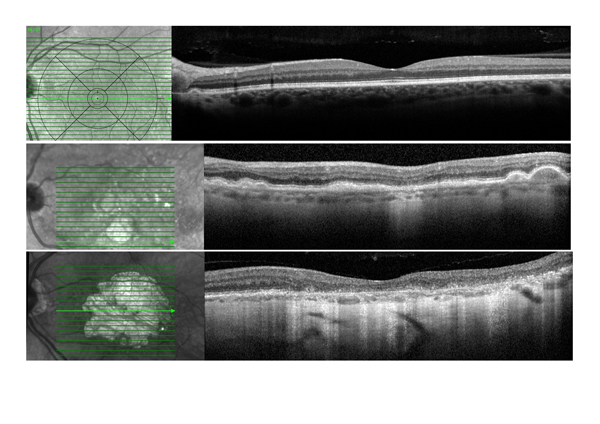
Small and intermediate sized drusen (< 125 μm – approximately the width of a retinal vein as it crosses the optic disc margin) have no visual consequence but herald the beginning of degeneration in the retina. Drusen can become large ( ≥ 125 μm), coalesce, and even lift the RPE forming a localised drusenoid pigment epithelial detachment (PED), see figure 1. Small or intermediate sized drusen (< 125 μm) with pigmentary abnormalities have a low risk of developing advanced AMD after 5 years (1.3%), whereas large ( ≥ 125 μm) extensive drusen with any atrophy have an 18% risk.5
Reticular pseudodrusen, or subretinal drusenoid deposits, are located above the RPE as a cone shaped intrusion into the neuroretina.4 These do not enlarge or coalesce but can be numerous and are associated with advanced forms of AMD. Rod photoreceptor dysfunction has been implicated in their formation.
Pigment changes, atrophy and geographic atrophy
As the RPE becomes increasingly disturbed focal hyperplasia or pigment deposits can occur on the macula, so called pigment clumps. Similarly, atrophic changes start to occur in the outer retina and RPE. Once the atrophy of the outer retina and RPE is complete this is termed geographic atrophy and any function is lost from this area.
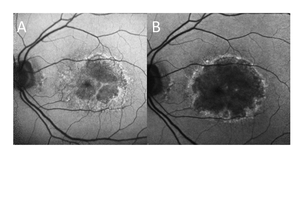
Patients may be aware of scotomas from parafoveal geographic atrophy, but once it progresses to involve the centre of the fovea useful central vision is lost, see figure 2. Geographic atrophy tends to expand until most of the central macula is affected. Rates of progression vary widely (at least six-fold) between individuals, but the size of the atrophic area is the strongest predictor of rate of progression.6 Patients’ vision deteriorates more rapidly at the specific time when the central fovea becomes affected.
Vitelliform lesion
A subfoveal lipofuscin deposit termed a vitelliform lesion can occur in younger patients as a macular dystrophy, but also in AMD. The lesion tends to flatten over months or years with resulting retinal atrophy and vision loss.
Choroidal and intraretinal neovascularisation
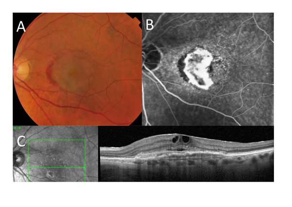
Neovascularisation usually starts in the choroid (choroidal neovascularisation; CNV) and can proliferate under the RPE (fibrovascular PED; type 1 CNV) or between the RPE and neurosensory retina (classic CNV; type 2 CNV) or both. Alternatively, it may start in the retina (retinal angiomatous proliferation) and proliferate into the same potential spaces. Finally, choroidal proliferation can be associated with polyp-like lesions which is more common in East Asian patients (polypoidal choriovasculopathy). When neovascularisation occurs leaking vessels lead to intraretinal fluid/macular oedema, subretinal fluid and macular haemorrhage which affect vision, see figure 3.
Neovascularisation disrupts tissue, causes fibrosis and without treatment will progress to a macular scar with a mixture of fibrosis and atrophy and consequent central vision loss. Sometimes this is referred to as a disciform scar but is more accurately termed a fibroatrophic macula.
Presentation and referral
AMD is a cause of painless, gradual loss of vision. Dry AMD symptoms are usually non-specific but reading tends to be affected more than distance vision. Asymptomatic aging changes or dry AMD may be noted by an optometrist at a routine visit. As no treatment is needed NICE does not recommend referral except when vision is substantially affected.7
NvAMD distorts the retinal architecture so its archetypal symptom is distortion of vision: ‘bending or kinking of straight lines’. If a patient over 55 years spontaneously reports new distortion, then NvAMD should be suspected (irrespective of macular signs). A central blob in the vision (scotoma) is another symptom but patients may only complain of general problems with vision.
An Amsler grid is a grid of lines which can highlight distortion to patients when they are asked to look at the centre with each eye individually. It is not a useful test for detecting NvAMD at its first use, as patients with dry AMD also report distortion on an Amsler grid. It can be useful for monitoring (e.g. weekly) for new onset distortion which is a useful marker of NvAMD onset, but no more than checking any straight lines.
Suspected NvAMD should be referred within one working day to ophthalmology, preferably to a dedicated AMD or macular service. There should be a clear local pathway for suspected NvAMD which can be accessed directly by optometrists or GPs. An optometrist report stating NvAMD is suspected and fulfilling referral guidelines should be fast tracked in the same way.
For direct referral by optometrists, patients should have visual loss or spontaneously report distortion or have a central scotoma; with macular haemorrhage or subretinal fluid or exudate on fundoscopy.8
GPs should suspect NvAMD in patients over 55 years who spontaneously report new distortion or deterioration in central vision over weeks or days. Patients with suspected NvAMD should be seen by ophthalmology within two weeks, and if the diagnosis and progressive disease are confirmed, proceed to treatment within the same timeframe.7
Dry AMD can be referred routinely when vision is substantially affected, in order to access low vision services, and for registration as sight impaired or severely sight impaired. Although AMD affects central vision, peripheral vision is retained.
Patients with macular aging changes are sometimes diagnosed with dry AMD by their optometrist and conclude that they are on a fast track to blindness. These patients need realistic information and reassurance which often requires an ophthalmologist’s assessment.
Investigation
Early and dry AMD diagnosed by fundoscopy, including by an optometrist, does not require further investigation.7
Suspected NvAMD is investigated with optical coherence tomography (OCT). OCT uses low-coherence near-infrared light to produce cross-sectional images through the macula with resolution of a few microns.
OCT demonstrates intraretinal and subretinal fluid, subretinal neovascular material and PED, and their absence excludes NvAMD. Their presence is not specific, so to diagnose NvAMD angiography is required. Increasingly OCT scans are provided by optometrists but expertise in interpreting abnormalities will vary.
Fluorescein angiography identifies neovascularisation because unlike the normal retinal vasculature these vessels leak the intravenously injected sodium fluorescein. It can enable differentiation of NvAMD from branch retinal vein occlusion, central serous chorioretinopathy, dry AMD and other differentials. Advancement of OCT technology has produced OCT angiography. However, this gives no information on the functional integrity of vessels and its interpretation with regard to NvAMD remains controversial.
Indocyanine green angiography utilises a larger fluorescent molecule which is protein bound to allow visualisation of the choroidal vasculature. This is required to diagnose polypoidal choriovasculopathy, and may add information to that discerned from fluorescein angiography.
The lipofuscin normally occurring in RPE cells fluoresces at a low level itself. This is used in fundus autofluorescence imaging to give a grey background to the fundus image. In geographic atrophy RPE cells die, so affected areas lose all autofluorescence and become black. Areas of geographic atrophy are often surrounded by areas of increased autofluorescence due to the accumulation of lipofuscin in distressed RPE cells. OCT scans show geographic atrophy as loss of the outer retinal layers and RPE, collapse of the remaining inner retinal layers, with a window defect through into the choroid.
Ophthalmologists often use multiple imaging modalities to assess macular disease fully, encompassing some or all of the above techniques, see box 2.
Risk factors
Increasing age and genetic predisposition are the biggest risk factors for AMD. Smoking is the only modifiable risk factor. A first-degree relative with AMD increases the risk of progression to advanced AMD in five years by 1.5 (95% CI: 1.2-1.8) and smoking by 2.0 (95% CI: 1.6-2.6).9
High degrees of short sight (myopia ≥ 6 dioptres) increase the risk of macular degeneration and can cause it in younger people. Any highly myopic person with distortion of vision should be seen in ophthalmology within two weeks.
Treatment
Dry AMD
There is no treatment for dry AMD. High dose vitamins/mineral/carotenoid pigment supplements (at significantly higher doses than recommended daily intake) are heavily promoted to reduce AMD progression but the evidence of any effect is weak and subject to bias. The Age-Related Eye Disease Study (AREDS), a randomised controlled trial investigating this in the USA, did not meet its primary endpoints.5
In a post hoc subgroup analysis of participants with more advanced AMD features, the estimated probability of progression to central geographic atrophy or NvAMD over five years was 20% in patients on the AREDS formulation of vitamins and minerals, compared with 28% on placebo.
In this underpowered subgroup, AREDS supplements reduced the odds of visual acuity loss, odds ratio (OR) 0.73 (99% CI: 0.54-0.99) just meeting statistical significance, and equating to a number needed to treat of 33.10 There have been no other large, randomised trials, but an industry has grown up based on retailing AREDS formulation supplements.
The original AREDS formulation included beta-carotene which can increase lung cancer in smokers.
AREDS 2 was a study which investigated removing beta-carotene, reducing zinc, adding carotenoid pigments (lutein and zeaxanthin) and/or omega-3 fatty acids to the original formulation.11 None of these individual manipulations had a significant effect in a study population at high risk of AMD progression; and with no untreated arm, AREDS 2 was not informative.
AREDS formulation supplements are not recommended by NICE but patients should be encouraged to increase the amount of fresh fruit and vegetables, especially green leafy vegetables, in their diet.
NvAMD
There is effective treatment for NvAMD: intravitreal anti-vascular endothelial growth factor (anti-VEGF) agents cause regression of neovascularisation and reduce macular oedema. Best vision outcomes are achieved if anti-VEGF therapy is started early in the disease course of neovascularisation before vision deteriorates substantially.12
However, not all NvAMD is progressive and NICE recommendations require that signs of progression are present. Treatment often has to be continued for three or more years to sustain benefits as neovascularisation can incompletely regress or recur.
NICE found no difference in safety and efficacy between anti-VEGF agents, aflibercept, bevacizumab and ranibizumab.7 However, NICE has not recommended bevacizumab although it is used by specialists off-label for AMD.
Treatment regimens usually start with three intravitreal injections over 3 months. Thereafter injections can be continued 1-2 monthly on fixed intervals, or what is termed a ‘treat and extend regimen’ where treatment intervals are extended to 3 or 4 months provided the disease is stable. Alternatively, treatment can be given as needed, but this requires continued 1-2 monthly monitoring.
Injections are given through the conjunctiva, sclera and pars plana into the vitreous with anaesthetic eye drops and a fine gauge needle in a dedicated treatment room.
While undoubtedly unpleasant and stressful for the patient, the treatment is mostly well tolerated and serious complications are rare.13 A small number of patients cannot tolerate intravitreal injections due to procedure related pain or anxiety.
Anti-VEGF treatment induces new vessel regression and reduces oedema, but treatment does not prevent the progression of degenerative and atrophic changes. Hence all studies following treated NvAMD patients for five years or more show a decline in average vision.14
Conclusion
AMD is a spectrum of aging changes in the macula from asymptomatic drusen to blinding macular atrophy and fibrosis.
Dry AMD is the most common form and causes visual loss mostly through untreatable geographic atrophy. In NvAMD, subretinal new vessels can rapidly damage the macula, but can be treated with anti-VEGF injections. Treatment requires regular intraocular injections over years, so there is much interest in developing longer acting treatment methods. Early treatment for NvAMD has better visual results, so patients with suspected NvAMD should be referred urgently for treatment within two weeks. Treatments are needed to slow the progression of dry AMD with candidate medications for geographic atrophy so far failing in clinical trials.
REFERENCES
1 Reidy A, Minassian DC, Vafidis G et al. Prevalence of serious eye disease and visual impairment in a north London population: population based, cross sectional study. BMJ 1998;316(7145):1643-46
2 Wilde C, Poostchi A, Mehta RL et al. Prevalence of age-related macular degeneration in an elderly UK Caucasian population-The Bridlington Eye Assessment Project: a cross-sectional study. Eye 2017;31(7):1042-50
3 Li JQ, Welchowski T, Schmid M et al. Prevalence and incidence of age-related macular degeneration in Europe: a systematic review and meta-analysis. Br J Ophthalmol 2020;104(8):1077-84
4 Brunk UT, Terman A. Lipofuscin: mechanisms of age-related accumulation and influence on cell function. Free Radic Biol Med 2002;33(5):611-19
5 Age-Related Eye Disease Study Research Group. A randomized, placebo-controlled, clinical trial of high-dose supplementation with vitamins C and E, beta carotene, and zinc for age-related macular degeneration and vision loss: AREDS report no. 8. Arch Ophthalmol 2001;119(10):1417-36
6 Fleckenstein M, Mitchell P, Freund KB et al. The progression of geographic atrophy secondary to age-related macular degeneration. Ophthalmology 2018;125(3):369-90
7 National Institute for Health and Care Excellence. NG82. Age-related macular degeneration. NICE. London. 2018 www.nice.org.uk/guidance/ng82
8 Downes CM, L Jenkins, SL Watson. AMD electronic referral form London: Royal College of Ophthalmologists
9 Klein ML, Francis PJ, Ferris FL et al. Risk assessment model for development of advanced age-related macular degeneration. Arch Ophthalmol 2011;129(12):1543-50
10 Kansagara D, Gleitsmann K, Gillingham M et al. Nutritional supplements for age-related macular degeneration: A systematic review. VA Evidence-based Synthesis Program Reports. Washington (DC). 2012
11 Age-Related Eye Disease Study 2 Research Group. Lutein + zeaxanthin and omega-3 fatty acids for age-related macular degeneration: the Age-Related Eye Disease Study 2 (AREDS2) randomized clinical trial. JAMA 2013;309(19):2005-15
12 Lee AY, Lee CS, Butt T et al. UK AMD EMR users group report V: benefits of initiating ranibizumab therapy for neovascular AMD in eyes with vision better than 6/12. Br J Ophthalmol 2015;99(8):1045-50
13 Gualino V, Fourmaux E, Grenet T et al. Patient experience of anti-vegf intravitreal injection. J Fr Ophtalmol 2020;43(10):1047-53
14 Rofagha S, Bhisitkul RB, Boyer DS et al. Group S-US. Seven-year outcomes in ranibizumab-treated patients in ANCHOR, MARINA, and HORIZON: a multicenter cohort study (SEVEN-UP). Ophthalmology 2013;120(11):2292-29
ARTICLE IN PDF
 Download this article in PDF format
EXTERNAL WEBLINKS
Patient support
Eye Clinic Liaison Officers
These may or may not be NHS funded, but most eye departments have one to support patients with the emotional and practical impact of vision loss
Low Vision Assessment
This is usually provided by optometrists in eye departments to assess and provide patients with magnifiers and other aids
Active patient support group and charity for AMD with helpline and local branches
Royal National Institute of Blind People (RNIB)
The charity provides a range of support including assistance with technology, an online shop for helpful products and free talking books
Certificate of visual impairment
Qualifying patients can be registered sight impaired or severely sight impaired by an ophthalmologist to be eligible for certain benefits, and access services provided, or contracted, by local councils