SEARCH

Millar L, Sharma S. Diagnosis and management of inherited cardiomyopathies. Practitioner 2014;258 (1775):21-25
Diagnosis and management of inherited cardiomyopathies
23 Oct 2014
AUTHORS
Dr Lynne Millar MB BCh BAO MRCP, Clinical Research Fellow Cardiac Risk in the Young
Professor Sanjay Sharma BSc (Hons) MBChB MD FRCP, Professor of Clinical Cardiology
St George’s University of London, Cardiac and Vascular Sciences, and University Hospital Lewisham, London, UK
Article
While coronary artery disease is the most common cause of sudden cardiac death in people over the age of 35, inherited heart conditions are the most common cause in those under 35. Furthermore, sudden cardiac death is the leading cause of non-traumatic death in young athletes, see figure 1.1
The classification of cardiomyopathies is shown in table 1. Many of these conditions follow an autosomal dominant pattern of inheritance. Therefore if someone dies in the family from a cardiomyopathy it is essential that all first-degree relatives are referred for screening to detect others who may be at risk.
Hypertrophic cardiomyopathy
Hypertrophic cardiomyopathy (HCM) is the most common inherited heart disease affecting 1 in 500 of the population.2 Previously it was referred to as hypertrophic obstructive cardiomyopathy but the term obstructive has been largely dropped as 75% of patients do not actually have left ventricular outflow tract obstruction at rest.3 Although many are aware of the classical asymmetrical septal hypertrophy form of the condition, see figure 2a and b, any pattern of myocardial hypertrophy is possible.
The disease is heterogeneous with respect to its clinical manifestation and its natural history. Whereas some patients may exhibit severe left ventricular hypertrophy, others may show nothing more than an abnormal ECG. Left ventricular hypertrophy most commonly manifests in the second decade of life.
Large QRS voltages are common but rarely occur alone; most patients have accompanying ST segment depression, T wave inversion, extreme, left axis deviation or pathological Q waves, see figure 2c.
The ECG pattern is the earliest manifestation of the disease and may be abnormal up to five years before the development of hypertrophy.
Sudden death is rare and usually affects patients in the first three decades whereas older patients present with heart failure, atrial fibrillation and stroke. Contrary to popular belief the overall prognosis of HCM is good with an annual mortality of 0.5-1% per annum.
The mainstay of diagnosis is based on the ECG and echocardiogram.
A 24-hour ECG and an exercise stress test are performed to identify patients who may be at high risk of sudden death.
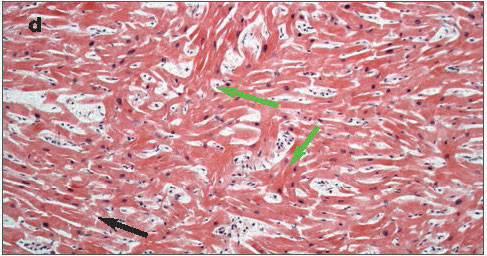
Microscopically the condition is characterised by myocyte disarray and fibrosis, see figure 2d.4
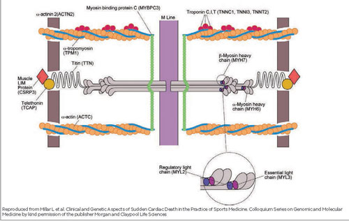
Up to 70% of cases of HCM are caused by mutations within genes encoding sarcomeric contractile proteins with the two most common mutations affecting myosin heavy chain 7 (MYH7) and myosin-binding protein C3 (MYBPC3).
Other gene mutations implicated include those affecting cardiac troponin, actin and titin, see figure 3.2 There have been several non-sarcomeric mutations described but these are less common. In addition there are a few rarer metabolic disorders which may mimic HCM such as Fabry’s disease, Danon disease and PRKAG-related cardiomyopathy.
Arrhythmogenic right ventricular cardiomyopathy
Arrhythmogenic right ventricular cardiomyopathy (ARVC) is a rare autosomal dominant heart muscle disorder which affects between 1 in 1,000 and 1 in 5,000 of the population.2,6
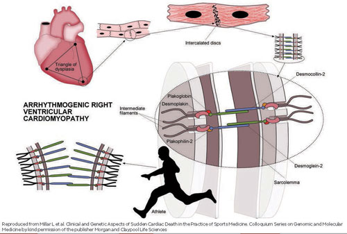
The term ARVC is a misnomer since the left ventricle may also be affected and the term arrhythmogenic cardiomyopathy may be more appropriate. The condition is caused by gene mutations encoding cardiac desmosomes which usually help bind the cardiomyocytes together. The most common desmosomal mutations associated with ARVC include those of desmoplakin, plakophilin-2, desmoglein-2 and desmocollin-2, see figure 1.
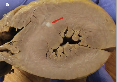
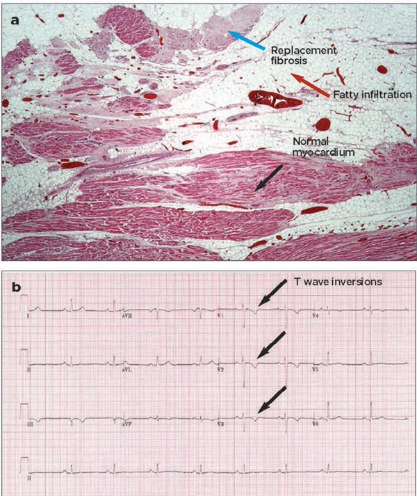
The current hypothesis is that abnormal desmosomal proteins lead to myocyte detachment during exertion and promote arrhythmias. Subsequent repair is abnormal leading to fibrofatty replacement of cardiac muscle, see figure 4a.
This fibrofatty replacement can interfere with the conduction pathways and hence predispose to cardiac arrhythmias.
Furthermore, it can weaken the myocardium predisposing to dilatation and aneurysm formation. ARVC is the most common cause of death in young sportsmen in Italy.7
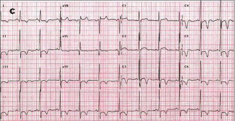
Typically the 12-lead ECG shows T wave inversions in the right precordial leads, V1-V3, see figure 4b, although T wave inversions in the lateral leads are suggestive of left ventricular involvement. The QRS complexes are often small and ventricular extra-systoles are common. The diagnosis of ARVC is based on the revised ARVC Task Force criteria.8 A plethora of electrical and imaging tests is usually required before the diagnosis is established.
Dilated cardiomyopathy
Dilated cardiomyopathy (DCM) is characterised by a dilated left ventricle with impaired function that cannot be explained by ischaemic heart disease, hypertension or valvular heart disease.
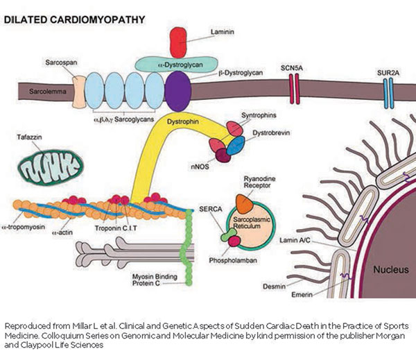
At least 25% of cases of DCM are familial and caused by a diverse spectrum of genetic mutations affecting the sarcomeric contractile proteins or the nuclear envelope. Lamin A/C mutations are the most common genetic abnormalities seen in patients with a familial DCM; however more than 30 gene mutations have been linked to this condition, see figure 5.
Dilated cardiomyopathy may be associated with multisystem conditions such as muscular dystrophy. Chemotherapy and certain other drugs, alcohol abuse and myocarditis may also lead to a dilated and poorly contracting left ventricle.
Restrictive cardiomyopathy
Restrictive cardiomyopathy is the least common form of the inherited cardiomyopathies and accounts for no more than 5% of all cardiomyopathies. The condition is characterised by stiffened ventricular walls associated with raised ventricular filling pressures. Mutations in genes encoding sarcomeric contractile proteins are often implicated. Patients are usually very symptomatic from pulmonary and peripheral congestion. The mainstay of treatment is loop diuretics.
Unclassified cardiomyopathies
Of the unclassified cardiomyopathies, left ventricular non-compaction (LVNC) cardiomyopathy is the most common.
LVNC is a heart muscle disorder which is characterised by marked left ventricular trabeculation and deep inter-trabecular recesses. It is unclear whether it is truly an independent cardiomyopathy as this phenomenon is seen in other cardiomyopathies such as HCM, DCM and in many neuromuscular disorders associated with cardiac involvement.
Several mutations have been implicated which also overlap with those causing HCM and DCM.
The condition is characterised by progressive left ventricular dysfunction, a predilection to ventricular arrhythmias and systemic thromboembolism.
The disease is heterogeneous and some patients may be completely asymptomatic.
Presentation
For many people the first manifestation of an inherited cardiomyopathy can be a sudden cardiac arrest.
GPs can play a key role in providing psychological support to the family during this difficult time. It might be helpful to put the family in touch with bereavement charities such as Cardiac Risk in the Young, see Useful information box.
Other presentations include chest pain or breathlessness during exertion, palpitations and syncope. Exercise-induced syncope or pre-syncope is a very ominous symptom and should be taken very seriously.
If an ECG or echocardiogram performed in the community is suggestive of an underlying cardiomyopathy, the patient should be referred to a cardiologist with a specialist interest in this field.
Diagnosis
In many of the cardiomyopathies, the diagnosis can be made with a standard ECG and echocardiogram; however if the diagnosis is not certain or the cardiologist wishes to look at the heart structure in greater detail, a cardiac MRI may be performed.
Cardiac MRIs have become a lot more commonplace as they provide more in-depth information about the heart structure, particularly some parts of the right ventricle, the lateral free wall of the left ventricle and the left ventricular apex. The main advantage of cardiac MRI over echocardiography is the ability to detect myocardial fibrosis which is often seen in the inherited cardiomyopathies and is a recognised substrate for ventricular arrhythmias.
Management
In inherited cardiomyopathies such as HCM and ARVC, if patients are stable they are usually reviewed annually at a cardiology clinic. Along with a general symptom review, the patient’s risk
of sudden cardiac death is assessed.
If the risk stratification deems them low risk they continue to be reviewed annually; however if they are felt to be high risk, an implantable cardioverter-defibrillator is recommended.
For symptomatic patients with HCM a beta-blocker is the first-line treatment and a calcium channel blocker such as verapamil can be used if a beta-blocker is contraindicated. For those with a significant left ventricular outflow gradient disopyramide can be added.
Atrial fibrillation may not be well tolerated in HCM and therefore cardioversion is preferred to rate control if possible. Beta-blockers are first-line treatment for ventricular extrasystoles or episodes of non-sustained ventricular tachycardia in ARVC but if the patient remains symptomatic despite these, amiodarone can be added. For those with a high ectopic burden, catheter ablation may be considered.
Dilated cardiomyopathy is treated with conventional heart failure treatment usually with a beta-blocker and an ACE inhibitor in the first instance.
Additional therapy such as an aldosterone antagonist can be added in if the patient deteriorates despite this and loop diuretics are recommended for the treatment of symptomatic pulmonary or peripheral oedema.
In most instances patients with cardiomyopathy will be managed in a specialist unit. They will not require regular monitoring in primary care with the exception of measuring electrolytes following the introduction of diuretics or a dose increment of an ACE inhibitor, angiotensin II receptor blocker or aldosterone antagonist.
Patients treated with amiodarone should have six monthly liver function and thyroid function tests. Angina and breathlessness can be managed by initiating or increasing the dose of beta-blocker or diuretic but an early review by a specialist should be requested.
We would consider the development of syncope or rapid palpitations a medical emergency.
Conclusion
Inherited cardiomyopathies are rare, but it is likely that GPs will encounter affected patients at some stage in their clinical practice. Sudden cardiac death is not an infrequent first presentation, so it is important to be vigilant for red flag symptoms such as exercise-induced syncope and certain ECG manifestations of cardiomyopathies.
Prompt referral of first-degree relatives of patients with inherited cardiomyopathies to a cardiologist with an interest in inherited heart disease is imperative to detect others who may be at risk.
Charities such as Cardiac Risk in the Young and the Cardiomyopathy Association, see Useful information box below, can be an extremely useful source of support to bereaved families and those adjusting to a new diagnosis of a cardiomyopathy.
Acknowledgement
With thanks to Professor Mary Shepphard, Institute of Cardiovascular Pathology, St George's, University of London for providing the cardiac pathology specimens.
REFERENCES
1 Maron BJ, Doerer JJ, Haas TS et al. Sudden death in young competitive athletes: analysis of 1866 deaths in the United States, 1980-2006. Circulation 2009;119(8): 1085-92
2 Jacoby D, McKeena WJ. Genetics of inherited cardiomyopathy. Eur Heart J 2012;33(3):296-304
3 Spirito P, Seidman CE, McKenna WJ et al. The management of hypertrophic cardiomyopathy. N Engl J Med 1997;336(5):349-50
4 Varnava AM, Elliott PM, Baboonian C et al. Hypertrophic cardiomyopathy: histopathological features of sudden death in cardiac troponin T disease. Circulation 2001;104(12):1380-4
5 Millar L, Sheikh N, Sharma S. Clinical and Genetic Aspects of Sudden Cardiac Death in the Practice of Sports Medicine. Colloquium Series on Genomic and Molecular Medicine. Morgan and Claypool Life Sciences. New Jersey. 2012
6 Gollob MH, Blier L, Brugada R et al. Recommendations for the use of genetic testing in the clinical evaluation of inherited cardiac arrhythmias associated with sudden cardiac death: Canadian Cardiovascular Society/Canadian Heart Rhythm Society joint position paper. Can J Cardiol 2011; 7(2):232-45
7 Corrado D, Thiene G, Nava A et al. Sudden death in young competitive athletes: clinicopathologic correlations in 22 cases. Am J Med 1990;89(5):588-96
8 Marcus FI, McKenna WJ, Sherrill D et al. Diagnosis of arrhythmogenic right ventricular cardiomyopathy/ dysplasia: proposed modification of the task force criteria. Circulation 2010;13: 1533-41
ARTICLE IN PDF
 Download this article in PDF format
CLICK ON IMAGES
PRESET SEARCHES
PubMed
NICE evidence
Immediate CPD prompts
These handy templates* prompt immediate drafting of personal learning, reflection and planning. Save your record straight into your personal development folder on your device or Cloud storage system.
*Adobe reader needed for iPad