SEARCH

Onida S, Lane TRA, Davies AH. Improving the management of varicose veins. Practitioner 2013; 257 (1766):21-24
Improving the management of varicose veins
05 Dec 2013
AUTHORS
Miss Sarah Onida BSc MBBS MRCS, Core Surgical Trainee, North West Thames Rotation, London, UK
Mr Tristan RA Lane BSc MBBS MRCS, Clinical Research Fellow in Vascular Surgery, Imperial College Healthcare, NHS Trust, London, UK
Professor Alun H Davies MA DM FRCS FEBVS FACPh, Professor of Vascular Surgery, Imperial College London and Honorary Consultant Vascular Surgeon, Imperial College Healthcare NHS Trust, London, UK. Chair of the NICE guideline development group on varicose veins
Article
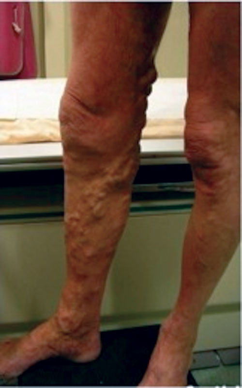
Varicose veins are a common manifestation of chronic venous disease and are defined as tortuous, dilated subcutaneous veins most commonly involving the lower limbs. The reported incidence is variable but it is thought that up to 30% of the UK population are affected.1
The aetiology of varicose veins is unclear. They are a manifestation of increased venous pressure in the lower limb caused by impaired venous return. Primary varicosities result from poor drainage from the superficial to the deep venous system. This is secondary to valvular incompetence at the site of connections between the two systems, most notably the saphenofemoral and saphenopopliteal junctions. There may also be incompetent valves within perforator veins that cause reflux from the deep to the superficial venous system.
Secondary varicosities arise as a result of underlying pathology impeding venous drainage, such as deep venous thrombosis (DVT) or increased intra-abdominal pressure caused by a mass, pregnancy or obesity.
Chronic venous insufficiency can be defined as the full spectrum of morphological and functional abnormalities of the venous system from telangiectasiae to venous ulcers.2
Chronic venous insufficiency and varicose veins present with a variety of signs and symptoms that have an important effect on patients’ quality of life.3 The sequelae of untreated chronic venous insufficiency can result in severe complications, including superficial thrombophlebitis, DVT, progressive skin changes, ulceration and potentially life-threatening bleeding.4 It is therefore important to recognise patients at risk and refer them to a vascular service.
NICE published a clinical guideline on the diagnosis and management of varicose veins in the legs in July 2013.5 The key recommendations on referral, assessment and treatment, management in pregnancy and information for patients, are listed in table 1.
Referral
Patients with bleeding varicose veins should be referred to a vascular service immediately.
Referral is also indicated in the following cases:
• Symptomatic primary or recurrent varicose veins
• Lower limb skin changes, such as pigmentation or eczema, thought to be caused by chronic venous insufficiency
• Superficial vein thrombosis (characterised by the appearance of hard, painful veins) and suspected venous incompetence
• A venous leg ulcer (a break in the skin below the knee that has not healed within two weeks)
• A healed venous leg ulcer
Assessment and investigation
Imaging is crucial in the assessment of the superficial and deep venous system to enable assessment of venous competence. The gold standard technique is colour duplex ultrasonography, which not only enables assessment of the anatomy of the superficial and deep venous systems, but also real time observation of the direction of flow and site of incompetence.
The Guidelines for the Society for Vascular Surgery and the American Venous Forum, published in 20116 and, most recently the NICE guidance, both recommend that duplex ultrasound should be used to confirm the diagnosis of varicose veins and the extent of truncal reflux, and to plan treatment for patients with suspected primary or recurrent varicose veins.
Venography may be used in cases with inconclusive duplex results, while magnetic resonance or computed tomographic venography are helpful when assessing abdominal and pelvic veins.
Treatment
Surgical treatment for varicose veins has been carried out for centuries, with superficial vein ligation, phlebectomy and stripping being the mainstay of treatment. However, these techniques are not without complications, including risk of DVT, damage to cutaneous nerves, pain and scarring.
In recent years, new techniques have been developed that are minimally invasive, enabling treatment of superficial venous incompetence with reduced morbidity.
NICE recommends that endothermal ablation, in the form of radiofrequency or laser treatment, should be offered as treatment for patients with confirmed varicose veins and truncal reflux.
Endothermal ablation
Endothermal ablation utilises heat energy within the vein to ablate the incompetent vessel. There are two main systems to deliver this energy: radiofrequency ablation (RFA) and endovenous laser ablation (EVLA).
Radiofrequency ablation
This technique employs radiofrequency waves to deliver thermal energy via a radiofrequency catheter, which is inserted into the superficial vein (usually long or short saphenous) via the Seldinger technique following ultrasound guided venous puncture. The catheter tip is placed about 2 cm from the junction (saphenofemoral or saphenopopliteal) in order to reduce the risk of the heat energy being delivered to the junction/deep vein, increasing the risk of DVT.
The heat is delivered via the catheter tip, which is 7 cm long and reaches temperatures of 85-120°C, damaging the venous endothelium. As the treatment is delivered, the catheter is sequentially withdrawn, ensuring the vein is treated throughout its length.
To protect the vein from thermal damage, tumescent anaesthesia is instilled under ultrasound guidance. This is a mixture of cold saline and local anaesthetic, which coats the vein, separating it from the surrounding structures and reducing the risk of heat energy being transmitted to other tissues.
RFA can be performed as a day surgery procedure under local anaesthesia; a meta-analysis has shown no significant difference in recurrence rates compared with open surgery7 but a quicker return to work in the RFA group. There is evidence suggesting patients tolerate it well, have a shorter recovery period and improved quality of life scores.8
Patients should be counselled about the risk of bleeding, infection, damage to the saphenous or sural nerves (secondary to thermal energy), DVT and superficial burns.
Endovenous laser ablation
EVLA works in a similar way to RFA in that it employs heat to damage the intima of the vein and therefore requires tumescent anaesthesia.
Unlike radiofrequency ablation, heat is generated by laser energy, reaching temperatures of up to 800°C at the very tip of the catheter, which is withdrawn at a constant speed during the treatment.
EVLA is also a day surgery procedure. Outcomes are comparable with RFA and open surgery,9,11 with the advantage of reduced postoperative pain and quicker return to work. EVLA enables the surgeon to treat a variety of veins; the catheter is smaller than that used for RFA and so it can be used to ablate very small veins.
Other minimally invasive treatments
There are a number of other minimally invasive treatments available, including steam therapy, cryoablation (using liquid nitrogen to freeze the vein), or mechano-chemical ablation, which combines direct trauma to the venous endothelium by means of a specially designed catheter tip and liquid sclerosant instillation.
This hybrid technique obviates the need for tumescent anaesthesia as no heat energy is delivered. Randomised controlled trials are being performed to assess the effectiveness of this method; preliminary results from prospective trials have shown that this is a safe, effective option to treat varicose veins.10
None of these treatments are recommended in the NICE guidance. However, it is important for clinicians to be aware of their existence.
Ultrasound guided foam sclerotherapy
In cases where endothermal ablation is unsuitable (e.g. very tortuous segments/truncal veins, small diameter veins), NICE recommends the use of ultrasound guided foam sclerotherapy (UGFS).
UGFS involves inserting a small volume of sclerosant into a vein followed by compression, allowing the endothelial surface to fibrose and the vein to seal. Sodium tetradecyl sulphate and polidocanol are the sclerosants most commonly used in the UK. Sclerotherapy can be delivered by an endovenous catheter (as in RFA/EVLA) or via direct ultrasound guided puncture with needle and syringe.
The sclerosant solution is deactivated by contact with blood; it is therefore aereated via the Tessari technique, which converts the liquid solution into foam, displacing blood and improving contact with the endothelium.
Results for foam sclerotherapy are heterogeneous but studies have shown occlusion rates comparable with open surgery at one year.8Complications include DVT, skin pigmentation, allergy to the sclerosant and skin necrosis in the case of extravasation.
Surgery
Where both endovenous ablation and UGFS are unsuitable or unsuccessful, surgery should be offered. The basic principle is disconnection of the refluxing superficial venous system from the deep system. This is achieved by saphenofemoral or saphenopopliteal junction ligation, entailing a groin or popliteal fossa incision, dissection to the junction, isolation and ligation of tributaries and transfixion of the superficial vein. To reduce recurrence rates, the superficial vein can be removed by stripping. Targeted ultrasound-guided cutdown can be performed on incompetent perforator veins requiring ligation.
Open venous procedures are performed under general or local anaesthetic as a day case. Effectiveness is comparable with RFA, EVLA and sclerotherapy;8 however patients have a prolonged return to work and increased postoperative pain.
Complications include bleeding, infection, haematoma formation, scarring, recurrence and damage to superficial nerves (saphenous, sural), particularly in the case of stripping.
Phlebectomy
The NICE guideline does not cover the use of phlebectomy. However, concomitant phlebectomy for superficial varicosities may be considered in patients receiving ablation/surgery for truncal disease with large varicose veins.
Phlebectomies are performed under local anaesthetic, with small stab incisions enabling access to the varicose segment with a phlebectomy hook. The veins are disconnected and removed. Studies have shown that phlebectomies alone can improve reflux and symptoms of venous disease.12
Compression
Compression therapy has been used since biblical times. The theory behind this technique is reduction of venous stasis and pressure in the skin by the application of graded external pressure to the leg (tightest at the ankle, reducing up the calf and thigh), compressing the varicose veins in the skin.
The literature in this field is extremely heterogeneous, with equivocal evidence with regards to the actual benefit of the technique.13 Patient reports are subjective, non-adherence rates are high and comparison between studies is difficult.
Importantly, compression should not be used in patients with arterial peripheral vascular disease and ankle brachial pressure index (ABPI) < 0.9. NICE recommends that compression hosiery should not be offered unless interventional treatment is unsuitable or should be temporarily avoided, e.g. in the case of pregnancy.
Conclusion
Varicose veins in the legs are a common cause of patient morbidity and affect a large proportion of patients in the community. The recent NICE clinical guideline provides evidence-based recommendations that will help improve the assessment and management of patients with this condition.
REFERENCES
1 Evans CJ, Fowkes FG, Ruckley CV, Lee AJ. Prevalence of varicose veins and chronic venous insufficiency in men and women in the general population: Edinburgh Vein Study. J Epidemiol Community Health 1999;53:149-153
2 Bergan JJ (Ed). The Vein Book. Elsevier Academic Press. Edinburgh. 2007
3 Darvall KA, Bate GR, Adam DJ, Bradbury AW. Generic health-related quality of life is significantly worse in varicose vein patients with lower limb symptoms independent of CEAP clinical grade. Eur J Vasc Endovasc Surg 2012;44(3):341-4
4 Ampanozi G, Preiss U, Hatch G et al. Fatal lower extremity varicose vein rupture; Leg Med (Tokyo) 2011;13(2):87-90
5 National Institute for Health and Care Excellence CG168. Varicose veins in the legs: The diagnosis and management of varicose veins. NICE. London. 2013
6 Gloviczki P, Comerota AJ, Dalsing MC et al. The care of patients with varicose veins and associated chronic venous diseases: clinical practice guidelines of the Society for Vascular Surgery and the American Venous Forum. J Vasc Surg 2011:53(5 Suppl):2S-48S. doi: 10.1016/j.jvs.2011.01.079
7 Brar R, Nordon IM, Hinchliffe RJ et al. Surgical management of varicose veins: meta-analysis. Vascular 2010;18:205-20
8 Rasmussen LH, Lawaetz M, Bjoern L et al. Randomized clinical trial comparing endovenous laser ablation, radiofrequency ablation, foam sclerotherapy and surgical stripping for great saphenous varicose veins.
Br J Surg 2011;98:1079-87
9 Carradice D, Mekako AI, Mazari FA et al. Clinical and technical outcomes from a randomized clinical trial of endovenous laser ablation compared with conventional surgery for great saphenous varicose veins. Br J Surg 2011;98:1117-23 doi 10:1002/bjs.7615
10 Boersma D, van Eekeren RR, Werson DA et al. Mechanochemical endovenous ablation of small saphenous vein insufficiency using the Clarivein device: one year results of a prospective series. Eur J Endovasc Surg 2013;45(3):299-303
11 Rasmussen L, Lawaetz M, Bjoern L et al. Randomized clinical trial comparing endovenous laser ablation and stripping of the great saphenous vein with clinical and duplex outcome after 5 years. J Vasc Surg 2013;58(2):421-6
12 Pittaluga P, Chastanet S, Rea B, Barbe R. Midterm results of the surgical treatment of varices by phlebectomy with conservation of a refluxing saphenous vein. J Vasc Surg 2009;50:107-8 doi:10.1016/j.jvs.2008.12.067
13 Palfreyman SJ, Michaels JA. A systematic review of compression hosiery for uncomplicated varicose veins. Phlebology 2009;24(suppl 1):13-33
USEFUL INFORMATION
Various versions of NICE CG168. Varicose veins in the legs, including information for patients are available from:
ARTICLE IN PDF
 Download this article in PDF format
PRESET SEARCHES
Immediate CPD prompts
These handy templates* prompt immediate drafting of personal learning, reflection and planning. Save your record straight into your personal development folder on your device or Cloud storage system.
*Adobe reader needed for iPad