SEARCH

Reynolds J, Tosounidou S, Gordon C. Optimising the management of systemic lupus erythematosus. Practitioner May 2019;263(1826):17-222
Optimising the management of systemic lupus erythematosus
22 May 2019
AUTHORS
Dr John Reynolds PhD MRCP, Senior Clinical Lecturer in Rheumatology, Rheumatology Research Group, Institute of Inflammation and Ageing, University of Birmingham, Consultant Rheumatologist, Sandwell and West Birmingham Hospitals NHS Trust, Birmingham, UK
Dr Sophia Tosounidou MRCP(UK), Consultant Rheumatologist, Sandwell and West Birmingham Hospitals NHS Trust, Birmingham, UK
Professor Caroline Gordon MD FRCP, Emeritus Professor of Rheumatology, Rheumatology Research Group, Institute of Inflammation and Ageing, University of Birmingham, Honorary Consultant Rheumatologist, Sandwell and West Birmingham Hospitals NHS Trust, Birmingham, UK
Competing interests
Professor Caroline Gordon has undertaken consultancies and received honoraria from Bristol-Myers Squibb, GlaxoSmithKline, Merck Serono, and UCB, has been a member of the speakers’ bureau for GlaxoSmithKline, UCB and Lilly and has received research grant support from UCB. Dr Sophia Tosounidou has been a member of the speakers’ bureau for UCB. Dr John Reynolds has no competing interests.
Article
Abstract
Systemic lupus erythematosus (SLE or lupus) is a multisystem, autoimmune disease. It can present in women or men at any age but is most common in women of reproductive age, especially those of African-Caribbean or South Asian descent. Lupus affected nearly 1 in 1,000 of the UK population in 2012 with a mean age at diagnosis of 49 years. Lupus nephritis occurs in a third of lupus patients in the UK and is associated with significant mortality and morbidity. Lupus mainly affects women and causes fatigue, rashes (often associated with photosensitivity), hair loss and inflammatory arthritis. However, lupus can also affect men in whom it is often more severe with a greater likelihood of renal involvement and a greater degree of organ damage. Constitutional (e.g. fever and lymphadenopathy), cardio-respiratory (e.g. pleurisy and pericarditis), GI (e.g. gall bladder vasculitis) and hepatic (e.g. autoimmune hepatitis) features may occur in up to 60% of patients. An autoantibody screen should be performed when there is a clinical suspicion of SLE. Around 95% of lupus patients are ANA positive. However, ANA is not specific for lupus. According to the BSR guideline, the diagnosis of SLE requires a combination of clinical features and the presence of at least one relevant immunological abnormality. Biopsy proven lupus nephritis in the presence of ANA or anti-dsDNA antibodies may also be classified as lupus without the need for other criteria.
Systemic lupus erythematosus (SLE or lupus) is a multisystem, autoimmune disease that can present in women or men at any age but is most common in women of reproductive age especially those of African-Caribbean and South Asian descent.1 Lupus affected nearly 1 in 1,000 of the population in the UK in 2012.1
The mean age at diagnosis was 48.9 years1 but was lower in those of African ancestry.1 Patients of African origin are at increased risk of lupus nephritis which occurs in about one third of lupus patients in the UK.
End-stage renal disease occurred in 20% of lupus nephritis patients in a UK cohort study. The mean age at death for all patients with lupus nephritis was 40.3 years with an average interval of 7.5 years between the development of lupus nephritis and death.2
In another UK cohort study, 382 patients were followed up for up to 21 years during which time 37 patients (9.7% of the cohort) died. Those who died had a mean age at death of 53.7 years.3 Most deaths were due to infection or cardiovascular disease and active lupus rarely caused death in the UK.3,4
Lupus causes considerable morbidity as it may present with slowly or rapidly progressive active disease that can lead to the accumulation of chronic damage if not promptly diagnosed, appropriately treated and regularly monitored.5,6
To optimise management and improve outcomes in this variable and potentially life-threatening disease the British Society for Rheumatology (BSR) published a NICE-accredited evidence-based guideline for the management of SLE in adults in 2018.7,8
Presentation
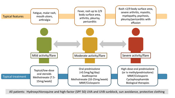
Lupus can present with a wide range of clinical features affecting any body system, so diagnosis can be challenging.9-11 Delays in diagnosis are well recognised and remain a concern.12 Some of the most typical mild, moderate and severe features are shown in figure 1.
It is important to ensure that the diagnosis of lupus is accurate before considering treatment.10,11 Lupus should be considered particularly, but not exclusively, in individuals at increased risk of the disease with African, South Asian and Chinese heritage.13
Lupus causes fatigue, skin rashes often associated with photosensitivity, hair loss and inflammatory arthritis mainly in women. However, it is important to remember that lupus can affect men as well. In men it often results in severe disease including renal involvement and more chronic scarring (damage) compared with women in many studies, possibly because of delayed diagnosis.3,4,14 Renal and neurological involvement cause considerable morbidity and mortality.3,4 Constitutional (e.g. fever and lymphadenopathy) cardio-respiratory (e.g. pleurisy and pericarditis), gastrointestinal (e.g. gall bladder vasculitis) and hepatic (e.g. autoimmune hepatitis) features may occur in up to 60% of patients but are often not recognised as being caused by lupus, and need to be distinguished from infection, drug side effects and other comorbid conditions including lymphoma.8 Depression occurs in about 20% of lupus patients but is not usually caused directly by autoimmune disease mechanisms. Eye involvement, other than sicca symptoms, is rare but potentially sight threatening and requires prompt referral to an experienced ophthalmologist.15
Causes and immunological manifestations
The clinical features of acute lupus are mostly due to the formation of immune complexes involving autoantibodies that induce complement activation and consumption resulting in inflammatory processes that affect a variety of organs.
Thrombosis associated with antiphospholipid antibodies may contribute to the pathogenesis in some patients.5,9 The autoantibodies are produced as a result of various mechanisms including defective clearance of apoptotic cells, excessive B cell activation and proliferation, altered T cell signalling and function, and changes in cytokine profiles with an increasing role for interferon-alpha in recent research.5,16
There is evidence for underlying genetic susceptibility involving an increasing number of loci (50-100 genes) and pathways with environmental triggers including infections, UV light, hormones such as oestrogen, and cigarette smoking.
Drug-induced lupus is usually resolved by withdrawal of the offending drug, e.g. tetracyclines, isoniazid, sulphonamides, propylthiouracil, hydralazine, procainamide, phenytoin and anti-TNF therapy.5,17,18 Anti-TNF may induce autoantibodies such as antinuclear antibody (ANA) without inducing lupus disease, however these drugs may also trigger full SLE in susceptible people.
The importance of environmental influences is shown by the observation that in only one in four twin pairs, where one twin has SLE, will the other twin develop lupus over time.
An autoantibody screen should be performed when there is a clinical suspicion of SLE. Most lupus patients are ANA positive (> 95%). If the patient is ANA negative, there is a low clinical probability of SLE. However, a minority have anti-double stranded (ds)DNA or anti-Ro antibody positivity without ANA that will be found when assessed on clinical grounds by a specialist.19
ANA tests are not specific for the diagnosis of lupus. ANA positive results may occur in other conditions including Sjögren’s syndrome, systemic sclerosis, dermatomyositis, viral infections such as infectious mononucleosis and cancer.11 The ANA test result can become negative in treated patients and the results can vary with different assays. Anti-dsDNA and anti-Sm antibodies are rare in other conditions but are less sensitive than ANA.9,19,20,21 Anti-dsDNA antibodies rise and complement C3 and C4 levels fall with active disease and are useful in monitoring disease as well as diagnosis, see box 1.7,8
Anti-Ro and anti-La antibodies are associated with photosensitivity, subacute cutaneous lupus rashes, Sjögren’s syndrome and neonatal lupus syndrome including congenital complete heart block in babies.11,22 Anti-RNP antibodies occur in overlap conditions such as mixed connective tissue disease with myositis and lung involvement.11
Lupus patients should be tested for antiphospholipid antibodies which confer an increased risk of arterial/venous thrombosis and adverse pregnancy outcomes such as three or more first trimester miscarriages, late pregnancy losses and pre-eclampsia before 34 weeks gestation.23,24
Confirmatory tests for antiphospholipid syndrome are positive lupus anticoagulant, anticardiolipin antibodies (IgG, IgM), and/or anti-beta2 glycoprotein I (IgG, IgM) antibodies on two occasions at least 12 weeks apart.23,24
The lupus anticoagulant test is the most specific of the three tests and is associated with a higher positive predictive value. Triple positivity characterised by positive lupus anticoagulant, anticardiolipin, and anti-beta2 glycoprotein I antibodies is associated with a cumulative incidence of thrombosis of 37.1% after ten years.25
Classification criteria
When considering a diagnosis of SLE, a detailed clinical history and examination is required to identify clinical features supported by renal and haematological assessment. A low CRP with a raised ESR is typical of active lupus unless infection or effusions are present. According to the BSR guideline7,8 the diagnosis requires a combination of clinical features and the presence of at least one relevant immunological abnormality as discussed above. A positive ANA occurs in approximately 5% of adults and has poor diagnostic value in the absence of clinical features of lupus or other autoimmune disease.
The American College of Rheumatology (ACR)26,27 and Systemic Lupus International Collaboration Clinics (SLICC)28 (see table 1) classification criteria are not diagnostic criteria but may be helpful when considering the diagnosis. However, they do not cover all the possible clinical manifestations of lupus.
Based on the ACR (previously the American Rheumatism Association) revised criteria for SLE published in 198226 and the 1997 modification,27 patients may be classified as having SLE if they have had four or more of 11 criteria at any time. However, not all patients with these criteria have lupus and not all patients diagnosed clinically with lupus have four or more of these criteria.
The SLICC group devised alternative classification criteria for lupus to overcome these and related issues (see table 1, p18).28 The critical difference is a requirement for at least one clinical and one immunological criterion and two other criteria from an expanded list of items compared with the traditional ACR criteria.27
Biopsy proven lupus nephritis in the presence of ANA or anti-dsDNA antibodies may also be classified as lupus without the need for other criteria.28
Low complement levels (C3 and/or C4) are included in the serological criteria as they reflect complement consumption due to formation of immune complexes in active lupus disease.
Although the SLICC criteria are more intuitive and include evidence for the immunopathological basis of lupus, diagnosis should not be restricted to patients that meet these criteria.
According to the BSR guideline for SLE7,8 a diagnosis of SLE can also be made in the presence of other lupus manifestations in the appropriate serological context and should not be made if features are better explained by an alternative diagnosis (such as a different systemic autoimmune disease, infection or cancer).
Clinical assessment
Clinical assessment should include a detailed history and review of systems, ideally with full clinical examination including urinalysis in order to determine whether symptoms and signs may be caused by lupus disease activity, accumulated damage due to the effects of the disease or its treatment such as lung fibrosis or atherosclerosis, drug toxicity and/or the presence of comorbidity.
Fatigue is very common but nonspecific as it can be hard to distinguish whether the cause is lupus itself or if there is another autoimmune condition such as hypothyroidism, anaemia due to iron deficiency (not haemolysis from lupus), fibromyalgia which can coexist with lupus in up to 30% of patients, depression and/or physical deconditioning.
Haematological, biochemical, immunological and imaging investigations help to determine whether new disease features are caused by active inflammation, thrombosis or comorbidity such as infection. This is an important distinction as this will affect the need for specialist review and future treatments. Antiphospholipid antibodies are associated with thrombotic events, damage and adverse outcomes in pregnancy and should be re-evaluated prior to pregnancy or surgery, and when there is a new severe manifestation or vascular event even if the tests were negative previously. Patients may need referral for skin or renal biopsies to ensure that lupus is the cause of the manifestations before starting definitive treatment.
Advice about further assessment and treatment should be sought from relevant specialists such as rheumatologists, nephrologists, haematologists and specialist obstetricians depending on the extent of disease activity and whether planning pregnancy or not.
Mild disease activity is clinically stable lupus with no life-threatening organ involvement (see figure 1). Those patients with moderate disease activity have more serious manifestations requiring more intense immunosuppression and severe disease is defined as organ or life threatening.
With an increase in disease activity (lupus flare) it can be helpful to consider triggers such as sunlight, concurrent or recent infection, hormonal changes, surgery and any recent drug change as this will guide further investigation, treatment and future monitoring.
Monitoring
Patients with lupus should be monitored regularly for disease features, drug toxicity and comorbidities.7,8 Depending on the complexity of the disease and the drug therapy this may need to be performed by a specialist e.g. rheumatologist, nephrologist, dermatologist or by the GP or shared care between the relevant teams.
Patients with active disease should be reviewed at least every 1-3 months with blood pressure, urinalysis, renal function, liver function tests, anti-dsDNA antibodies, complement levels and full blood count with further tests as necessary (see box 1). ESR is usually high and CRP low with active lupus so an increase in CRP should prompt an infection screen. Patients with stable low disease activity or in remission may be reviewed less frequently such as 6 to 12 monthly.
As lupus disease activity and complications such as atherosclerotic disease, infection and drug side effects can develop at any time, patients should be monitored indefinitely.7,8 Anti-Ro and anti-La antibodies are associated with neonatal lupus (including congenital heart block) and should be checked prior to pregnancy but are not helpful for assessing disease activity (see box 1).
A detailed guide to the interpretation of haematological, renal and other biochemical parameters is included in the BSR guideline on the management of lupus and is available online.8
Patients with lupus are at increased risk of infection, atherosclerotic disease, malignancy, osteoporosis and avascular necrosis.
Patients should be screened for modifiable risk factors including hypertension, dyslipidaemia, diabetes, high body mass index and smoking at least annually.7,8 They should have their vaccination status reviewed and be immunised as appropriate for example against influenza and pneumococcal pneumonia if they are on immunosuppressants (see table 2, left).
Complications of lupus including chronic fatigue, cardiovascular risk, osteoporosis, infection, and cancer risk should be managed according to national and international guidelines.7,8
Treatment
Until recently only prednisolone and hydroxychloroquine were licensed for lupus.5 There were relatively few trials until the past 15 years but in 2011, belimumab became the first drug to be licensed for the treatment of active lupus for more than 50 years.7,29,30
Much of the evidence supporting the use of drug therapies off licence in lupus comes from observational studies. This evidence is reviewed in the BSR guideline which divides the treatment of lupus into three sections covering mild, moderate and severe lupus7,8 (see figure 1). There is only summary advice about the use of drugs in the management of pregnant lupus patients as there is already a BSR guideline on the use of drugs in rheumatic diseases in pregnancy and breastfeeding.31,32
High factor UVA and UVB sunscreen are strongly recommended in the treatment and prevention of UV induced cutaneous lesions supported by good evidence. There is also some evidence that this strategy prevents other lupus manifestations. Patients should be advised about sun avoidance and protective clothing.7,8
There is high quality evidence for the management of mild lupus using the disease modifying drugs hydroxychloroquine and methotrexate. These drugs reduce the need for corticosteroids.7,8 Prednisolone doses up to 7.5 mg/day may be required for maintenance therapy but topical preparations for cutaneous manifestations and intra-articular injections for arthritis are preferred although there is less evidence for their use. Short courses of NSAIDs may be used for symptomatic benefit but the evidence is weak and the risks are considerable especially in patients with renal disease in whom they are contraindicated.7,8
The management of moderate lupus requires specialist advice and will usually involve the use of immunosuppressive, steroid-sparing drugs such as azathioprine, mycophenolate mofetil, calcineurin inhibitors (ciclosporin and tacrolimus) or methotrexate with corticosteroids, in addition to hydroxychloroquine and avoidance of UV radiation.7,8 The aim is to reduce disease activity, prevent further flares and lower the risk of damage accrual due to disease and corticosteroids in patients with features such as arthritis, cutaneous disease, serositis, vasculitis, or cytopenias not controlled by hydroxychloroquine.
Initial doses of prednisolone up to 0.5 mg/kg/day or the use of intramuscular or intravenous doses of methylprednisolone may be required to control active disease but prednisolone should be reduced steadily to a baseline of 7.5 mg/day or less and withdrawn later if possible.7,8
Some patients are refractory to these drugs, and these cases should be discussed with and/or seen by a regional specialist lupus centre.
The biologic drugs belimumab and rituximab may be considered if the patients meet the appropriate criteria.7,8,33,34
Organ- or life-threatening severe lupus disease, for example lupus nephritis or neuropsychiatric lupus, requires treatment by a specialist with intensive immunosuppression followed by prolonged maintenance therapy to prevent relapse.7,8 This usually involves intravenous methylprednisolone initially but may consist of high-dose oral prednisolone (up to 1 mg/kg/day) to induce remission with mycophenolate mofetil or cyclophosphamide, followed by steroid taper over several months.
Belimumab and rituximab are considered if patients fail conventional immunosuppressive drugs due to inefficacy or intolerance as described above. Intravenous immunoglobulin and plasmapheresis are rarely used. In some cases there may be a thrombotic component to the clinical features that requires anticoagulation. The evidence is best for the management of lupus nephritis, weaker for neuropsychiatric disease and is sparse for other specific organ-specific manifestations.7,8
Organisation of care
GPs should refer patients with suspected lupus to a physician with experience of managing lupus so that they can confirm the diagnosis, assess disease activity and provide advice on treatment and monitoring of lupus, its complications and side effects of therapy. Immunosuppressive therapies are challenging to use in patients with lupus because of the risk of adverse events including infection, difficulties with attribution of cytopenias to lupus or cytotoxic drugs, and the need to distinguish disease activity from damage and comorbid conditions.8
Shared care arrangements may be put in place once patients are stable on therapy. Patients should have access to a multidisciplinary team comprising a variety of specialists including rheumatologists, nephrologists, dermatologists, haematologists, cardiologists, chest physicians, neurologists, obstetricians, nurse specialists, physiotherapists, as well as podiatrists, occupational therapists and clinical psychologists working as part of collaborative clinical networks involving regional specialist centres, local hospitals and GPs.8
Patients should be monitored to ensure that they achieve a low level of disease activity if not remission35,36 using hydroxychloroquine, immunosuppressants and the minimum amount of corticosteroids, in order to reduce accumulating damage from the disease and its treatment.35 If drug treatment is not working within the expected timeframe, it is important to consider adherence and to get patients reviewed so that drug therapy can be optimised to promote disease control and to reduce the risk of further morbidity and premature death.
It is important for GPs to be involved in ensuring use of sunscreen and sun avoidance, adequate vitamin D3 intake given the inability to synthesise vitamin D in the skin without UV light exposure, weight control, exercise, smoking cessation and other measures to reduce atherosclerotic and osteoporosis risk factors, as well as cancer screening, contraception and pregnancy planning when the disease is under good control on appropriate treatment (see box 2).8
REFERENCES
1 Rees F, Doherty M, Grainge M et al. The incidence and prevalence of systemic lupus erythematosus in the UK, 1999-2012. Ann Rheum Dis 2016;75(1):136-41
2 Croca SC, Rodrigues T, Isenberg DA. Assessment of a lupus nephritis cohort over a 30-year period. Rheumatology (Oxford) 2011; 50(8):1424-30
3 Yee CS, Su L, Toescu V et al. Birmingham SLE cohort: outcomes of a large inception cohort followed for up to 21 years. Rheumatology (Oxford) 2015;54(5):836-43
4 Chambers SA, Allen E, Rahman A, Isenberg D. Damage and mortality in a group of British patients with systemic lupus erythematosus followed up for over 10 years. Rheumatology (Oxford) 2009;48(6):673-75
5 Lisnevskaia L, Murphy G, Isenberg D. Systemic lupus erythematosus. Lancet 2014;384(9957):1878-88
6 Bruce IN, O'Keeffe AG, Farewell V et al. Factors associated with damage accrual in patients with systemic lupus erythematosus: results from the Systemic Lupus International Collaborating Clinics (SLICC) Inception Cohort. Ann Rheum Dis 2015;74(9):1706-13
7 Gordon C, Amissah-Arthur MB, Gayed M et al. The British Society for Rheumatology guideline for the management of systemic lupus erythematosus in adults: Executive Summary. Rheumatology (Oxford) 2018; 57(1):14-18
8 Gordon C, Amissah-Arthur MB, Gayed M et al. The British Society for Rheumatology guideline for the management of systemic lupus erythematosus in adults. Rheumatology (Oxford) 2018;57(1):e1-e45
9 Cervera R, Khamashta MA, Hughes GR. The Euro-lupus project: epidemiology of systemic lupus erythematosus in Europe. Lupus 2009;18(10):869-74
10 Smith PP, Gordon C. Systemic lupus erythematosus: clinical presentations. Autoimmun Rev 2010; 10(1):43-45
11 Goldblatt F, O'Neill SG. Clinical aspects of autoimmune rheumatic diseases. Lancet 2013;382(9894):797-808
12 Lateef A, Petri M. Unmet medical needs in systemic lupus erythematosus. Arthritis Res Ther 2012;14 Suppl 4:S4
13 Kumar K, Chambers S, Gordon C. Challenges of ethnicity in SLE. Best Pract Res Clin Rheumatol 2009; 23(4):549-61
14 Amaral B, Murphy G, Ioannou Y, Isenberg DA. A comparison of the outcome of adolescent and adult-onset systemic lupus erythematosus. Rheumatology (Oxford) 2014;53(6):1130-35
15 Papagiannuli E, Rhodes B, Wallace GR et al. Systemic lupus erythematosus: an update for ophthalmologists. Surv Ophthalmol 2016;61(1):65-82
16 Wahren-Herlenius M, Dorner T. Immunopathogenic mechanisms of systemic autoimmune disease. Lancet 2013;382(9894):819-31
17 Almoallim H, Al-Ghamdi Y, Almaghrabi H, Alyasi O. Anti-tumor necrosis factor-alpha induced systemic lupus erythematosus. Open Rheumatol J 2012;6:315-19
18 Garcia-De LI, Garcia-Valladares I. Antinuclear antibody (ANA) testing in patients treated with biological DMARDs: is it useful? Curr Rheumatol Rep 2015;17(4):23
19 Ippolito A, Wallace DJ, Gladman D et al. Autoantibodies in systemic lupus erythematosus: comparison of historical and current assessment of seropositivity. Lupus 2011;20(3):250-55
20 Isenberg D. Thirty years, five hundred patients: some lessons learned from running a lupus clinic. Lupus 2010;19(6):667-74
21 Arroyo-Avila M, Santiago-Casas Y, McGwin G, Jr et al. Clinical associations of anti-Smith antibodies in PROFILE: a multi-ethnic lupus cohort. Clin Rheumatol 2015;34(7):1217-23
22 Brucato A, Cimaz R, Caporali R et al. Pregnancy outcomes in patients with autoimmune diseases and anti-Ro/SSA antibodies. Clin Rev Allergy Immunol 2011;40(1):27-41
23 Pierangeli SS, de Groot PG, Dlott J et al. 'Criteria' aPL tests: report of a task force and preconference workshop at the 13th International Congress on Antiphospholipid Antibodies, Galveston, Texas, April 2010. Lupus 2011;20(2):182-90
24 Ruiz-Irastorza G, Cuadrado MJ, Ruiz-Arruza I et al. Evidence-based recommendations for the prevention and long-term management of thrombosis in antiphospholipid antibody-positive patients: report of a task force at the 13th International Congress on antiphospholipid antibodies. Lupus 2011;20(2):206-18
25 Pengo V, Ruffatti A, Legnani C et al. Incidence of a first thromboembolic event in asymptomatic carriers of high-risk antiphospholipid antibody profile: a multicenter prospective study. Blood 2011;118(17):4714-18
26 Tan EM, Cohen AS, Fries JF et al. The 1982 revised criteria for the classification of systemic lupus erythematosus. Arthritis Rheum 1982;25(11):1271-77
27 Hochberg MC. Updating the American College of Rheumatology revised criteria for the classification of systemic lupus erythematosus. Arthritis Rheum 1997;40(9):1725
28 Petri M, Orbai AM, Alarcon GS et al. Derivation and validation of the Systemic Lupus International Collaborating Clinics classification criteria for systemic lupus erythematosus. Arthritis Rheum 2012; 64(8):2677-86
29 Navarra SV, Guzman RM, Gallacher AE et al. Efficacy and safety of belimumab in patients with active systemic lupus erythematosus: a randomised, placebo-controlled, phase 3 trial. Lancet 2011;377(9767):721-31
30 Furie R, Petri M, Zamani O et al. A phase III, randomized, placebo-controlled study of belimumab, a monoclonal antibody that inhibits B lymphocyte stimulator, in patients with systemic lupus erythematosus. Arthritis Rheum 2011;63(12):3918-30
31 Flint J, Panchal S, Hurrell A et al. BSR and BHPR guideline on prescribing drugs in pregnancy and breastfeeding-Part I: standard and biologic disease modifying anti-rheumatic drugs and corticosteroids. Rheumatology (Oxford) 2016;55(9):1693-97
32 Flint J, Panchal S, Hurrell A et al. BSR and BHPR guideline on prescribing drugs in pregnancy and breastfeeding-Part II: analgesics and other drugs used in rheumatology practice. Rheumatology (Oxford) 2016;55(9):1698-1702
33 National Institute for Health and Care Excellence. TA397. Belimumab for treating active autoantibody-positive systemic lupus erythematosus. NICE. London. 2016 www.nice.org.uk/guidance/TA397
34 NHS England. 2013 interim clinical commissioning policy statement for rituximab in adult SLE patients www.england.nhs.uk/wp-content/uploads/2013/09/a13-psa.pdf
35 van Vollenhoven RF, Mosca M, Bertsias G et al. Treat-to-target in systemic lupus erythematosus: recommendations from an international task force. Ann Rheum Dis 2014;73(6):958-67
36 van VR, Voskuyl A, Bertsias G et al. A framework for remission in SLE: consensus findings from a large international task force on definitions of remission in SLE (DORIS). Ann Rheum Dis 2017;76(3):554-61
ARTICLE IN PDF
 Download this article in PDF format
EXTERNAL WEBLINKS
British Society for Rheumatology guidelines
Patient information on lupus