SEARCH

Taylor S, Lobo AJ. Diagnosis and treatment of inflammatory bowel disease. Practitioner 2016;260(1795)19-23
Diagnosis and treatment of inflammatory bowel disease
01 Aug 2016
AUTHORS
Dr Sarah Taylor MB ChB MRCP, Specialist Registrar
Professor Alan J Lobo MD FRCP, Professor of Gastroenterology
Gastroenterology Unit, Royal Hallamshire Hospital, Sheffield Teaching Hospitals, and University of Sheffield, Sheffield, UK
Article
Abstract
Patients with inflammatory bowel disease (IBD) may previously have received a diagnosis of irritable bowel syndrome and there may be a delay in making the correct diagnosis. This is particularly the case in patients with ileal Crohn’s disease and those under 40. Diagnosis of IBD involves endoscopy and biopsy. Histology may not be available – for example in small bowel Crohn’s disease – and in this situation, typical radiological appearances help make the diagnosis. Crohn’s disease can affect any part of the gastrointestinal tract – most commonly the terminal ileum or colon. Perianal involvement is also common. Intestinal inflammation in Crohn’s disease can extend transmurally. Ulcerative colitis almost always affects the rectum with a variable proximal extent and continuous distribution. Inflammation affects the mucosa only. Approximately 25% of people with IBD will have extra-intestinal manifestations of the disease, involving skin, eyes, joints or the liver. There is an increased risk of colorectal cancer in patients with ulcerative colitis estimated at 2% at 10 years, 8% at 20 years and 18% at 30 years.It is important to be aware of infection risk in IBD both due to the disease itself and the impact of treatment. Flu vaccination should be offered to patients with IBD.
Inflammatory bowel disease (IBD) includes Crohn’s disease and ulcerative colitis. Both are chronic disorders of unknown aetiology, which characteristically run a relapsing and remitting course.
Data suggests that there are at least 115,000 people in the UK with a diagnosis of Crohn’s disease1 and around 146,000 with a diagnosis of ulcerative colitis.2 Most studies show a peak incidence of ulcerative colitis and Crohn's disease in the second to fourth decades, with a possible, second, later peak reported in fewer studies.3
Crohn’s disease can affect any part of the gastrointestinal tract – most commonly the terminal ileum or colon. Perianal involvement is also common. Intestinal inflammation in Crohn’s disease can extend transmurally. Ulcerative colitis almost always affects the rectum with a variable proximal extent and continuous distribution. Inflammation affects the mucosa only.
Genome wide association studies have shown that more than 200 genes are linked to Crohn’s disease, ulcerative colitis, or both.4 Abnormalities in intestinal immune responses and intestinal microbiota have also been demonstrated. Smoking is an important risk factor, doubling the risk of Crohn's disease.5 Smoking is also associated with poorer outcomes following medical treatment and surgery.
Clinical features
Lower gastrointestinal symptoms are common in the general population. Hence, patients with IBD may previously have received a diagnosis of irritable bowel syndrome6 and there may be a delay in making the correct diagnosis.6 This is particularly the case in patients with ileal Crohn’s disease and those under 40.6
The presence of weight loss, nocturnal diarrhoea and abnormal initial laboratory investigations (see below) make irritable bowel syndrome less likely. Typical symptoms of ulcerative colitis and Crohn’s disease are listed in table 1.
The severity of an attack of ulcerative colitis is usually described using the criteria of Truelove and Witts,2 (see table 2).
Initial investigations
IBD may be associated with a number of laboratory abnormalities:
• Anaemia: Anaemia may be caused by iron deficiency or chronic disease.
It may also be a reflection of severe disease. Iron deficiency is frequently under-recognised as a cause of symptoms such as malaise and fatigue in IBD.
• Thrombocytosis: A raised platelet count is part of an acute phase reaction caused by active inflammatory disease but may also reflect functional hyposplenism in IBD.
• Acute phase response: Active inflammation is represented by raised CRP or ESR. A low albumin is also a reflection of inflammatory activity.
The extent of the rise in CRP or ESR is greater in Crohn’s disease than ulcerative colitis, where these markers may be normal in those with mild or moderately severe inflammation. Raised inflammatory markers also help identify those that are more likely to respond to therapies such as anti-TNF-α agents.
• Stool culture: Evidence of infection should always be sought when patients present initially and, in those with established IBD, during a severe flare, if symptoms flare following foreign travel or if clinical features suggest infection.
• Faecal calprotectin: Faecal calprotectin is a protein produced by intestinal neutrophils as part of an inflammatory response. Faecal calprotectin testing helps to distinguish between IBD and irritable bowel syndrome7 with the aim of reducing the need for invasive investigations. It has a low specificity for IBD.
Testing for faecal calprotectin should not be carried out if bowel cancer is suspected or the diagnosis of IBS is unlikely, for example in those with weight loss, rectal bleeding, nutrient deficiency, nocturnal diarrhoea, perianal inflammation or sepsis. These patients should be referred for investigation or specialist opinion. NSAIDs should be stopped for four weeks before testing.
Diagnosis
NICE quality standards for IBD suggest that a delay in assessment and diagnosis in those suspected of having IBD may be associated with complications and a negative effect on quality of life and therefore that the interval from referral to specialist assessment should be no more than four weeks.8
More urgent assessment or even emergency admission may be needed in those with severe symptoms or complications.
Diagnosis of IBD involves endoscopy and biopsy. Histology may not be available – for example in small bowel Crohn’s disease – and in this situation, typical radiological appearances help make the diagnosis.
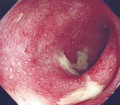
Colonoscopy allows a diagnosis of ulcerative colitis and Crohn’s disease of the colon to be made and the extent of inflammation to be assessed, which in turn helps guide treatment, see figure 1.
Small bowel involvement in Crohn’s disease can be assessed by MR enterography, small bowel barium meal/follow-through or capsule endoscopy according to local availability and practice.
Complications of Crohn’s disease, for example intra-abdominal abscess formation or intestinal obstruction caused by stricturing, may be best assessed by initial CT scan.
Treatment:Ulcerative colitis
Because of the relapsing and remitting nature of IBD, treatment requires phases to induce and then maintain remission. Some agents can be used in both phases of treatment.
Treatment to induce remission in ulcerative colitis depends on the extent of disease and its severity,2 see table 3.
Acute severe ulcerative colitis should be regarded as a medical emergency and requires admission to hospital for assessment and intensive intravenous corticosteroid therapy.
Fluids and blood transfusion may be necessary. Prophylaxis against venous thromboembolism is essential.
If patients do not settle rapidly on this regimen, escalation to treatment with ciclosporin or infliximab may be necessary. Approximately 20% of patients with acute severe ulcerative colitis do not settle with medical therapy and require colectomy.9
Maintenance of remission
Aminosalicylates are the mainstay of maintenance treatment. Once daily dosing helps improve adherence and is as effective as split dose regimens. There is a small risk of nephrotoxicity and so patients should have renal function checked while on these medications.
Thiopurines are used for patients with steroid-dependent disease – defined as a requirement for two or more courses of systemic steroids in 12 months. Following a severe flare of symptoms, the risk of a further severe flare is high and azathioprine would normally be used to try to prevent this.7
In the UK, azathioprine (which is subsequently metabolised to mercaptopurine) is usually used initially and mercaptopurine may be used in those who are intolerant of azathioprine. Prior testing of thiopurine methyl transferase status is essential to avoid life-threatening severe leukopenia that may occur in 1:300 patients. Ongoing blood monitoring, particularly for milder, and usually reversible, leucopenia is also essential.7
Treatment: Crohn’s disease
Again, disease site and the severity of symptoms should be taken into account when deciding on treatment.1 At present, gastroenterologists still adopt a step-up approach to therapy. There is debate as to whether a more aggressive top-down approach with earlier use of anti-TNF-α agents and immunosuppressants may result in fewer long-term sequelae from tissue damage.
Controlled ileal release budesonide refers to locally released preparations of budesonide, which undergo extensive first pass metabolism in the liver.
This results in fewer side effects than systemically available corticosteroids such as prednisolone – although with some reduction in comparative efficacy. They are effective in terminal ileal and ascending colonic Crohn’s disease.
Systemic steroids such as prednisolone are effective for active small and large bowel Crohn’s disease but associated with more side-effects than budesonide.
Methotrexate is effective for induction of remission and subsequent maintenance. It is usually administered intramuscularly or subcutaneously initially before switching to oral administration.
When prescribing, and consulting patients on, methotrexate it is essential to be clear about the dosing regimen (once per week). It is good practice to record the day of the week on which the dose is administered, to help prevent prescription errors.
Drug interactions should also be carefully checked. Serious interactions occur with trimethoprim.10 Local arrangements for monitoring blood count and liver function should be followed. The drug is teratogenic and is absolutely contraindicated in pregnancy.
Perianal Crohn’s disease
Perianal manifestations of Crohn’s disease include abscesses and fistulae, which can cause significant morbidity, impaired quality of life and result in frequent admissions. A multimodal approach is essential with close liaison between GPs, gastroenterology teams and specialist surgeons to allow rapid assessment (MRI scan and examination under anaesthetic), intervention and treatment.
Localised sepsis must be cleared and drained. Setons, usually silastic loops placed through a fistula track, help to ensure drainage continues and prevent abscesses recurring. They are usually placed as part of an examination under anaesthetic. Uncontrolled abscess formation leads to the development of complex, ramifying perianal fistulae.
A seton allows infection to drain before an abscess develops by deliberately keeping the fistula open and allows control of sepsis to be achieved. Multiple setons can be placed.
Once it is clear that sepsis is fully drained, anti-TNF-α treatment may be started with the aim of healing the fistula - and at that point, the seton(s) may be removed. Some patients achieve such good control of their symptoms with setons, that they elect to leave them in place as long-term treatment.
Maintenance of remission
The evidence for benefit from aminosalicylates in Crohn’s disease is limited and so they are less widely used. Thiopurines and methotrexate may be used for steroid-dependent disease.
Anti-TNF-α agents also maintain remission. A number of monoclonal antibody therapies are currently available or will become available in the near future. Infliximab is administered by intravenous infusion, at 8 weekly intervals in the maintenance phase following induction therapy. Adalimumab and golimumab are given by subcutaneous injection (golimumab is currently approved for ulcerative colitis only).
The agents are effective for IBD refractory to systemic steroids and/or immunosuppressants such as azathioprine. They are also widely used for perianal Crohn’s disease. Their use is associated with a number of positive and important outcomes including healing of inflamed and ulcerated intestinal mucosa, improved quality of life and reduced hospitalisation and need for surgery in patients with severe or refractory disease.
The optimal duration of therapy is uncertain. NICE guidance11 is that withdrawal is considered in those in established remission at 12-month intervals. However, withdrawal of anti-TNF-α therapy is also associated with a relapse rate of approximately 30-40% within 12 months.
Side effects include infusion reactions which can sometimes be delayed, increased risk of infection (especially reactivation of latent TB), demyelinating disease and worsening of heart failure. There is also a rare association with aggressive hepatosplenic T-cell lymphoma, particularly in young males.
Combined therapy with anti-TNF-α and immunosuppressive therapies can be considered. While this offers reduced immunogenicity, there is some concern about increased risk of infection and possible increased risk of non-Hodgkin’s lymphoma. However, the absolute rate of these events remains low and should be weighed against the benefits associated with treatment. 1
Vedolizumab is an intravenously administered anti-integrin agent which is now approved for use in both ulcerative colitis and Crohn’s disease.
At present, it is most frequently used after patients have failed treatment with anti-TNF-α agents. It is slower to take effect than anti-TNF-α agents and so other concomitant treatment may be needed initially.13
Extra-intestinal manifestations
Approximately 25% of people with IBD will have extra-intestinal manifestations of the disease, involving skin, eyes, joints or the liver. Furthermore, it is felt that having one extra-intestinal manifestation may increase the risk of having a second.14 Common extra-intestinal manifestations are listed in table 4, below.14
Cancer risk
There is an increased risk of colorectal cancer in patients with ulcerative colitis estimated at 2% at 10 years, 8% at 20 years and 18% at 30 years.17 This risk starts to rise from about 10 years after disease onset and is more likely in those patients with ulcerative colitis and the following characteristics:
• Extensive disease (left-sided or greater). There is no increased risk compared with the general population in isolated proctitis
• Persistent inflammation
• Disease onset during childhood/adolescence
• Primary sclerosing cholangitis
• Positive family history – a first-degree relative developing colorectal cancer under the age of 50
In Crohn’s disease affecting the whole colon, the risk is similar to those with ulcerative colitis and pancolitis.
Regular therapy with aminosalicylates, mercaptopurine or azathioprine may reduce cancer risk. Patients with extensive disease should be offered colonoscopic surveillance.18
Infection
It is important to be aware of infection risk in IBD both due to the disease itself and the impact of treatment.
Factors include:
• Malnutrition due to malabsorption
• Potential related hyposplenism
• Treatment with prolonged courses of corticosteroids
• Treatment with immunosuppressive and anti-TNF-α agents
Live vaccines are contraindicated in patients on immunosuppressive therapy, however, flu vaccination should be offered to patients.
Patients should undergo screening for evidence of latent TB (chest X-ray, interferon gamma release assay and history of TB exposure), HIV and hepatitis B prior to anti-TNF-α therapy.
Organisation of care
The care of patients with IBD should be undertaken by a multidisciplinary team that includes gastroenterologists with particular expertise in the management of IBD, nurse specialists in IBD, colorectal surgeons and specialist radiologists and histopathologists.8
The organisation of care should also include defined pathways for patients with suspected IBD to allow rapid access for those who experience a flare of their symptoms. IBD services and general practice should increasingly be looking at ways to support self-management programmes.
REFERENCES
1 National Institute for Health and Care Excellence. CG152. Crohn’s disease: management. NICE. London. 2012 (updated 2016) www.nice.org.uk/Guidance/cg152
2 National Institute for Health and Care Excellence. CG166. Management of acute severe ulcerative colitis. NICE. London. 2013
3 Molodecky NA, Soon IS, Rabi DM et al. Increasing incidence and prevalence of the inflammatory bowel diseases with time, based on systematic review. Gastroenterology 2012;142:46-54
4 McGovern DP, Kugathasan S, Cho JH. Genetics of inflammatory bowel diseases. Gastroenterology 2015;149:1163-1176.e2
5 Mahid SS, Minor KS, Soto RE et al. Smoking and inflammatory bowel disease: a meta-analysis. Mayo Clin Proc 2006;81:1462-71
6 Mozdiak E, O'Malley J, Arasaradnam R. Inflammatory bowel disease. BMJ 2015;351:h4416
7 National Institute for Health and Care Excellence. DG11. Faecal calprotectin diagnostic tests for inflammatory diseases of the bowel. NICE. London. 2013 www.nice.org.uk/Guidance/dg11
8 National Institute for Health and Care Excellence. QS81. Inflammatory bowel disease. NICE. London. 2015. www.nice.org.uk/Guidance/qs81
9 Laharie D, Bourreille A, Branche J et al. Ciclosporin versus infliximab in patients with severe ulcerative colitis refractory to intravenous steroids: a parallel, open-label randomised controlled trial. Lancet 2012;380:1909-15
10 British National Formulary. Drug interactions- Trimethoprim. BNF. July 2016
11 National Institute for Health and Care Excellence. TA329. Infliximab, adalimumab, and golimumab for treating moderately to severely active ulcerative colitis after the failure of conventional therapy. NICE. London. 2015 www.nice.org.uk/Guidance/ta329
12 Brooks AJ, Sebastian S, Cross SS et al. Outcome of elective withdrawal of anti-tumour necrosis factor-α therapy in patients with Crohn's disease in established remission. J Crohn’s Colitis DOI: http://dx.doi.org/10.1016/j.crohns.2014.09.007 jjv000 First published online: 18 May 2015
13 National Institute for Health and Care Excellence. TA342. Vedolizumab for treating moderately to severely active ulcerative colitis. NICE. London. 2015 www.nice.org.uk/Guidance/ta342
14 Levine JS, Burakoff R. Extraintestinal manifestations of inflammatory bowel disease. Gastroenterol Hepatol 2011;(4):235-241
15 Yüksel I, Başar O, Ataseven H et al. Mucocutaneous manifestations in inflammatory bowel disease. Inflamm Bowel Dis 2009;15:546-50
16 Lebwohl M, Lebwohl O. Cutaneous manifestations of inflammatory bowel disease. Inflamm Bowel Dis 1998;4:142-48
17 Eaden JA, Abrams KR, Mayberry JF. The risk of colorectal cancer in ulcerative colitis: a meta-analysis. Gut 2001;48(4):526-35
18 Cairns RJ, Scholefield JH, Steele RJ et al. Guidelines for colorectal cancer screening and surveillance in moderate and high risk groups (Update from 2002). Gut 2010; 59:666-89
ARTICLE IN PDF
 Download this article in PDF format
PRESET SEARCHES
PubMed
Inflammatory bowel disease review
Inflammatory bowel disease children
NICE evidence search
EXTERNAL WEBLINKS
For healthcare professionals
British Society of Gastroenterology
For patients