SEARCH

Keane MG, Pereira SP. Improving the detection and treatment of liver cancer. Practitioner 2013;257 (1763):21-26
Improving the detection and treatment of liver cancer
25 Jul 2013
AUTHORS
Dr Margaret G Keane BSc (Hons) MBBS MRCP, Academic Clinical Fellow in Gastroenterology and Hepatology
Dr Stephen P Pereira BSc (Hons) PhD FRCP, Reader in Hepatology and Gastroenterology
UCL Institute for Liver and Digestive Health, University College London, London, UK
Article
Liver cancer is the sixth most common cancer worldwide with an estimated 750,000 new cases every year.1
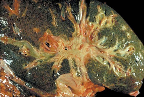
The majority (75-90%) of all primary liver tumours are hepatocellular carcinomas (HCC), arising from the liver parenchyma. Other primary liver cancers include cholangiocarcinomas (CC), which make up 10-25% of liver cancers, see figure 1. These tumours arise from cells lining the biliary tree. Angiosarcomas and hepatoblastomas are very rare primary liver tumours.2
CCs may arise from the intra- or extrahepatic bile ducts. Management of intra- and extrahepatic lesions varies considerably. This article will focus on the management of HCC and intrahepatic CC.
The incidence of liver cancer varies significantly worldwide. Chronic viral hepatitis and in particular hepatitis B virus (HBV) remains the most important risk factor for the development of HCC.3 Areas with high prevalence such as SE Asia, China and sub-Saharan Africa see the highest rates of HCC.
The prevalence of CC also varies globally. The highest rates are seen in Thailand where liver fluke infestation is endemic. In the UK the incidence of CC is 1-2 cases per 100,000 population, but rates are increasing4 and in the mid-1990s CC overtook HCC as the most common cause of liver cancer-related death.5
Over the past decade survival in liver cancer has been steadily improving as a result of developments in surgery, transplantation and the introduction of a number of novel local, ablative and molecular targeted therapies, as outlined below.
Presentation
Symptoms that have been associated with HCC include fatigue, weight loss, abdominal pain, pruritus and jaundice, which may also be caused by the underlying liver disease. However, HCCs are often relatively asymptomatic until late in the disease. Unlike CC where 70% of tumours occur sporadically, in HCC 90% of cases are associated with one or more risk factors, see table 1. Hence most high-risk individuals are followed up as part of a surveillance programme and are diagnosed via this pathway.12,22
Currently the European Association for the Study of the Liver (EASL) recommends HCC surveillance in the following groups:
• Cirrhotic patients, Child-Pugh stage A and B (see table 2)
• Cirrhotic patients, Child-Pugh stage C (see table 2) awaiting liver transplantation
• Non-cirrhotic HBV carriers with active hepatitis or a family history of HCC
• Non-cirrhotic patients with chronic hepatitis C and advanced liver fibrosis
The British Society of Gastroenterology (BSG) and EASL recommend that surveillance should be performed by abdominal ultrasound every 6 months.23,22
In addition, the BSG guidelines, published 10 years ago, recommend that surveillance ultrasound should be combined with measuring serum alpha fetoprotein (AFP).24
Classical symptoms of CC include jaundice, dark urine, clay-coloured stools and pruritus and occur as a result of progressive bile duct obstruction by the tumour.25,26
Screening and vaccination of individuals at high risk of HBV is recommended by the WHO and EASL. It is also recommended that children born to mothers who are HBsAg positive should be vaccinated against HBV at birth to prevent transmission.27,28
Diagnosis
Blood tests
AFP is the most widely used and tested biomarker for the diagnosis of HCC. As a diagnostic tool AFP is only useful when it is found to be markedly elevated; an AFP > 400 ng/ml is considered to be diagnostic (normal range 10-20 ng/ml). In surveillance for HCC in high-risk groups the current BSG guidelines recommend undertaking an AFP six monthly in combination with an abdominal ultrasound.24 However, only 10-20% of small tumours will have an abnormal AFP level and its overall role in surveillance is debated.24,29
In suspected CC, CA 19-9 is used frequently in combination with imaging or other diagnostic tools. However, its overall sensitivity and specificity for diagnosis and surveillance are too low to be recommended in clinical practice.30
Routine liver function tests are usually abnormal in both cancers.30
Ultrasound
Abdominal ultrasound is the most widely available imaging technique for the liver. Sensitivity is 58-89% for detecting HCC and CC in high-risk groups. However, the test is operator dependent, may miss small tumours and cannot define distal spread.
Cross-sectional imaging
Contrast-enhanced CT or MRI is considered the optimal imaging technique for suspected HCCs.
These tumours demonstrate unique radiological criteria taking up contrast during the arterial phase and producing washout during the venous phase.31 The American Association for the Study of Liver Disease (AASLD) has recommended that these findings are diagnostic of HCC and in nodules > 2 cm histological confirmation is not required.32
For CC contrast-enhanced MRI/MRCP (magnetic resonance cholangiopancreatography) is recommended to delineate biliary anatomy, tumour extent and parenchymal disease.
A staging CT (chest, abdomen and pelvis) is undertaken in both tumours to assess for evidence of metastatic disease, see figure 2.
Liver biopsy
Although the need for liver biopsy in HCC is debated because of the small risk of tumour seeding and post-procedure bleeding (2 cm without diagnostic radiological criteria and of lesions 1-2 cm in which two different forms of imaging do not demonstrate typical radiology findings.23
A liver biopsy allows differentiation between benign, premalignant nodules and early HCC so correct surveillance intervals can be defined and treatments instigated.33
Cholangiography
MRCP has been shown to be superior to endoscopic retrograde cholangiopancreatography (ERCP) for differentiating between CC and benign biliary strictures.34 However, ERCP allows for tissue acquisition, essential for patients undergoing chemotherapy and/or radiotherapy, and stent placement to relieve obstructive jaundice when present.
The development of new diagnostic adjuncts such as cholangioscopy have enabled targeted biopsies which improve the diagnostic accuracy.35
Treatment
Treatment of primary liver tumours is dependent on a number of factors such as the stage of the tumour, the position of the tumour, involvement of major vessels and the general health of the patient including the presence of cirrhosis which is defined using the Child-Pugh scoring system, see table 2.
To decide on the most appropriate treatment option for HCC, several staging criteria have been developed but the Barcelona Clinic Liver Cancer (BCLC) staging and treatment strategy is most frequently applied, see figure 3. CC staging is based on the tumour node metastasis system.
Surgery
In patients with primary liver tumours and Child-Pugh A cirrhosis or a normal liver, resection is the first-line treatment. Perioperative mortality is 2-3% and five-year survival can be up to 80% in well selected patients.37 Neoadjuvant therapies have not been shown to improve prognosis. Currently only 5% of patients diagnosed with HCC in the UK are suitable for surgical resection. If tumours recur following surgery they can be restaged and managed in accordance with the BCLC algorithm.38
Transplantation
Historically liver transplantation for HCC has been associated with low survival rates (20-36%) which limited its use in liver cancer. However, following the introduction of the Milan criteria, see table 3, p24, which restricted the indication for transplantation to small HCCs, survival improved dramatically.37
HCC is now one of the most common reasons for liver transplantation in many transplant centres worldwide. The upper limit of tumour size for transplantation is controversial and several expanded criteria such as UCSF, see table 3, have been proposed, but without impact on overall survival.
Transplantation is not routinely recommended for CC but there is emerging evidence that when combined with neoadjuvant therapy, it can be associated with up to a 70% five-year survival in highly selected patients.41,42
Chemotherapy
Recent randomised trials of chemotherapy in HCC have shown response rates of less than 20%, with limited improvements in survival.43,44 Chemotherapy is therefore rarely used for HCC and not recommended in European guidelines for the management of HCC.23 However, in locally advanced and metastatic CC the ABC-01 and ABC-02 randomised trials showed that 24 weeks of cisplatin plus gemcitabine improved overall survival from 8.1 to 11.7 months (P < 0.001).45,46 There is emerging evidence that neoadjuvant chemotherapy may also be able to downstage some unresectable CC patients to resectability.47
Biologic therapies
Sorafenib is an oral multi-tyrosine kinase inhibitor which inhibits blood vessel development and tumour cell proliferation by inhibiting the Raf cascade, vascular endothelial growth factor and platelet-derived growth factor receptors in tumour cells. It has been shown to improve survival from 7.9 to 10.7 months (P < 0.001) in patients with advanced HCC.48 However, because of its high cost, the treatment is currently not approved by NICE.49
Local and ablative treatments
Local and/or ablative therapies are considered to be first-line treatment in patients with early HCC who are not suitable for surgical resection or transplantation, see table 4.
In intrahepatic CC they can be used similarly to HCC but because of the rarity of intrahepatic CC fewer large studies have been conducted to evaluate these treatments.
Conclusion
Most patients with HCC are diagnosed with advanced disease. Current guidelines recommend high-risk groups should be surveyed every six months by abdominal ultrasound with or without measurement of serum AFP.
Recent advances in surgery and extension of transplant criteria have meant more patients are now amenable to curative therapy. There have been significant advances in the development of targeted therapies, which individually or in combination, can prolong survival and have the potential to downstage patients so they are eligible for curative treatment.
Treatments are now available for all patients at any stage of liver cancer and should be tailored to account for the degree of liver dysfunction and other comorbidities.
REFERENCES
1 Ferlay J, Shin HR, Bray F et al. GLOBCAN 2008, cancer incidence and mortality worldwide: IARC CancerBase No.10. 2010. Lyon, France: International Agency for Research on Cancer. 2010. Available from: http://globocan.iarc.fr/
2 London WT, McGlynn KA. Liver cancer. In Cancer epidemiology and prevention. 3rd edition. pp 763-86.
Eds. D Schottenfeld, JF Fraumeni Jr. Oxford University Press. New York. 2006
3 Parkin DM Bray F, Ferlay J, Pisani P. Global cancer statistics 2002. CA Cancer J Clin 2005;55:74-108
4 Khan SA, Toledano MB, Taylor-Robinson SD. Epidemiology, risk factors, and pathogenesis of cholangiocarcinoma. HPB (Oxford) 2008;10(2):77-82
5 Khan SA, Taylor-Robinson SD, Toledano MB et al. Changing international trends in mortality rates for liver, biliary and pancreatic tumours. J Hepatol 2002;37:806-13
6 Shaib Y, El-Serag HB. The epidemiology of cholangiocarcinoma. Semin Liver Dis 2004;24:115-25
7 Marcellin P, Peguignot F, Delarocque-Astagneau E et al. Mortality related to chronic hepatitis B and chronic hepatitis C in France: evidence for the role of HIV coinfection and alcohol consumption. J Hepatol 2008;48:200-207
8 Claessen MM, Vleggaar FP, Tygat KM et al. High lifetime risk of cancer in primary sclerosing cholangitis. J Hepatol 2009;50:158-64
9 Deugnier YM, Guyader D, Crantock L et al. Primary liver cancer in genetic haemochromatosis: a clinical, pathological, and pathogenic study of 54 cases. Gastroenterology 1993;104:228-34
10 Perlmutter DH. Pathogenesis of chronic liver injury and hepatocellular carcinoma in alpha-1-antitrypsin deficiency. Pediatr Res 2006;60:233-8
11 Sangiovanni A, Prati GM, Fasani P et al. The natural history of compensated cirrhosis due to hepatitis C virus: a 17-year cohort study of 214 patients. Hepatology 2006;43:1303-10
12 Tyson GL, El-Serag HB. Risk factors for cholangiocarcinoma. Hepatology 2011;54(1):173-84
13 Marrero J, Fontana RJ, Fu S et al. Alcohol, tobacco and obesity are synergistic risk factors for hepatocellular carcinoma. J Hepatol 2005;42:218-24
14 Trichopoulos D, Bamia C, Lagiou P et al. Hepatocellular carcinoma risk factors and disease burden in a European cohort: a nested case-control study. J Natl Cancer Inst 2011;103:1686-95
15 Groopman JD, Scholl P, Wang JS. Epidemiology of human aflatoxin exposures and their relationship to liver cancer. Prog Clin Biol Res 1996;395:211-22
16 El-Serag HB, Richardson PA, Everhart JE. The role of diabetes in hepatocellular carcinoma: a case-control study among United States Veterans. Am J Gastroenterol 2001;96:2462-7
17 Gorayski P, Thompson CH, Subhash HS, Thomas AC. Hepatocellular carcinoma associated with recreational anabolic steroid use. Br J Sports Med 2008;42(1):74-5
18 Jones DE, Metcalf JV, Collier JD et al. Hepatocellular carcinoma in primary biliary cirrhosis and its impact on outcomes. Hepatology 1997;26(5):1138-42
19 Adenugba A, Khan SA, Taylor-Robinson SD et al. Polychlorinated biphenyls in bile of patients with biliary tract cancer. Chemosphere 2009;76:841-6
20 Miki Y, Swensen J, Shattuck-Eidens D, Futreal PA et al. A strong candidate for the breast and ovarian cancer susceptibility gene BRCA1. Science 1994;266:66-71
21 Tanabe KK, Lemoine A, Finkelstein DM et al. Epidermal growth factor gene functional polymorphisms and the risk of hepatocellular carcinoma in patients with cirrhosis. JAMA 2008;299:53-60
22 Llovet JM, Burroughs A, Bruix J. Hepatocellular carcinoma. Lancet 2003;362:1907-1917
23 EASL. EASL-EORTC Clinical Practice Guidelines: Management of hepatocellular carcinoma. J Hepatol 2012;56:908-43
24 Ryder S. Guidelines for the diagnosis and treatment of hepatocellular carcinoma (HCC) in adults. Gut 2003;52(Suppl III):iii1-iii8
25 Pitt HA, Dooley WC, Yeo CJ, Cameron JL. Malignancies of the biliary tree. Current Problems in Surgery 1995;32:1-90
26 deGroen PC, Gores GJ, LaRusso NF et al. Biliary Tract Cancers. N Engl J Med 1999;341:1368-78
27 EASL. EASL Clinical Practice Guidelines: management of chronic hepatitis B. J Hepatol 2009;50:277-42
28 WHO. Hepatitis B vaccines. Weekly epidemiological record of the World Health Organization 2009;84:405-20
29 Yamashita T, Forques M, Wang W et al. EpCAM and alpha-fetoprotein expression defines novel prognostic subtypes of hepatocellular carcinoma. Cancer Res 2008;68: 1451-61
30 Khan SA, Davidson BR, Goldin RD et al. Guidelines for the diagnosis and treatment of cholangiocarcinoma: an update. Gut 2012;61:1657-69
31 Bruix J, Sherman M. Management of hepatocellular carcinoma. Hepatology 2005;42:1208-36
32 Bruix J, Sherman M. Management of hepatocellular carcinoma: an update. Hepatology 2011; 53:1020-2
33 Stigliano R, Marelli L, Yu D et al. Seeding following percutaneous diagnostic and therapeutic approaches for hepatocellular carcinoma. What is the risk and the outcome? Seeding risk for percutaneous approach of HCC. Cancer Treat Rev 2007;33:437-47
34 Ashok K. Role of MRCP versus ERCP in bile duct cholangiocarcinoma and benign stricture. Biomed Imaging Interv J 2007;3:e12-545
35 Chen YK, Parsi MA, Binmoeller KF et al. Single-operator cholangioscopy in patients requiring evaluation of bile duct disease or therapy of biliary stones (with videos). Gastointest Endosc 2011;74:805-14
36 Angermayr B, Cejna M, Karnel F et al. Child-Pugh versus MELD score in predicting survival in patients undergoing transjugular intrahepatic portosystemic shunt. Gut 2003;52(6):879-85
37 Mazzaferro V, Bhoori S, Sposito C et al. Milan criteria in liver transplantation for HCC: an evidence-based analysis of 15 years experience. Liver Transpl 2011; 17:S44-57
38 Llovet JM, Bru C, Bruix J. Prognosis of hepatocellular carcinoma: the BCLC staging classification. Semin Liver Dis 1999;19:329-38
39 Mazzaferro V, Regalia E, Doci R et al. Liver transplantation for the treatment of small hepatocellular carcinomas in patients with cirrhosis. N Engl J Med 1996;334(11):693-9
40 Yao FY, Ferrell L, Bass NM et al. Liver transplantation for hepatocellular carcinoma: expansion of the tumour size limits does not adversely impact survival. Hepatology 2001;33(6):1394-403
41 Darwish Murad S, Kim WR, Hamois DM et al. Efficacy of neoadjuvant chemoradiation followed by liver transplantation for perihilar cholangiocarcinoma at 12 US centres. Gastroenterology 2012;143:88-98.e3
42 Rosen CB, Heimbach JK, Gores GJ. Surgery for cholangiocarcinoma: the role of liver transplantation. HPB (Oxford) 2008;10:186-9
43 Yeo W, Mok TS, Zee B et al. A randomized phase III study of doxorubicin versus cisplatin/interferon alpha-2b/doxorubicin/fluorouracil (PIAF) combination chemotherapy for unresectable hepatocellular carcinoma. J Natl Cancer Inst 2005; 97(20):1532-8
44 Gish RG, Porta C, Lazar L et al. Phase III randomized controlled trial comparing the survival of patients with unresectable hepatocellular carcinoma treated with nolatrexed or doxorubicin. J Clin Oncol 2007;25(21):3069-75
45 Valle J, Wasan H, Palmer DH et al. Cisplatin plus gemcitabine versus gemcitabine for biliary tract cancer. N Engl J Med 2010;362:1273-81
46 Valle JW, Wasan H, Johnson P et al. Gemcitabine alone or in combination with cisplatin in patients with advanced or metastatic cholangiocarcinomas or other biliary tract tumours: a multicentre randomised phase II study: the UK ABC-01 Study. Br J Cancer 2009;101:621-7
47 Donahue TR, Isacoff WH, Hines OJ et al. Downstaging chemotherapy and alteration in the classic computed tomography/magnetic resonance imaging signs of vascular involvement in patients with pancreaticobiliary malignant tumors: Influence on patient selection for surgery. Arch Surgery 2011;146(7):836-43
48 llovet JM, Ricci S, Mazzaferro V et al. Sorafenib in advanced hepatocellular carcinoma. N Engl J Med 2008; 359(4): 378-90
49 National Institute for Clinical Excellence. TA189. Sorafenib for the treatment of advanced hepatocellular carcinoma. NICE. London. 2010
50 Khan KN, Yatsuhashi H, Yamasaki K et al. Prospective analysis of risk factors for early intrahepatic recurrence of hepatocellular carcinoma following ethanol injection. J Hepatol 2000;32:269-78
51 Lencioni R, Bartolozzi C, Caramella D et al. Treatment of small hepatocellular carcinomas with perculaneous ethanol injection. Analysis of prognostic factors in 105 Western patients. Cancer 1995;76:1737-46
52 Livraghi T, Giorgio A, Marin G et al. Hepatocellular carcinoma and cirrhosis in 746 patients: long-term results of percutaneous ethanol injection. Radiology 1995; 197:101-8
53 Lin SM, Lin CJ, Lin CC et al. Randomised controlled trial comparing percutaneous radiofrequency thermal ablation, percutaneous ethanol injection and percutaneous acetic acid injection to treat hepatocellular carcinoma of 3cm or less. Gut 2005;54:1151-6
54 Shiina S, Teratani T, Obi S et al. A randomised controlled trial of radiofrequency ablation versus ethanol injection for small hepatocellular carcinoma. Gastroenterology 2005;129:122-30
55 National Institute for Clinical Excellence. IPG2. Radiofrequency ablation of hepatocellular carcinoma. NICE. London. 2003
56 Steel AW, Postgate AJ, Khorsandi S et al. Endoscopically applied radiofrequency ablation appears to be safe in the treatment of malignant biliary obstruction. Gastrointest Endosc 2011;73(1):149-53
57 National Institute for Clinical Excellence. IPG 214. Microwave ablation of hepatocellular carcinoma. NICE. London. 2007
58 Shibata T, Iimuro Y, Yamamoto Y et al. Small hepatocellular carcinoma: comparison of radio-frequency ablation and percutaneous microwave coagulation therapy. Radiology 2002;223:331-7
59 Orlacchio A, Bazzocchi G, Pastorelli D et al. Percutaneous cryoablation of small hepatocellular carcinoma with US guidance and CT monitoring: initial experience. Cardiovasc Intervent Radiol 2008;31:587-94
60 Wang S, Keltner L, Winship J, Lee W. A phase 1/2 safety and efficacy study of intratumoural light activated drug therapy using talporfin sodium in patients with inoperable hepatocelluar carcinoma. J Clin Oncol 2009: 27A.
61 Ng KK, Poon RT, Chan SC et al. High intensity focused ultrasound for hepatocellular carcinoma: a single-centre experience. Ann Surg 2011;253:981-7
62 Oliveri RS, Wettersley J, Gluud C. Transarterial (chemo)embolisation for unresectable hepatocellular carcinoma. Cochrane Database Syst Rev 2011;16(3): CD004787
63 Salem R, Lewandowski RJ, Mulcachy MF et al. Radioembolisation for hepatocellular carcinoma using Yttrium-90 microspheres: a comprehensive report of long-term outcomes. Gastroenterology 2010;138:52-64
64 Kulik LM, Carr BI, Mulcahy MF et al. Safety and efficacy of 90Y radiotherapy for hepatocellular carcinoma with and without portal vein thrombosis. Hepatology 2008;47:71-81
65 Hilgard P, Hamami M, Fouly AE et al. Radioembolisation with yttrium-90 glass microspheres in hepatocellular carcinoma: European experience on safety and long-term survival. Hepatology 2010;52:1741-9
ARTICLE IN PDF
 Download this article in PDF format


EXTERNAL WEBLINKS
US National Cancer Institute
PDQ® (Physician Data Query) is NCI's cancer database of peer-reviewed, regularly updated evidence-based, referenced summaries
Adult primary liver cancer
Liver Cancer screening
Childhood liver cancer
NICE
British National Formulary
Malignant disease and immunosuppression
BMJ Learning CPD modules
Alcohol related liver disease 1 hour
Liver function tests: interpreting abnormal results 1 hour
PubMed
PubMed is a database of the US National Library of Medicine/National Institutes of Health
Palliative care at home