SEARCH

The Practitioner 2010; 254 (1730): 25–30
Managing unstable angina and non-ST elevation MI
22 Jun 2010
AUTHORS
Dr Mohammed Majid Akhtar MBBS BSc
Foundation year 2, general medicine, Southend University Hospital NHS Trust
Dr Yassir Hassan Iqbal MBBS BSc
Foundation year 2, cardiothoracic surgery, St George's Healthcare NHS Trust
Professor Juan Carlos Kaski MD DSc FRCP FESC FACC
Professor of cardiovascular science, Head, Cardiovascular Sciences Research Centre, Division of Clinical Sciences, St George's, University of London
Article
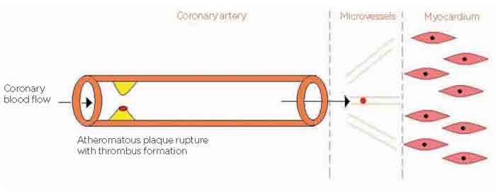
Acute coronary syndrome (ACS), encompassing unstable angina (UA), non-ST elevation myocardial infarction (NSTEMI), see figure 1 above, and ST-elevation myocardial infarction (STEMI), is often the result of an acute thrombotic occlusion of the coronary vessels, associated with atheromatous plaque rupture or erosion.
ACS is associated with a severely impaired prognosis and requires prompt and efficient specialist treatment.
Patients presenting with STEMI most often show a complete occlusion of one or more of the epicardial coronary arteries.1 NSTEMI and UA - collectively known as non-ST elevation ACS (NSTE-ACS) - share a similar pathogenesis with acute coronary thrombosis reducing the lumen of the coronary artery and severely restricting myocardial blood supply.
The thrombus in NSTE-ACS is often platelet rich and sub-occlusive as opposed to the fibrin-rich red clot that completely occludes the coronary artery in patients with STEMI. Distal embolisation of platelets and other components of the thrombus often takes place in NSTEMI, resulting
in the occlusion of the coronary microcirculation and further myocardial ischaemia and/or necrosis.
Diagnosing unstable angina and NSTEMI
The clinical presentation may be identical across all three components of ACS. Establishing an accurate diagnosis without delay is of paramount importance to start treatment promptly, see figure 2, p26. Patients with suspected ACS need to be referred immediately to A&E. Important features pointing towards a diagnosis of ACS include:
- typical characteristics of chest pain
- presence of risk factors
- ECG changes suggestive of myocardial ischaemia, see table 1, p26.
Chest pain
Chest discomfort in patients with ACS typically occurs at rest, is anginal in character and can range from mild tightness to central crushing chest pain. This is not infrequently associated with symptoms such as nausea, dyspnoea or diaphoresis. The chest pain may radiate to the arms, back or jaw and is often >20 minutes in duration. Atypical chest pain at rest is relatively common in the elderly and in patients with diabetes mellitus.
Extra-cardiac causes of chest pain at rest mimicking angina pectoris should be identified but it should be remembered that these may coexist with ACS and not necessarily be the cause of the chest pain that brings the patient to the attention of the physician.
It is important to remember that although in most cases angina can be easily recognised, patients may occasionally present with atypical symptoms that make the diagnosis of ACS difficult; this is particularly common in patients with cognitive impairment, diabetes mellitus and the elderly.1
ECG changes
In all patients presenting with chest pain suggestive of myocardial ischaemia a 12-lead ECG should be recorded. This is of paramount importance for the diagnosis of ACS and should be obtained as soon as possible, preferably within 10 minutes of first contact with a healthcare professional. Serial ECGs are useful to look for dynamic changes of the ST segment and to assess the ECG response to the administration of treatment. Previous ECGs, if and when available, are useful for comparison.
In ACS patients with persistent ST segment elevation (i.e. STEMI), primary percutaneous coronary intervention (PCI) or fibrinolytic therapy if PCI is not available should be carried out without delay. There
is no need to wait for the results of blood tests, i.e. cardiac troponin levels, to proceed to intervention in these patients. Management of STEMI, however, is beyond the scope of this article.
Approximately 5% of patients with NSTE-ACS have normal ECGs.1 In the presence of a history suggestive of ACS, these patients with normal or equivocal ECGs should be transferred immediately to hospital for specialist assessment and treatment. 1
In NSTE-ACS patients, the diagnosis of UA (typical symptoms and ECG changes, without elevation of markers of necrosis) vs NSTEMI (positive troponin results) may not be possible until troponin measurements are carried out in the hospital setting. The 12-lead ECG should be repeated on a regular basis in stable patients (i.e. every 6-24 hours) and every time there is a recurrence of chest pain or other cardiovascular symptoms.
Risk stratification in ACS
Despite a relatively similar presentation, prognosis may differ in different patients. Several risk scores, involving clinical, biochemical and ECG characteristics, have been proposed, which help in the risk stratification of ACS patients and represent the basis to guide treatment; in particular, coronary intervention,2 see table 2, below.
Despite the usually serious nature of ACS, some patients are at a relatively low risk of developing cardiovascular events and may be considered for inpatient or outpatient investigations to characterise their condition further. Patients in the moderate-risk group however, should have their coronary anatomy assessed within 72-96 hours, and those falling in the high-risk bracket need urgent angiographic investigation and intervention.
There are several scoring systems which may be useful for the assessment of risk of further major adverse cardiac events (MACE). The three most widely used risk stratification systems in the clinical setting are:
- Thrombolysis In Myocardial Infarction (TIMI) risk score 2
- Global Registry for Acute Cardiac Events (GRACE)3
- Platelet Glycoprotein IIb-IIIa in Unstable Angina: Receptor Suppression Using Integrilin Therapy (PURSUIT) 2
These risk stratification systems are based on findings in large population registries encompassing the full spectrum of ACS patients.1 The GRACE score uses different variables to predict the risk of in-hospital death and 6-month mortality.1 The GRACE risk score involves more complex calculations than some of the other risk stratification systems, as it requires the assessment of many variables, some of which may not be available in primary care. The GRACE investigators have divided patient risk into three categories i.e. low, intermediate and high. These are, as expected, associated with different mortality risk both in-hospital and post-discharge.
The TIMI risk scoring system was previously more widely used than the GRACE scoring system due to its reliance on fewer variables to assess patient outcome. However, for this reason it is perhaps less accurate than the GRACE scoring system.1 The TIMI risk index, however, allows clinically useful prediction of 30-day and 1-year mortality risk in ACS patients.2 The PURSUIT risk stratification score is also of interest, as it allows the differentiation between NSTEMI and UA regarding 30-day mortality risk.2
The evaluation of risk in a given individual is a dynamic process that needs constant updating, as the clinical situation evolves, often very rapidly.
Evidence-based treatment options
Prompt institution of treatment is essential in ACS and patients with suspected ACS should be transferred without delay to specialist centres or hospital emergency departments. Current treatment is based on tackling the pathophysiological mechanisms responsible for the condition and includes:
- antiplatelet drugs
- antithrombotic agents
- angina drugs
- analgesic interventions
- PCI
Initial treatment, the prehospital phase, should be initiated rapidly.
It includes the use of aspirin, nitrates, morphine and oxygen (if hypoxic), see figure 5, left.
Pharmacotherapy
Pharmacological treatment options are listed in table 3, p28.
Antiplatelet therapy
Aspirin is the first choice antiplatelet agent; in the absence of contraindications, an initial dose of 300 mg should be given immediately, orally (rectally or via a nasogastric tube are alternative routes) in all cases of suspected ACS. If there is a clear contraindication to aspirin, clopidogrel can be used as an alternative. In addition to aspirin, a loading dose of clopidogrel, 300 mg, should be given to optimise antiplatelet treatment, followed by 75 mg daily, standard dose, for 12 months. This is recommended especially in patients who have a 6-month mortality risk exceeding 1.5% and in those without contraindications i.e. risk of profound bleeding.4
Prasugrel has been recently added to the list of antiplatelet agents recommended for use in patients with ACS. High-risk patients with UA or NSTEMI, including those with ongoing chest pain and dynamic changes on the ECG who are likely to undergo angiography within 96 hours of admission, should be considered for GP IIb/IIIa inhibitors (i.e. eptifibatide or tirofiban). The GP IIb/IIIa antagonist abciximab is used as an adjunct to PCI for patients at intermediate or high risk.
These agents are administered in the hospital setting once blood tests and accurate risk assessment have been carried out.4
Angina treatment
Sublingual nitrates (spray or tablets) should be given immediately. A nitrate infusion in the form of 50 mg glyceryl trinitrate (GTN) in 50 ml of saline is given at a starting rate 1-2 ml/h to provide continuous nitrate therapy. Careful blood pressure monitoring is required to prevent the development of hypotension, which could further reduce myocardial perfusion. Symptoms need to be assessed regularly to determine whether nitrate tolerance develops. This is best achieved by asking the patient to score their discomfort regularly on a scale of 1-10.
Beta-blockers are needed to achieve further control of symptoms in NSTE-ACS. These agents have anti-ischaemic effects, which depend on their actions on increased sympathetic tone, as well as negative chronotropic and inotropic actions that help reduce myocardial oxygen demand. 3
Antithrombotic agents
Patients with UA or NSTEMI should be given a subcutaneous injection of LMWH. Fondaparinux is offered to patients without high risk of bleeding unless angiography is planned within 24 hours of admission. Unfractionated heparin is given to patients with creatinine concentrations >265 µmol/L.
Analgesia
Relieving ischaemia and chest pain is of paramount importance. Patients who continue to experience chest pain despite initial nitrate therapy, should be offered intravenous opioids,
i.e. morphine. Morphine has potent effects as an analgesic and anxiolytic and helps in providing haemodynamic stability. It also reduces systolic blood pressure and heart rate thus reducing myocardial oxygen demand.2
Optimal management after discharge
The GP plays a major role in ensuring optimal patient management after hospital discharge and secondary prevention of cardiovascular events. Patients should be reviewed regularly in order to be offered the following:
- lifestyle advice e.g. smoking cessation, healthy diet, regular exercise
- regular assessment of cardiovascular risk e.g. Framingham Score/University of Manchester Charts
- monitoring of serum fasting lipids and assessment of beneficial effects and side effects of cholesterol- and triglyceride-lowering drugs
- monitoring of serum fasting glucose and arranging oral glucose tolerance tests if appropriate. In patients with diabetes, ensure good glycaemic control and monitoring HbA1C
- monitoring blood pressure regularly and assessment of effects/side effects of antihypertensive agents
It is also important to ensure that patients are receiving the following treatment where appropriate:
- antiplatelet therapy
- angina therapy and doses are
up-titrated if necessary
- heart failure medications
- post MI patients - drugs shown to improve prognosis e.g. ACE inhibitors and beta-blockers
- healthcare education
Conclusion
ACS is a serious condition which requires immediate attention and prompt management. Risk stratification is an imperative process in the assessment of ACS to allow efficient delivery of specialist care.
The treatment options available for patients with ACS are based on addressing the pathophysiological mechanisms leading to myocardial ischaemia and necrosis, preventing and minimising further thrombotic occlusion of the epicardial coronary arteries and reducing myocardial damage.
REFERENCES
1 Bassand JP, Hamm CW, Ardissino D et al. Guidelines for the diagnosis and treatment of non-ST-segment elevation acute coronary syndromes: The Task Force for the Diagnosis and Treatment of Non-ST-Segment Elevation Acute Coronary Syndromes of the European Society of Cardiology. Eur Heart J (2007) 28, 1598-1660
2 National Institute for Health and Clinical Excellence. CG 95. Chest pain of recent onset - assessment and diagnosis of recent onset chest pain or discomfort of suspected cardiac origin. NICE. London. 2010
3 National Institute for Health and Clinical Excellence. CG 94. Unstable angina and NSTEMI: The early management of unstable angina and non-ST elevation myocardial infarction. NICE. London. 2010
4 Global Registry of Acute Coronary Events.www.outcomes-umassmed.org
5 Granger CB, Goldberg RJ, Dabbous O et al. Predictors of hospital mortality in the global registry of acute coronary events. Arch Intern Med 2003; 163:2345-53
ARTICLE IN PDF
 Download this article in PDF format