SEARCH

Stollery N. Photoguide:Papulosquamous conditions. Practitioner 2016;260(1796)28-29
Papulosquamous conditions
23 Sep 2016
AUTHORS
Dr Nigel Stollery MB BS DPD, GPwSI Dermatology, Kibworth, Leicestershire, UK
Article
Pityriasis rosea
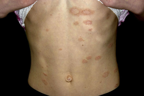
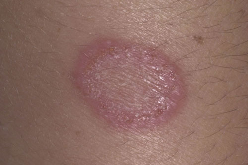
Pityriasis rosea is seen more commonly in the autumn and winter. The seasonal variation has led to the suggestion of a viral aetiology but this remains unproven. Typically, a single annular lesion 2-5 cm in diameter, the so-called herald patch, is seen first with a fine collarette of scale. This is then followed by the appearance of multiple smaller round scaly lesions typically on the torso and in a Christmas tree distribution. The rash usually lasts 2-10 weeks and resolves spontaneously without treatment. If itching is present a moderately potent topical steroid can be helpful. UVB or sunlight may hasten the resolution.
Discoid lupus erythematosus
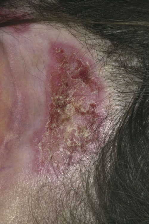
The cause of discoid lupus erythematosus (DLE) is unknown but ultraviolet light is thought to be a factor. Plaques are found on the skin, ranging from one or two to multiple lesions in different areas. DLE is characterised by scale, follicular plugging and erythema. Scarring and atrophy may also be present. As in psoriasis the plaques are well demarcated, but occur mostly on the sun-exposed areas of the scalp, face and ears. Where hair loss occurs as a result of the plaques, scarring may mean that this is permanent, and the white atrophic appearance may persist. Although DLE is a skin condition progression to the systemic form (SLE) may occur. Treatment involves potent or very potent topical steroids which may need to be used on areas such as the face where they are usually avoided. Unresponsive areas may require intralesional steroid injections or systemic treatments such as the oral antimalarial drug hydroxychloroquine.
Lichen planus
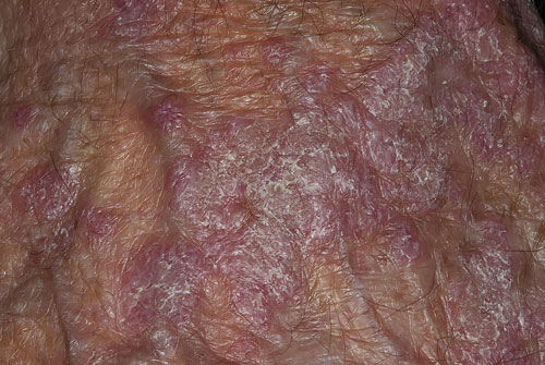
There are various forms of lichen planus: annular, bullous, hypertrophic and ulcerative. Lichen planus affects the skin, genitals and mouth. The cause is unknown. Lesions characteristically have a lilac or violet colour and occur as flat-topped papules. Closer inspection may reveal a white streaky lace-like pattern on the surface known as Wickham’s striae. The rash is often itchy and if the skin is scratched the rash may spread along the lines of trauma (the Koebner phenomenon). If the diagnosis is in doubt a biopsy can be helpful. The condition can last from a few months to more than a year and in one in six cases will recur. A lichenoid drug reaction can mimic this condition and if a drug is suspected it should be stopped. Treatment can be difficult. Potent topical steroids can reduce the itching and flatten the lesions. In more severe cases systemic treatments or PUVA may be required.
Seborrhoeic dermatitis
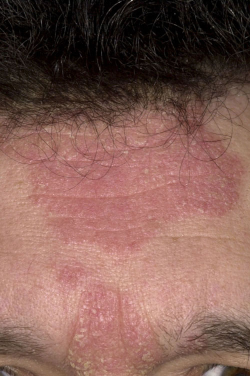
Seborrhoeic dermatitis is not related to seborrhoea but does occur predominantly in sebum-rich areas. It can run in families and some believe it is related to an overgrowth of the Malassezia yeast which although is not the cause can be linked to T-cell depression and activation of the alternative complement pathway. Severity varies from the more common dandruff to the rare exfoliative erythroderma. Affected areas may burn and itch and have a typical red scaly appearance. It is more common over the winter and will often improve in the summer; this is thought to be related to the effect of sunlight on Malassezia. Diagnosis is usually made clinically based on distribution and history. Treatments include short-term topical steroids, antifungal creams, calcineurin inhibitors and in severe cases oral antifungals such as fluconazole.
Tinea corporis
Tinea corporis (ringworm) is a common dermatophyte infection causing an itchy, scaly annular rash which is more pronounced at the periphery. At this leading edge vesicles or pustules may be present and the diameter will slowly increase with time as the fungus advances outwards across the epidermis. There are three main genera of fungi responsible: Trichophyton, Microsporum and Epidermophyton. Other areas that can be affected include the scalp, nails, groin, axillae and toe webs. If the diagnosis is in doubt, skin scrapings can be sent to the laboratory for mycology or examination under a Wood’s lamp which may demonstrate fluorescence of the affected area. Treatment with a topical antifungal is usually all that is needed, but in more severe or resistant cases systemic therapy may be required.
Plaque psoriasis
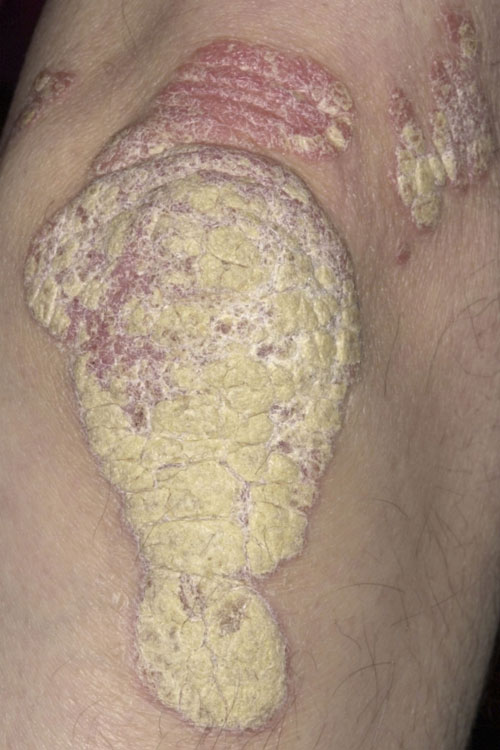
Chronic in nature, plaque psoriasis presents typically with well demarcated plaques over the extensor surfaces of the elbows and knees with thick silvery adherent scales. The scalp, flexures, genitals, nails and in some cases the whole body can be affected. Hyperproliferation of epidermal keratinocytes and an increased cell turnover rate means that the skin is produced and shed at an accelerated rate. Although no cure is known recent advances in biologics have made a significant improvement to the treatments available. Biologics are normally only prescribed if the patient has failed to respond to standard systemic therapies such as methotrexate, ciclosporin or PUVA, or these treatments are contraindicated or not tolerated, and has a PASI score of ≥ 10 and a DLQI of >10.
ARTICLE IN PDF
 Download this article in PDF format