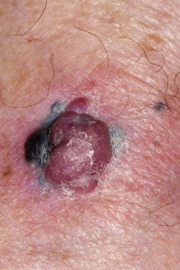
Nodular malignant melanoma
Although numerous adjuvant therapies are being investigated and trialled for the treatment of malignant melanoma, early detection and excision remains crucial for long-term survival. In general, the thicker a lesion at the time of excision the poorer the prognosis. Nodular melanomas such as this one have a poor prognosis. New adjuvant therapies include: interferon alpha; radiation therapy; retinoid therapy; vitamin therapy or biologic therapies, but as yet none of these have been found to increase overall survival. NICE is currently considering the use of ipilimumab in combination with dacarbazine for previously untreated unresectable stage III or IV melanoma.
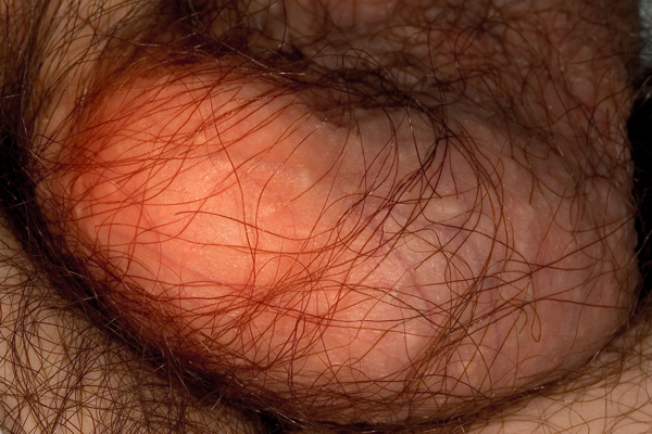
Hydrocoele
A hydrocoele is a collection of serous fluid that has accumulated via a defect in the tunica vaginalis of the scrotum. There are three types: congenital (communicating); non-communicating; and a hydrocoele of the cord. They affect approximately 1% of men, with 7-10% occurring bilaterally. Hydrocoeles can occur following trauma such as radiotherapy, infections or injury to the area and may affect fertility. Apart from the congenital form, hydrocoeles usually occur after the age of 40. The majority are small and asymptomatic, but some may cause enlargement of the scrotum leading to pain which may be referred to the back. Unlike inguinal scrotal hernias, which may be similar in appearance, hydrocoeles have no bowel sounds and transilluminate. However, the two conditions may coexist especially on the right side. It is important to differentiate between a hydrocoele and an acute scrotum (ischaemic testicle, torsion or haemorrhage) and ultrasound can be useful here. Surgical intervention is not always necessary but can be undertaken for large hydrocoeles especially if they threaten the circulation of the scrotum.
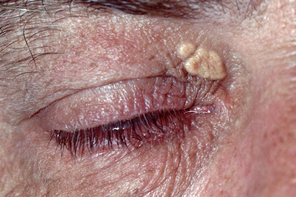
Xanthelasma
The term xanthelasma is derived from the Greek xanthos (yellow) and elasma (beaten metal plate) and describes yellow plaques that occur most frequently on the eyelids. They tend to be symmetrical and may progress and coalesce with time. Women are more commonly affected than men, with a peak incidence in the fourth and fifth decades. In 50% of cases the plaques are associated with elevated lipid levels, but they usually fail to improve when cholesterol levels are reduced with medication. They are generally asymptomatic, but may cause ptosis if lesions are extensive. Management approaches include: surgical excision; laser therapy; diathermy; cryotherapy or chemical cauterisation with chlorinated acetic acids.
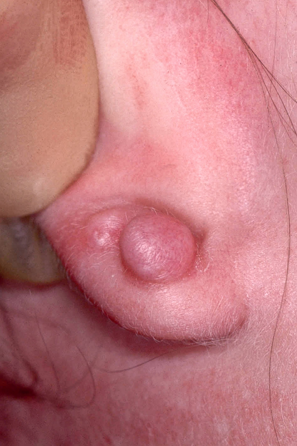
Keloid scarring
The difference between keloid scars and hypertrophic scars is that keloid scars extend beyond the borders of the original wound, do not spontaneously resolve and recur after excision. Autosomal dominant and recessive forms have been described. Keloid scars are rarely found in newborns, and have a peak incidence between the ages of 10 and 30. Histologically, keloids contain dull, pink bundles of collagen which are not present in hypertrophic scars. Keloids can occur anywhere on the body and are more common in certain ethnic groups, affecting 16% of black Africans. They are especially troublesome when they occur secondary to conditions such as acne or in exposed areas, for example, following piercing on the head and neck. Management can be difficult. Treatment options include: intralesional steroids; cryotherapy; laser therapy; interferon alpha; doxorubicin; bleomycin; verapamil; tamoxifen; retinoic acid; 5-fluorouracil; imiquimod 5% cream; tacrolimus; and botulinum toxin.
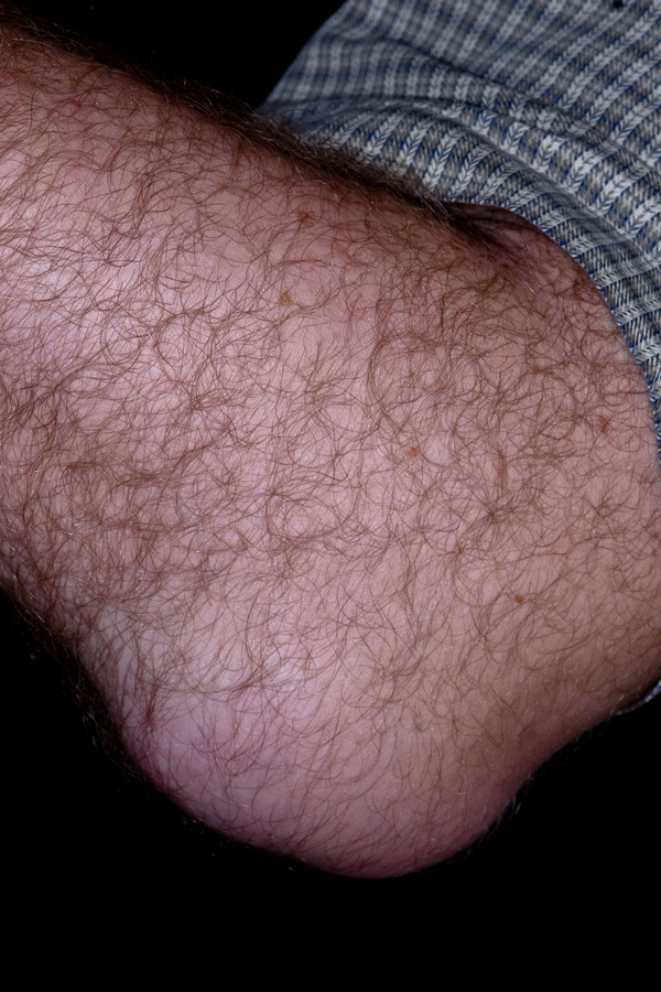
Olecranon bursitis
This condition occurs when there is inflammation and swelling of the bursa which overlies the olecranon process at the proximal aspect of the ulna. It is a relatively common condition that can occur secondary to trauma or, more rarely, following infection. The differential diagnosis includes gout, acute CPP crystal arthritis (pseudogout), olecranon fracture and triceps tendon avulsion. Treatment is not always necessary. In persistent cases, physiotherapy may help and where infection is present antibiotics should be prescribed. Aspiration can be undertaken but there is a risk of chronic (dripping tap) leakage if the aspiration point fails to heal which can then be very difficult to treat.
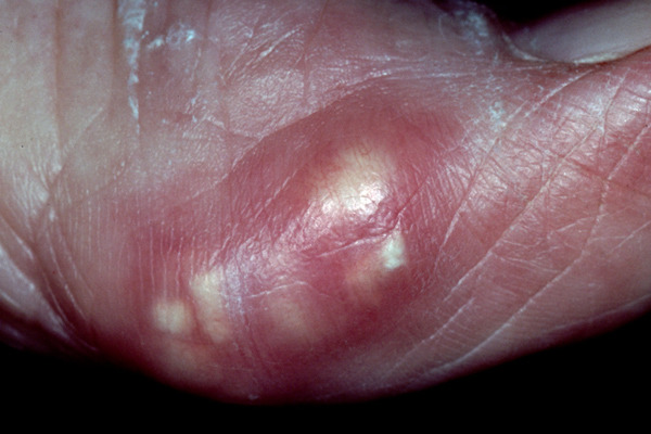
Tophaceous gout
In gout, deposits of monosodium urate monohydrate crystals form in the joints and soft tissues causing pain, and inflammation. When the condition is not diagnosed and treated early enough or when compliance with treatment is poor, chronic tophaceous gout can develop. Treatment of gout usually consists of three parts: management of acute attacks, generally with NSAIDs; prophylaxis to prevent recurrence; and lowering of excess stores of urate to prevent flares of gouty arthritis and the deposition of urate crystals. Where tophi do develop they can be excised surgically. Indications for this include joint deformity, recurrent infection, compression of adjacent structures e.g. the spinal cord or intractable pain.
 Download this article in PDF format