SEARCH

O’Sullivan M, Overton C. Tailor management to the patient with fibroids. Practitioner 2017;261(1802):19-22
Tailor management to the patient with fibroids
22 Mar 2017
AUTHORS
Miss Marie O’Sullivan MBChB MRCOG, Registrar in Obstetrics & Gynaecology
Mrs Caroline Overton MBBS MD FRCOG FHEA, Consultant Gynaecologist & Subspecialist in Reproductive Medicine & Laparoscopic Surgery
St Michael’s University Hospital, Bristol, UK
Article
Abstract
Fibroids are benign, hormone-dependent tumours of uterine smooth muscle and connective tissue. They are commonly asymptomatic, but can cause symptoms such as heavy menstrual bleeding and pelvic pressure symptoms. Between 20 to 30% of women with heavy menstrual bleeding have fibroids. Fibroids are most prevalent in women aged 30-50 years and there may be a genetic predisposition. They are more common in black women than white women. Other risk factors include obesity and nulliparity. Asymptomatic women should only be referred if their uterus is palpable abdominally, if fibroids distort the uterine cavity or the uterus is larger than 12 cm in length. Symptomatic women should be referred when heavy menstrual bleeding has not responded to medical treatment, if large fibroids are causing pressure symptoms or when fibroids are associated with fertility or obstetric problems. Malignant change (leiomyosarcoma) is rare in premenopausal women. Fast track referral is indicated for: women with rapid onset and progressive symptoms or rapidly enlarging fibroids, as these symptoms are suspicious of leiomyosarcoma; postmenopausal women presenting with enlarging fibroids or vaginal bleeding; and women with fibroids with any other features of cancer e.g. abnormal bleeding or weight loss.
Fibroids, also known as uterine myomas, fibromyomas or leiomyomas are benign, hormone-dependent tumours of uterine smooth muscle and connective tissue.
In a US study of 1,364 women aged 35-49, approximately 40% of white women had detectable fibroids by age 35 and 60% of black women. This increased to 70% of white women and 80% of black women by 49 years of age.1
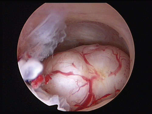
Fibroids can be single or multiple, and vary in size from that of a pip to a watermelon. They may be pedunculated, subserosal, intramural, submucosal (see figure 1), or cervical.
Fibroids are commonly asymptomatic but can cause symptoms such as heavy menstrual bleeding and pelvic pressure symptoms.
Fibroids are one of the main causes of heavy menstrual bleeding, 20-30% of women with heavy menstrual bleeding have fibroids. They may also be associated with reproductive problems such as subfertility and miscarriage. In response to the hormones of pregnancy, fibroids can enlarge and undergo red degeneration. In postmenopausal women they generally decrease in size.
Malignant change (leiomyosarcoma) is rare in premenopausal women, but can present with abnormal bleeding.
Fibroids are most prevalent in women aged between 30 and 50 and there may be a genetic predisposition. They are more common in black women than white women. Other risk factors include obesity and nulliparity.2
Presentation
Fibroids are generally slow growing tumours, enlarging by around 1 cm per year, so the history is usually of long-standing, rather than acute, symptoms. Acute pain is rare with fibroids and usually signifies degeneration, or possibly associated adenomyosis or endometriosis. Rarely a pedunculated fibroid can tort, presenting acutely with severe pain of sudden onset requiring admission.
Other presentations include:
• Incidental finding of a pelvic mass on examination or imaging
• Heavy menstrual bleeding
• Infertility or subfertility (see discussion on surgical treatment)
• Pressure symptoms due to the size of the fibroids - pelvic discomfort, bloating, pressure on the bladder with urinary frequency, incontinence or nocturia, pressure on the bowel with constipation, early satiety, indigestion and increasing girth
• Rarely acute urinary retention resulting from bladder-neck obstruction
• Massive haemorrhage caused by a fibroid prolapsing through the cervix
• Miscarriage and preterm labour
• Pain in pregnancy (red degeneration)
Examination and investigation
Abdominal and vaginal examinations should be carried out. A uterus palpable abdominally must be larger than 12 weeks’ size or 12 cm in length. Irregular bleeding or pelvic pain should prompt a vaginal examination and inspection of the cervix. A full blood count should be performed if periods are excessively heavy.
Endometrial biopsy should be undertaken if the woman is more than 45 years old with menorrhagia or irregular bleeding. An ultrasound scan can help to distinguish between a uterine and an ovarian mass.
Confirming diagnosis
Fibroids are echo dense on ultrasound and often obscure views of the endometrium and ovaries.
MRI can distinguish between uterine fibroids and ovarian fibromas or thecomas and is particularly useful for mapping before surgical or radiological procedures.
Indications for routine and fast track referral are listed in table 1.
Management
Management should focus on symptoms and how these affect the woman’s work and social life. Asymptomatic women require no treatment. The patient’s views on management and whether she wishes to retain her fertility are important considerations when deciding on the approach to treatment.
Management approaches are:
• Pharmacological: non-hormonal or hormonal
• Radiological: uterine artery embolisation (UAE) and MRI-guided high frequency focused ultrasound (MRgFUS)
• Surgical: endometrial ablation, myomectomy, hysterectomy
If a woman presents with heavy menstrual bleeding only, without any intermenstrual bleeding or pressure symptoms, then NICE recommends starting pharmacological management without carrying out a physical examination.3
Pharmacological treatment
Non-hormonal: Tranexamic acid is an antifibrinolytic and is an evidence-based treatment for heavy menstrual bleeding.3 A personal history of thromboembolic disease would be a contraindication to tranexamic acid. NICE recommends the use of tranexamic acid, or NSAIDs, for women with heavy menstrual bleeding and fibroids < 3 cm in diameter.
Hormonal: Suitable for women who are not actively trying to conceive and have heavy menstrual bleeding and fibroids < 3 cm in diameter.3
The combined oral contraceptive pill (COCP) and progesterone-only pill (POP) are used in the treatment of heavy menstrual bleeding. They may cause endometrial atrophy and reduce menstrual blood loss. The evidence for their use in the management of fibroids is inconclusive. Fibroids have oestrogen and progesterone receptors and the contraceptive pill can stimulate fibroid growth. Women should be assessed on their response to treatment after a short trial (the authors suggest three months).
NICE recommends the levonorgestrel intrauterine system (LNG-IUS) as the first-line treatment for women with fibroids < 3 cm in diameter and heavy menstrual bleeding,3 but there is conflicting evidence regarding change in uterine fibroid volume and expulsion rates. An LNG-IUS can be fitted in a woman with fibroids, provided that there is a way in through the cervix and that the cavity length does not exceed 11 cm. As a short-term option for controlling heavy menstrual bleeding norethisterone 5 mg three times a day orally on days 5-26 of the cycle can be used, or for the longer term, long-acting reversible contraception.
Gonadotrophin-releasing hormone (GnRH) agonists are well documented as preoperative treatment to reduce uterine volume and fibroid size. They stop periods and correct preoperative iron-deficiency anaemia.4 Reduction in uterine volume may be sufficient to allow a bikini line incision for surgical treatments, rather than a midline incision for very large fibroids.
Treatment is given by injection or nasal spray and started in the first five days of the menstrual cycle. Side effects of low oestrogen such as hot flushes are common. Non-hormonal, barrier methods of contraception should be used throughout the entire treatment period. Preoperative treatment is normally for three months and is only licensed for a total of six months because of a small reduction in bone density.
Ulipristal acetate is a selective progesterone receptor modulator.
It induces apoptosis within the fibroid and inhibits cellular proliferation.
A course of treatment is 12 weeks commenced during the first week of menstruation (5 mg orally once daily). Treatment results in cessation of menstruation and reduction in the size of fibroids.
Courses may be repeated, starting no sooner than the first week of the second menstruation following completion of the first course, with a maximum of four courses. Reduction in size is cumulative with each treatment course.
NICE3 recommends treatment with ulipristal acetate for women with fibroids ≥ 3 cm in diameter and pressure symptoms or anaemia (Hb ≤ 102 g/l). Non-hormonal, barrier methods of contraception should be used for the entire treatment period.
Radiological treatment
UAE involves cannulating the femoral artery through the groin and injecting an embolic agent into the uterine arteries, reducing the blood supply to the uterus and very vascular fibroids. Before treatment, women are assessed by a gynaecologist and an interventional radiologist. Most women will require an MRI to assess if their fibroids are suitable for embolisation.5
Treatment takes one to two hours and involves a hospital stay of one to two days. Recovery time is about two weeks. The majority of women will have complete or significant resolution of their symptoms for up to five years.
Pregnancy has been reported following UAE, but there is little data regarding pregnancy outcomes. Careful discussion regarding fertility before the procedure should include the risk of premature menopause (1 in 100 women) and hysterectomy (3 in 200 women) in the event of serious complications such as infection.5 Fertility may be better following surgical myomectomy compared with UAE but there has not been enough research involving a direct comparison of the procedures to determine any significant difference.
In a Cochrane review, there was no clear evidence of a difference between UAE and surgery in the risk of major complications, but UAE was associated with a higher rate of minor complications and an increased likelihood of requiring surgical intervention within two to five years of the initial procedure.6
Around 7% of women will need further surgery within two years of hysterectomy or myomectomy, however, between 15% and 32% will need further surgery within two years of UAE. This increase in surgical re-intervention may nullify initial cost advantages of UAE. Thus although UAE is a safe, minimally invasive alternative to surgery, patient selection and counselling are important.
MRgFUS uses MRI to locate and target fibroids and ultrasound energy to heat the fibroid to a temperature above 55°C for a few seconds. Heating of the tissue results in local coagulation and necrosis. Similar to the way a magnifying glass focuses light; ultrasound waves are directed through the patient's abdomen into the fibroid. Single applications of energy are called sonications and a typical treatment comprises multiple sonications. Sonications are repeated until the entire fibroid has been treated.
Women are assessed by a gynaecologist and radiologist before treatment. Pre-treatment MRI helps to select women with fibroids suitable for MRgFUS. Fibroids need to be accessible to the ultrasound beam without bowel loops between the beam and the fibroid. An abdominal incision in the way of the beam is a contraindication because skin burns have been reported.
Treatment takes 60-180 minutes with a day’s stay in hospital. Patients recover within one to three days. Minor complications occur in up to 10.4% of cases and include first- and second-degree skin burns, abdominal, transient lower back and leg pain, fever, mild subcutaneous or abdominal muscle oedema and vaginal spotting or discharge. The incidence of major complications is 0.4%, including bowel perforation, damage to sciatic nerves, endometritis, deep vein thrombosis, and prolonged heavy menstrual bleeding caused by necrotic submucosal fibroid remnant requiring hysteroscopic removal.7
Surgical treatment
Endometrial ablation involves treating the endometrial cavity to remove or destroy the endometrial lining. These techniques are suitable for women who have heavy menstrual bleeding and have completed their family, with fibroids < 3 cm in diameter that are not significantly distorting the uterine cavity.
Myomectomy is the surgical removal of fibroids. Intramural, subserous or pedunculated fibroids can be removed laparoscopically or by open surgery.
The operation carries a risk of bleeding and women should be counselled regarding the 1-2% risk of hysterectomy in the event of catastrophic bleeding. Intraoperative use of a tourniquet around the uterine vessels, use of intramyometrial vasoconstrictors such as diluted vasopressin and intravenous tranexamic acid all reduce intraoperative blood loss. Use of cell salvage can recycle blood lost for autotransfusion.
Currently, there is insufficient evidence from randomised controlled trials to evaluate the role of myomectomy
to improve fertility.8 Laparoscopic myomectomy is associated with less subjectively reported postoperative pain, lower postoperative fever and shorter hospital stays compared with open myomectomy. Current evidence from two randomised controlled trials suggests there is no significant difference between the laparoscopic and open approach in terms of fertility or recurrence. This evidence needs to be viewed with caution because of the small number of studies. More studies are needed to assess rates of uterine rupture, occurrence of thromboembolism, need for repeat myomectomy and hysterectomy at a later stage.9
To remove fibroids laparoscopically, they need to be reduced to fragments inside the abdomen to facilitate removal through the small incisions. This is called morcellation. There has been discussion about the safety of laparoscopic power morcellation, as these devices pose a risk of spreading unsuspected cancerous tissue (uterine sarcoma or rarely leiomyosarcoma) within the abdomen and pelvis, significantly worsening the patient’s long-term survival. Based on a US Food and Drug Administration (FDA) analysis of available data, it is estimated that 1 in 350 women undergoing surgical treatment for fibroids will be found to have an unsuspected uterine sarcoma, a type of uterine cancer that includes leiomyosarcoma.
Currently, there is no reliable method for predicting or testing whether a woman with fibroids may have a uterine sarcoma. Because of this risk and the availability of alternative options, the FDA is warning against the use of laparoscopic morcellators in the majority of women undergoing myomectomy or hysterectomy for treatment of fibroids.10 Techniques are being developed where the fibroid can be morcellated inside a bag within the abdomen.
Malignant change is rare in premenopausal women, but discussion about the route of surgery should include the possibility of unsuspected carcinoma.
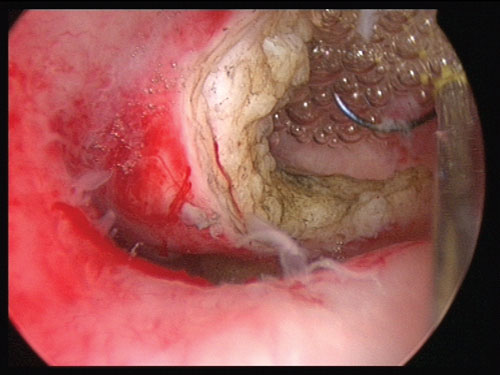
Submucosal fibroids can be hysteroscopically resected (see figure 2). If the fibroid is only partly in the endometrial cavity, it may require repeated procedures to resect it.
There is currently no evidence from randomised controlled trials regarding the effect of hysteroscopic myomectomy on fertility outcomes.7 The FDA has stated that the guidance on laparoscopic morcellators does not apply to hysteroscopic morcellators. It states: ‘When used in accordance with current indications and instructions for use, hysteroscopic morcellators do not pose the same risk as laparoscopic morcellation because any sarcomatous tissue present does not enter the peritoneal cavity.’10
If pharmacological and less invasive management options have failed women may wish to discuss the option of a hysterectomy. Depending upon the uterine size this would either be vaginal, laparoscopic or open (via a bikini line or midline incision). It is normal to conserve healthy ovaries in premenopausal women. NICE recommends the preoperative use of GnRH agonists or ulipristal for all women awaiting hysterectomy for fibroids.
Conclusion
Treatment of fibroids depends on whether women are symptomatic and on the woman’s desire for future childbearing. Asymptomatic fibroids need no treatment. Depending on their size, number and location, symptomatic fibroids can be managed by myomectomy or hysterectomy. Submucous fibroids can be removed by hysteroscopic resection. UAE or MRgFUS may also be used. Other treatments include endometrial ablation, using energy such as microwave or heat, which may be suitable for heavy menstrual bleeding associated with fibroids. Hormone-based treatments may be used on a short-term basis to relieve symptoms, or to reduce the size of the fibroids before surgery or other interventional treatment.
REFERENCES
1 Baird D, Dunson DB, Mill MC et al. High cumulative incidence of uterine leiomyoma in black and white women: ultrasound evidence. Am J Obstet Gynecol 2003;88:100-07
2 Younas K, Hadoura E, Majoko F, Bunkheila A. A review of evidence-based management of uterine fibroids. Obstetrician Gynaecologist 2016;18:3-42
3 National Institute for Health and Care Excellence. CG44. Heavy menstrual bleeding: assessment and management. NICE. London. 2007 (Last updated: August 2016)
4 Lethaby A, Vollenhoven B, Sowter M. Pre-operative GnRH analogue therapy before hysterectomy or myomectomy for uterine fibroids. Cochrane Database Syst Rev 2001;(2):CD000547
5 Royal College of Obstetricians and Gynaecologists and Royal College of Radiologists. Clinical recommendations on the use of uterine artery embolisation (UAE) in the management of uterine fibroids. 3rd edn. 2013. RCOG and RCR. London. 2013
6 Gupta JK, Sinha A, Lumsden M, Hickey M. Uterine artery embolization for symptomatic uterine fibroids. Cochrane Database Syst Rev 2014, Issue 12. Art. No.: CD005073. DOI: 10.1002/14651858.CD005073.pub
7 Pron G. Magnetic resonance–guided high-intensity focused ultrasound (MRgHIFU) treatment of symptomatic uterine fibroids: an evidence-based analysis. Ont Health Technol Assess Ser 2015;15(4):1-86
www.hqontario.ca/Portals/0/Documents/evidence/reports/eba-mrghifu-1503-en.pdf
8 Metwally M, Cheong YC, Horne AW. Surgical treatment of fibroids for subfertility. Cochrane Database Syst Rev 2012, Issue 11. Art. No.: CD003857. DOI: 10.1002/14651858.CD003857.pub
9 Bhave Chittawar P, Franik S, Pouwer AW, Farquhar C. Minimally invasive surgical techniques versus open myomectomy for uterine fibroids. Cochrane Database Syst Rev 2014 Oct 21;(10):CD004638. doi: 10.1002/14651858.CD004638.pub3
10 US Food and Drug Administration. Updated. Laparoscopic Uterine Power Morcellation in Hysterectomy and Myomectomy: FDA Safety Communication. www.fda.gov/medicaldevices/safety/alertsandnotices/ucm424443.htm (Link no longer active by 5/9/2018) Try: https://www.fda.gov/MedicalDevices/ProductsandMedicalProcedures/SurgeryandLifeSupport/ucm584463.htm
ARTICLE IN PDF
 Download this article in PDF format
PRESET SEARCHES
PubMed
Fibroids malignant change (rare)
National Cancer Institute (US)
NICE evidence search
Immediate CPD prompts
These handy templates*, associated with all articles, prompt drafting of personal learning, reflection and planning.
Save your record straight into your personal development folder on your device or cloud storage system.
*Adobe fillable PDFs. Adobe reader needed for iPad
Comment or question
If you, or your peer group, would like to comment on an article or have a question for the authors, write to:
Editor
Case report submissions
Do you have an interesting case that you would like to share with other readers?
Your article would need to be around 1,500 words long, include a brief review of the literature to put the case in context, and be fully referenced in the Vancouver style.
Submissions for consideration for publication should be sent to: [email protected]