SEARCH

Oliver A, Overton C. Detecting ovarian disorders in primary care. Practitioner 2014;258 (1769):15-19
Detecting ovarian disorders in primary care
20 Mar 2014
AUTHORS
Dr Abigail Oliver MBBS BSc MRCOG, Senior Registrar in Obstetrics & Gynaecology
Mrs Caroline Overton MBBS MD FRCOG FHEA, Consultant Gynaecologist, Subspecialist in Reproductive Medicine & Laparoscopic Surgery, St Michael's University Hospital, Bristol, UK
Article
Ovarian cysts are a common diagnosis. They occur more often in premenopausal than postmenopausal women, with a respective prevalence of 34.9% compared with 17.0%.1 It is estimated that up to 10% of women will have surgery for an ovarian cyst in their lifetime.2 The majority of these cysts will be benign, with the risk of malignancy increasing with age.
The risk of a symptomatic ovarian cyst in a premenopausal female being malignant is approximately 1:1,000 increasing to 3:1,000 at the age of 50.3
Ovarian cysts can be classified into:
• Benign - functional cysts, endometrioma, mature teratoma or dermoid cyst, mucinous and serous cystadenomas
• Malignant - epithelial carcinoma, germ cell tumour and sex cord tumour, see table 1.
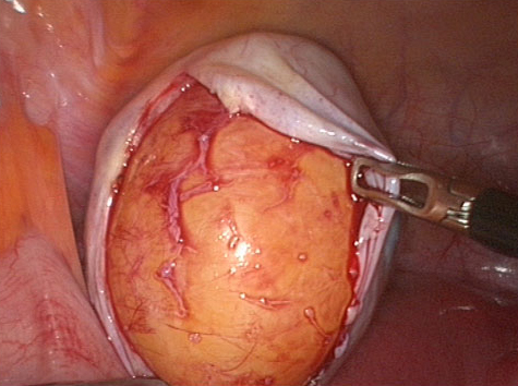
Functional cysts develop during the menstrual cycle (follicular cysts and corpus luteal cysts). These functional cysts particularly when they are < 5 cm diameter, usually resolve spontaneously without the need for intervention. Endometriomas (endometriotic cysts in the ovary) contain a thick brown liquid composed of altered blood, giving rise to the description ‘chocolate cyst’. Dermoid cysts, see figure 1 arise from germ cells and contain a variety of mature solid tissues, which may include teeth, hair and sebum. They are particularly prone to torsion. Mucinous cystadenomas make up one fifth of all ovarian cysts and can reach a massive size. Serous cystadenomas contain clear straw-coloured fluid.
Presentation
Ovarian cysts may be diagnosed clinically, but they can be difficult to detect on examination alone if they are of small or moderate size.
They may also be discovered incidentally in asymptomatic women during ultrasonography performed for different indications.
Ovarian cysts may be asymptomatic, but presenting symptoms include pelvic pain, pressure symptoms and discomfort and menstrual disturbance. Functional cysts in particular can be linked with irregular vaginal bleeding or menorrhagia. Deep dyspareunia, particularly when persistent and unilateral, may be linked to the presence of a cyst.
Endometriosis should be suspected when a woman has cyclical pelvic pain in combination with dysmenorrhoea, dyspareunia (painful intercourse) or dyschezia (painful defecation).
As a cyst enlarges, it exerts pressure effects. Frequency of micturition and an uncomfortable sensation of abdominal bloating are often reported, while some women may have a frequent urge to defecate or difficulty in bowel evacuation. Large cysts extending above the umbilicus can be associated with reduced appetite, nausea and indigestion.
Ovarian torsion
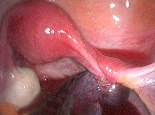
Although there are reports of torsion of normal ovaries, ovarian torsion is most common in the presence of an ovarian cyst, see figure 2, below. Dermoid cysts most commonly tort. Malignant ovarian cysts and endometriotic cysts are less likely to tort because of adhesions that fix the ovary to surrounding tissues.
The diagnosis of ovarian torsion is made clinically. The classic description is sudden onset of severe colicky unilateral pain radiating from groin to loin. There may be nausea and vomiting. It is often confused with ureteric colic where the pain is similar but radiates loin to groin and there is associated haematuria.
Torsion may be acute or subacute, with less severe episodes preceding admission. Clinical examination reveals generalised abdominal tenderness with localised rebound or guarding, sometimes associated with
a low-grade pyrexia, tachycardia and leucocytosis if the torted ovary has become necrotic.
Cyst rupture or haemorrhage into a cyst is associated with sudden onset of a sharp stabbing pain, which is not usually associated with a disturbance in basic observations or abnormal blood tests. Occasionally, such a cyst ‘accident’ may lead to tachycardia and hypotension secondary to substantial haemorrhage within the peritoneal cavity.
Ovarian malignancy
Symptoms which may be suggestive of a malignant ovarian cyst particularly in the over 50 age group include weight loss, persistent abdominal distension or bloating, early satiety, pelvic or abdominal pain and increased urinary urgency and frequency.
Public awareness of these symptoms has been promoted recently by women’s health charities, with an acronym that is easy to remember:
BEAT
Bloating that is persistent and does not come and go
Eating less and feeling fuller
Abdominal pain
Telling your GP
A flow chart for the management of women presenting with symptoms suggestive of ovarian malignancy is shown in figure 3.
Polycystic ovarian syndrome
Polycystic ovarian syndrome (PCOS) is characterised by menstrual disturbance, oligo-ovulation or anovulation leading to irregular periods, and symptoms of androgen excess dating from menarche.
The name suggests that the ovary has many cysts, but there are no large cysts present. The appearance of polycystic ovaries on ultrasound scan is the peripheral arrangement of multiple, small follicles in a string of pearls pattern. The ultrasound appearance is not essential for diagnosis, see table 3, p18.5
Polycystic ovaries do not increase in size and are not a cause of pelvic or abdominal pain. Patients with PCOS may present with oligomenorrhoea or amenorrhoea, unwanted facial and body hair, oily skin, acne, hair loss from the scalp and weight gain. PCOS is common, but it is important that other endocrinopathies such as Cushing’s syndrome or testosterone-producing tumours are considered and excluded.
A history and examination focused on the endocrine system may detect common symptoms and signs of hyperandrogenism. These include unusual headache, visual disturbance, alopecia, hirsutes, acne, virilisation (voice change), development of new striae, galactorrhoea, weight gain or menstrual disturbance.
The timing and progression of these symptoms may give an indication as to the underlying cause. Long-standing, slowly progressing hirsutism starting soon after puberty is typical in PCOS, while a sudden onset of excessive hair growth associated with a deepening of the voice is more suggestive of
a testosterone-producing adrenal tumour.6 Women with Cushing’s syndrome may present with irregular periods and unwanted hair growth. The presence of new pink or purple striae over the body and severe fatigue and myalgia support this diagnosis.
Primary care assessment
Risk factors for ovarian cancer should be identified. These include:
• Family history of breast or ovarian cancer
• Nulliparity
• Smoking (although probably too common to be discriminatory)
• Increasing age (most common after 55 years old)
Protective factors include a history of pregnancy, breastfeeding and oral contraceptive use.
Persistent abdominal distension, reduced appetite and an early feeling of fullness suggest ovarian malignancy. Symptoms of cyclical pelvic pain, dysmenorrhoea, dyspareunia and dyschezia in menstruating women suggest a diagnosis of endometriosis.7
Abdominal and pelvic examinations should be carried out to look for masses and ascites, although the detection rate of ovarian cysts on clinical examination alone is low.8 Pelvic ultrasound scan is the most effective way of evaluating an ovarian cyst and should be considered a first-line investigation in most cases.
CA125
The serum marker CA125 is not specific for ovarian cancer. Levels can be elevated in benign conditions including pelvic inflammatory disease, appendicitis, pregnancy, endometriosis, adenomyosis, fibroids, appendicitis, pancreatitis or liver disease.
NICE advises that CA125 levels should be checked in women who present with frequent bloating, feeling full quickly, loss of appetite, pelvic or abdominal pain or needing to urinate quickly or urgently.
CA125 should also be checked in women over 50 who present with symptoms which suggest irritable bowel syndrome such as bloating, abdominal pain or changes in bowel habit. A significant proportion of women with early stage ovarian cancer have a normal CA125.
A CA125 ≥ 35 IU/ml is considered elevated.4 If the CA125 is raised, an ultrasound scan of the abdomen and pelvis should be arranged with urgent referral if there are findings suggestive of ovarian cancer.4
Premenopausal women with a simple ovarian cyst < 5 cm do not require a CA125 measurement.3
Postmenopausal women with an ovarian cyst should have their CA125 levels checked, as they are used in combination with ultrasound appearance to calculate the likelihood of malignancy. The risk of malignancy index score will guide subsequent management.
Referral
Postmenopausal women
Asymptomatic women with simple unilateral cysts < 5 cm in size who have a normal CA125 level may be managed conservatively with a follow-up ultrasound scan after four months.9 Symptomatic women, those with any other type of ovarian cyst, or raised CA125 levels, should be referred to secondary care.
Features on ultrasound scan which should prompt an urgent two week wait referral include:
• Ascites
• An irregular solid mass
• Bilateral lesions
• Multilocular lesions
• Evidence of solid areas
• Very strong blood flow to the mass
Premenopausal women
Asymptomatic premenopausal women with simple cysts < 5 cm need not be referred and do not require follow-up, as these cysts are very likely to be physiological and usually resolve spontaneously over 2-3 menstrual cycles.
Simple cysts which measure ≥ 5 cm are less likely to resolve and need an annual ultrasound assessment as a minimum.
Women with simple cysts ≥ 7 cm should be referred to gynaecology for ongoing management, consideration of additional imaging and surgical intervention, as should women with ultrasound features suggestive of endometriomas or dermoid cysts.3
Urgent referral, under the two week wait rule, is appropriate for a woman of any age with ultrasound features suggestive of malignancy, see table 4.
Treatment
Simple cysts
Women with simple ovarian cysts may opt safely for conservative management with or without ultrasound surveillance as detailed above.
Ovarian cysts that persist or increase in size over a period of six months to
a year are unlikely to be functional and are more likely to be associated with complications such as torsion or rupture. Surgical management, usually in the form of laparoscopic ovarian cystectomy, is appropriate in these cases, although there is no evidence-based consensus on the size of cyst above which surgery should be considered.3
Ovarian cancer
The primary aim of treatment is to reduce the cancer volume by surgery and chemotherapy.
Primary surgery usually involves abdominal hysterectomy and bilateral salpingo-oophorectomy, appendicectomy and omentectomy. Patients should be treated within a specialised cancer unit and by a multidisciplinary team.
PCOS
Many women with PCOS are overweight, and have a higher prevalence of impaired glucose tolerance and type 2 diabetes.
Annual screening with a fasting blood glucose should be offered, or an oral glucose tolerance test arranged if there is a strong family history of diabetes or the woman is obese (BMI ≥ 30). Blood pressure should be monitored with appropriate treatment of hypertension. Lipid-lowering treatment is not currently recommended routinely, and referral should be made to a specialist in the presence of an abnormal profile.5
Women with PCOS may be more at risk of developing endometrial hyperplasia, particularly if the interval between menstrual bleeds is more than three months. To prevent this, the aim should be at least four bleeds per year naturally. If this is not occurring, giving ten days of progestogen
can induce a withdrawal bleed. Alternatively regular withdrawal bleeds can be achieved using the combined or progesterone-only contraceptive pill.
REFERENCES
1 Pavlik EJ, Ueland FR, Miller RW et al. Frequency and disposition of ovarian abnormalities followed with serial transvaginal ultrasonography. Obstet Gynecol 2013; 122 (2 Pt 1):210-217
2 Hilger WS, Magrina JF, Magtibay PM et al. Laparoscopic management of the adnexal mass.
Clin Obstet Gynecol 2006;49:535-548
3 Royal College of Obstetricians and Gynaecologists. RCOG Guideline No. 62. Management of suspected ovarian masses in premenopausal women. RCOG. London. 2011
4 National Institute for Health and Clinical Excellence. CG122. Ovarian cancer: The recognition and initial management of ovarian cancer. NICE. London. 2011
5 Royal College of Obstetricians and Gynaecologists. RCOG Guideline No. 33. Long term consequences of polycystic ovary syndrome. RCOG. London. 2007
6 Meek CL, Bravis V, Don A, Kaplan F. Polycystic ovary syndrome and the differential diagnosis of hyperandrogenism. The Obstetrician & Gynaecologist 2013;5:171–176
7 Park C, Overton CE. Endometriosis. More on the missed disease. BMJ 2010;341:163
8 Padilla LA, Radosevich DM, Milad MP. Accuracy of the pelvic examination in detecting adnexal masses. Obstet Gynecol 2000;96:593–598
9 Royal College of Obstetricians and Gynaecologists. RCOG Guideline No. 34. Ovarian cysts in postmenopausal women. RCOG. London. 2010
10 Timmerman D, Valentin L, Bourne TH et al. Terms, definitions and measurements to describe the sonographic features of adnexal tumours: a consensus opinion from the International Ovarian Tumour Analysis (IOTA) group. Ultrasound Obstet Gynaecol 2000;16:500-505
ARTICLE IN PDF
 Download this article in PDF format
PRESET SEARCHES
Immediate CPD prompts
These handy templates* prompt immediate drafting of personal learning, reflection and planning. Save your record straight into your personal development folder on your device or Cloud storage system.
*Adobe reader needed for iPad
EXTERNAL WEBLINKS
Ovacome
The charity provides information and advice on ovarian cancer for healthcare professionals and patients
Current evidence - Ovarian cancer
US NCI
PDQ® (Physician Data Query) is NCI's cancer database of peer-reviewed, regularly updated evidence-based, referenced summaries
Ovarian
PubMed
UK NICE
Ovarian
UK Risk Assessment Tools
QCancer
Qcancer women
UK Early diagnosis programmes
UK NCAT
Macmillan - early diagnosis
US NCI
PDQ® (Physician Data Query) is NCI's cancer database of peer-reviewed, regularly updated evidence-based, referenced summaries
Women
Breast
Ovarian
Endometrial screening
Endometrial
Cervical