SEARCH

Ralston SH, Fraser J. Optimising the assessment and management of osteoporosis. Practitioner March 2021;265(1846):17-21
Optimising the assessment and management of osteoporosis
22 Mar 2021
AUTHORS
Professor Stuart H Ralston MD FRCP
Professor of Rheumatology, Centre for Genomic and Experimental Medicine, University of Edinburgh, Western General Hospital, Edinburgh, UK
Dr Jamie Fraser BSc MB ChB MRCGP GP, Southside Surgery, Inverness, UK
Competing interests
Professor Stuart Ralston’s institution has received grants for participating in clinical research studies with romosozumab (UCB) and teriparatide (Eli Lilly). The institution has received medicinal products for trials with zoledronic acid in early Paget’s disease (Novartis) and for trials of teriparatide in osteogenesis imperfecta (Eli Lilly). Dr Jamie Fraser has no competing interests
Article
Osteoporosis is characterised by architectural deterioration of bone tissue, low bone mineral density (BMD) and an increased risk of fragility fractures. It affects around 40% of women and 13% of men at some point in their lives. While almost any bone can fracture as the result of osteoporosis, the most common sites are the wrist, spine, hip and humerus. The two most important risk factors for osteoporosis are increasing age and female gender. Other risk factors that are associated with reduced BMD and an increased risk of fragility fractures include chronic inflammatory diseases, malabsorption, chronic liver disease, chronic kidney disease and chronic neurological diseases. Drug treatments that predispose to osteoporosis include corticosteroids, thiazolidinediones, antiepileptic drugs, proton pump inhibitors and aromatase inhibitors. The presence of one or more of these clinical risk factors in individuals aged 50 and over is an indication for a fracture risk assessment. The most commonly used fracture risk calculators in the UK are FRAX and Qfracture. To make a diagnosis of osteoporosis DXA examination of the lumbar spine and hip should be carried out. The diagnosis can be confirmed if the BMD T-score value at either site is -2.5 or less. There is a strong evidence base for drug treatment in DXA proven osteoporosis and those with low trauma vertebral fractures. Bisphosphonates are the most commonly used drugs. If oral bisphosphonates are contraindicated or poorly tolerated, parenteral bisphosphonate therapy should be considered, typically with intravenous zoledronic acid.
Osteoporosis is a common condition characterised by architectural deterioration of bone tissue, low bone mineral density (BMD) and an increased risk of fragility fractures. Osteoporosis is defined as a BMD that lies 2.5 standard deviations or more below the average value for young healthy women.1 It has been estimated that the lifetime risk of a clinical fracture is about 40% for women and 13% for men.2 The proportion of individuals with fractures who have osteoporosis varies but in a recent study was around 23% of women and 10% of men aged 50 years and above.3
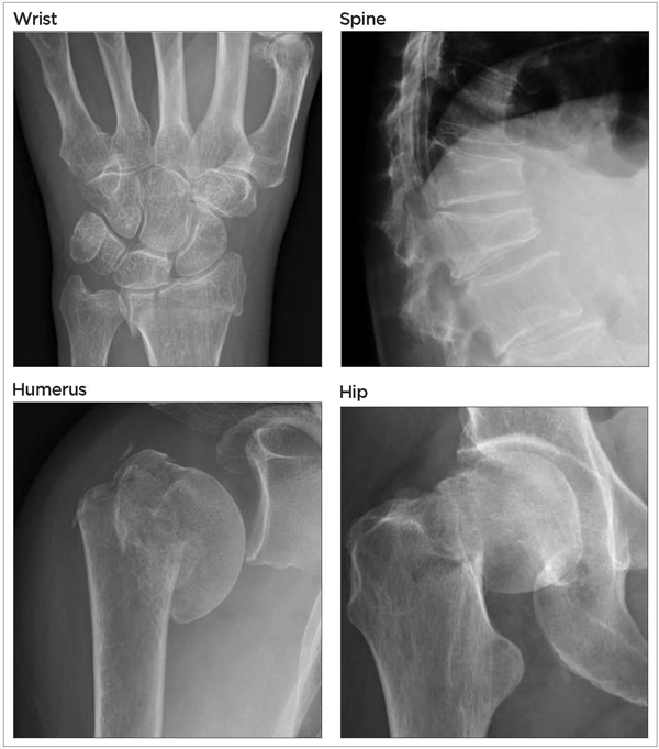
It is often clinically silent until a fracture occurs. While almost any bone can fracture as the result of osteoporosis, the most common sites are the wrist, spine, hip and humerus, see figure 1.
Although fractures are characteristic of osteoporosis, it is important to remember that many individuals with fractures do not have osteoporosis and a significant proportion have normal BMD. This reflects the fact that the occurrence of a fracture is not only attributable to the strength of the bone, which is impaired in osteoporosis but also the degree of trauma and severity of the fall.
Risk factors
The two most important risk factors for osteoporosis are increasing age and female gender. This is because bone is lost with age and women undergo an accelerated phase of bone loss after the menopause due to oestrogen deficiency.
Clinical risk factors that are associated with reduced BMD and increased risk of fragility fractures include chronic inflammatory diseases, malabsorption, chronic liver disease, chronic kidney disease and chronic neurological diseases, see table 1.
Drug treatments that predispose to osteoporosis include corticosteroids, thiazolidinediones, antiepileptic drugs, proton pump inhibitors and aromatase inhibitors.
Given the importance of age in osteoporosis it has been recommended that the presence of one or more of these clinical risk factors in an individual aged 50 years or over is an indication for a fracture risk assessment as discussed below. Fracture risk assessment may also be considered in younger people with very strong risk factors such as current or frequent recent use of oral or systemic glucocorticoids, untreated premature menopause or previous fragility fractures.
Quantifying fracture risk
The risk of fractures can be estimated on the basis of clinical risk factors and further refined by assessment of BMD by dual energy X-ray absorptiometry (DXA). The two most commonly used fracture risk calculators in the UK are FRAX (www.shef.ac.uk/FRAX) and QFracture (www.qfracture.org).
Both collect information on age, body weight and height, along with some of the clinical risk factors mentioned above.
The QFracture algorithm was developed by observations made on more than 3 million individuals from 535 general practices in the UK4 and was independently validated in a further cohort of 2.2 million individuals from 364 other practices in the UK.5 It is likely to be more accurate at predicting fractures in the UK population than FRAX.
The FRAX algorithm was developed by analysis of several cohorts in Europe (including the UK), the USA, Canada, Australia and Japan with a total sample size of 276,826.6
The QFracture calculator allows clinicians to enter more clinical risk factors than the FRAX algorithm and can provide calculations ranging from one- to ten-year risk. It can also be implemented within the EMIS software system. The FRAX calculator only provides ten-year estimates of risk.
An advantage of FRAX over QFracture is that it is possible to refine the estimated fracture risk calculated on the basis of clinical risk factors by adding information from DXA provided that hip BMD measurements have been taken.
Both the NICE guideline (CG146)7 and the recently updated SIGN guideline (SIGN 142)8 recommend that a fracture risk assessment with one of these tools should be carried out before DXA.
While NICE did not provide guidance on the fracture risk threshold that should be reached before ordering a DXA scan, the SIGN guideline suggests that DXA should be considered when ten-year fracture risk is 10% or more, based on the fact that most clinical trials of osteoporosis treatments had enrolled individuals with a ten-year fracture risk above this threshold.
Confirming diagnosis
Osteoporosis cannot be diagnosed by a fracture risk calculation alone. To make a diagnosis of osteoporosis DXA examination of the lumbar spine and hip should be performed. The diagnosis can be confirmed when the BMD T-score value at either site is -2.5 or less. If the DXA scan indicates osteoporosis it is usual to perform screens for secondary causes of osteoporosis. These include thyroid function tests, tTG for coeliac disease, kidney and renal function, serum calcium for hyperparathyroidism, myeloma and MGUS screen (FBC, ESR, immunoglobulins, Bence Jones protein) and in men testosterone and gonadotrophins.
Quite frequently DXA examination can reveal osteoporosis at one skeletal site but not at another. For example, it is quite common to encounter patients with osteoporosis of the spine but not the hip. This is thought to be because loss of bone occurs more rapidly at the spine with ageing as the vertebrae are rich in trabecular bone.
Genetic factors and mechanical loading can also influence susceptibility to osteoporosis at different skeletal sites. Paradoxically, it is possible to have osteoporosis of the spine with normal BMD values at this site due to coexisting diseases such as osteoarthritis, degenerative disc disease and aortic calcification which elevate measured BMD values. In this situation the diagnosis of spinal osteoporosis can be made by the presence of a vertebral fracture on imaging, provided of course that there has not been a history of major trauma.
Increasingly, a common mode of presentation for spinal osteoporosis is when vertebral fractures are detected by MRI or CT imaging performed for another reason. In these circumstances DXA imaging would be indicated.
As well as its role in the diagnosis of osteoporosis, DXA is also useful in assessing whether or not patients have responded to treatment and this is especially valuable in patients who fracture while on therapy.
Complications
Most fractures occur after a fall, but vertebral fractures may occur spontaneously. Kyphosis may occur in patients with multiple vertebral fractures and this in turn may cause respiratory symptoms by adversely affecting anatomy of the chest wall.
Management
There is a strong evidence base for drug treatment in DXA proven osteoporosis and those with low trauma vertebral fractures.
The National Osteoporosis Guideline Group (NOGG) guideline9 advocates starting treatment on the basis of a high fracture risk alone, without performing DXA. The effectiveness of osteoporosis treatments at preventing fractures using the NOGG cut-off points is largely unknown.
There is evidence that drug treatment may reduce the risk of hip fracture in elderly women with a ten-year fracture risk of ≥ 20% but prevention of other fractures using this approach has not been demonstrated.10
When discussing treatment options with patients it is important to explain that the benefits of treatments are modest, unless fracture risk is very high especially for non-vertebral, non-hip fractures.
A calculator has been developed to facilitate these calculations and is available online and as a smartphone app (see Useful information).
It is important to identify and treat any underlying causes, correct lifestyle factors where appropriate such as stopping smoking and reducing alcohol consumption, assessing falls risk and reducing or stopping drugs that may predispose to osteoporosis where possible.
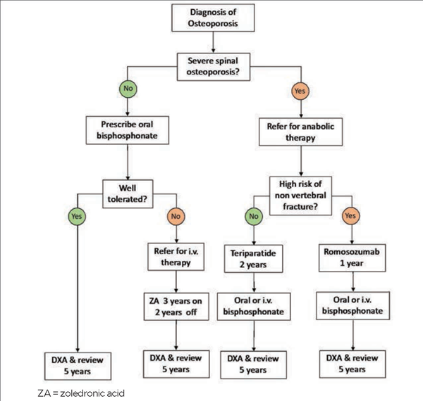
Options for drug treatment are summarised in figure 2.
Bisphosphonates
Bisphosphonates are the most commonly used drugs. They inhibit osteoclastic bone resorption and prevent bone loss. Bisphosphonate treatment also increases BMD, but this is mainly due to increased mineralisation of existing bone as opposed to a gain in bone mass. Usually, treatment is initiated with an oral bisphosphonate such as alendronic acid 70 mg weekly or risedronate 35 mg weekly.
Bisphosphonates may be combined with a calcium and vitamin D supplement if dietary calcium intake is less than 700 mg daily or with a standalone vitamin D supplement in those at risk of deficiency. The most common adverse effect is upper gastrointestinal (GI) upset but some patients who develop GI upset with one oral bisphosphonate can tolerate another perfectly well, so it is worth trying both.
If oral bisphosphonates are contraindicated or poorly tolerated, parenteral bisphosphonate therapy should be considered, typically with intravenous zoledronic acid. This is usually delivered in secondary care.
The most common adverse effects with bisphosphonates are GI upset with oral preparations and a transient flu-like reaction with intravenous agents.
Rare adverse effects include atypical femoral fractures with long-term treatment (80 per 100,000 patient-years of therapy) and osteonecrosis of the jaw (1 per 100,000 patient-years of therapy).11
Denosumab
Denosumab is an inhibitor of bone resorption with similar antifracture efficacy to zoledronic acid which is given by subcutaneous injection. Although this is an effective treatment, it has recently become apparent that when denosumab is stopped there is a rebound increase in bone remodelling and rapid bone loss which can be accompanied by vertebral fractures and even hypercalcaemia. Because of this denosumab needs to be continued on a long-term basis once started, or if treatment is stopped for any reason, it should be followed up with an oral or intravenous bisphosphonate.12
Teriparatide
Teriparatide is a bone anabolic drug which is particularly effective in the treatment of postmenopausal women with severe spinal osteoporosis.
It stimulates bone remodelling but enhances bone formation to a greater extent than bone resorption causing gain of bone.
Clinical trials have shown that teriparatide is superior to oral risedronate in preventing new vertebral fractures in patients with severe spinal osteoporosis13 and has also been found to be more effective than standard care in observational studies.14
The recently updated SIGN guideline recommends that teriparatide should be considered in preference to oral bisphosphonates in this patient group. The treatment course of teriparatide is two years and following completion of therapy patients should be transitioned to an antiresorptive drug to maintain the increase in BMD.14 Normally patients requiring teriparatide are referred to secondary care for initiation of treatment and follow-up.
Romosozumab
Romosozumab is a bone anabolic drug which is highly effective in patients with severe osteoporosis. It stimulates bone formation and at the same time inhibits bone resorption causing positive uncoupling of the bone remodelling process.
Treatment with romosozumab increases BMD substantially at both the spine and hip and has been shown to be superior to alendronic acid at preventing hip fractures, spine fractures and non-spine, non-hip fractures in postmenopausal women with severe osteoporosis.15
The treatment course is one year and following completion of therapy patients should be transitioned to an antiresorptive drug to maintain the increase in BMD.14
The SIGN guideline recommends romosozumab in preference to oral bisphosphonates for patients with severe spinal osteoporosis with vertebral fractures who are also at high risk of non-vertebral fractures.8 Romosozumab is usually initiated in secondary care.
Other treatments
Other treatments are used for osteoporosis less frequently than those mentioned above. Oral ibandronate (150 mg once a month) or intravenous ibandronate (3 mg 3-monthly) has been shown to reduce the risk of vertebral fractures in osteoporosis but the trial results for non-vertebral fractures were less convincing than with the bisphosphonates mentioned above.
Hormone replacement therapy can prevent postmenopausal bone loss, and fractures. It still has a role in postmenopausal women with osteoporosis and in those with an early menopause but is seldom used beyond the age of 65 due to concerns about an increased risk of cardiovascular disease and breast cancer with long-term use.
Monitoring
Repeated measurements of BMD are a useful means of monitoring the response to treatment but because changes in BMD with antiresorptive agents occur slowly, repeat BMD measurements should not generally be performed for at least two years after the initial DXA. If a patient is on anabolic drugs, repeat DXA should be done at the end of the course of treatment but then repeated after the patient transitions to antiresorptive therapy.
For antiresorptive drugs there is a concern that with very long-term treatment, there is inhibition of the normal renewal and repair process in bone and that this can actually increase bone fragility and impair tissue repair, leading to complications such as atypical femoral fractures and osteonecrosis of the jaw. Because of this the MHRA advises that the need for continued antiosteoporosis treatment be reviewed every five years. In most individuals, this involves a medication review and a repeat DXA.
In general patients who remain osteoporotic on repeat DXA after five years on oral bisphosphonates who are considered to be at high risk of vertebral fractures because of low spine BMD or preexisting vertebral fractures should be offered continued therapy for a further five years.
In those who no longer have osteoporosis, a treatment holiday can be offered given that these drugs remain in the skeleton for prolonged periods and continue to exert inhibitory effects on bone resorption and protect against fractures.16 For zoledronic acid, a common approach is to give treatment in cycles of three years on and two or three years off, as the rate of fracture prevention with three years’ zoledronic acid treatment has been shown to be similar to that following six years’ treatment.17 Most specialists advise that a treatment holiday should be given after ten years, although there is very little clinical evidence to support this recommendation.
Patients who fracture while on treatment
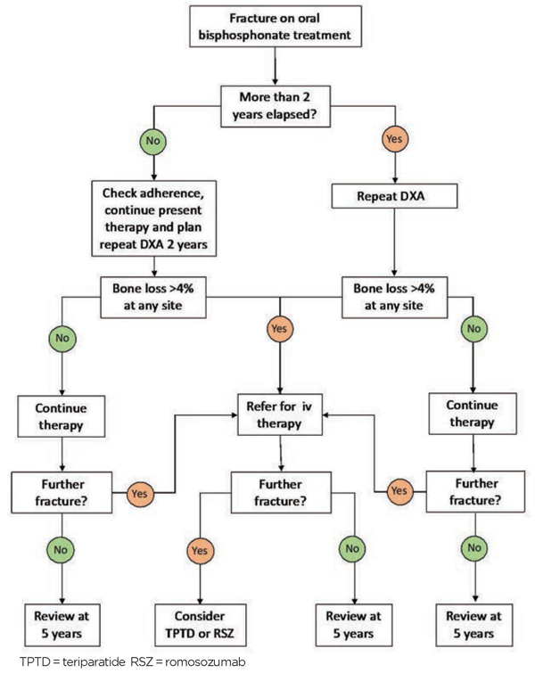
None of the treatments currently available are completely effective and so the occurrence of a fracture when a patient is on treatment does not necessarily mean the therapy is not working. A suggested approach for the management of patients who fracture on treatment is shown in figure 3.
To summarise, if a patient on oral bisphosphonates fractures on therapy and at least two years have elapsed since the baseline DXA a repeat scan should be requested. If the BMD is stable or has increased, treatment should be continued. If there is significant bone loss ( ≥ 4% at any site) then transition to an intravenous bisphosphonate should be considered. If a patient suffers more than two fractures over a five-year period on antiresorptive therapy, then a course of anabolic treatment should be considered.
Special groups
The management of male osteoporosis follows the same general strategy as postmenopausal osteoporosis but the evidence that treatments prevent fractures in men with osteoporosis, apart from vertebral fractures, is poor.
In glucocorticoid-induced osteoporosis, there is evidence that bisphosphonates and teriparatide are effective at preventing vertebral fractures, when combined with calcium and vitamin D supplements. However, there is no evidence that treatment prevents non-vertebral fractures.
There is evidence that fractures occur in glucocorticoid-induced osteoporosis at higher levels of BMD than in postmenopausal osteoporosis so therapy should be considered in patients with a BMD T-score of -1.5 or less.
REFERENCES
1 www.who.int/chp/topics/Osteoporosis.pdf
2 McLellan AR, Gallacher SJ, Fraser M, McQuillian C. The fracture liaison service: success of a program for the evaluation and management of patients with osteoporotic fracture. Osteoporos Int 2003;14:1028-34
3 Tei RMH, Ramlau-Hansen CH, Plana-Ripoll O et al. OFELIA: Prevalence of Osteoporosis in Fragility Fracture Patients. Calcif Tissue Int 2019;104:102-14
4 Hippisley-Cox J, Coupland C. Predicting risk of osteoporotic fracture in men and women in England and Wales: prospective derivation and validation of QFractureScores. BMJ 2009;339:b4229
5 Collins GS, Mallett S, Altman DG. Predicting risk of osteoporotic and hip fracture in the United Kingdom: prospective independent and external validation of QFractureScores. BMJ 2011;342:d3651
6 Kanis JA, Oden A, Johnell O et al. The use of clinical risk factors enhances the performance of BMD in the prediction of hip and osteoporotic fractures in men and women. Osteoporos Int 2007;18:1033-46
7 National Institute for Health and Care Excellence. CG146. Osteoporosis: assessing the risk of fragility fracture. NICE. London. 2012 Updated 2017
8 Scottish Intercollegiate Guidelines Network. SIGN 142. Management of osteoporosis and prevention of fragility fractures. SIGN. Edinburgh. 2015 Updated 2021
9 Compston J, Cooper A, Cooper C et al. National Osteoporosis Guideline Group. UK clinical guideline for the prevention and treatment of osteoporosis. Arch Osteoporos 2017;12:43
10 Shepstone L, Lenaghan E, Cooper C. Screening in the community to reduce fractures in older women (SCOOP): a randomised controlled trial. Lancet 2018;391:741-47
11 Brown JP. Antiresorptives: safety concerns - clinical perspective. Toxicol Pathol 2017;45(7):859-63
12Tsourdi E, Zillikens MC, Meier C et al. Fracture risk and management of discontinuation of denosumab therapy: a systematic review and position statement by ECTS. J Clin Endocrinol Metab 2021;106:264-81
13 Kendler DL, Marin F, Zerbini CAF et al. Effects of teriparatide and risedronate on new fractures in post-menopausal women with severe osteoporosis (VERO): a multicentre, double-blind, double-dummy, randomised controlled trial. Lancet 2018;391:230-40
14 Oswald AJ, Berg J, Milne G, Ralston SH. Teriparatide treatment of severe osteoporosis reduces the risk of vertebral fractures compared with standard care in routine clinical practice. Calcif Tissue Int 2014;94:176-82
15 Saag KG, Petersen J, Brandi ML et al. Romosozumab or alendronate for fracture prevention in women with osteoporosis. N Engl J Med 2017;377:1417-27
16 Schwartz AV, Bauer DC, Cummings SR et al. Efficacy of continued alendronate for fractures in women with and without prevalent vertebral fracture: the FLEX trial. J Bone Miner Res 2010;25:976-82
17 Black DM, Reid IR, Boonen S et al. The effect of 3 versus 6 years of zoledronic acid treatment of osteoporosis: a randomized extension to the HORIZON-Pivotal Fracture Trial (PFT). J Bone Miner Res 2012;27:243-54
ARTICLE IN PDF
 Download this article in PDF format
PRESET SEARCHES
PubMed
NICE Evidence search
EXTERNAL WEBLINKS
The University of Edinburgh Osteoporosis Risk Benefit Calculator
The smartphone app is available from the Apple Store and Google play