SEARCH

Negoescu A. Managing osteoporosis in postmenopausal women. Practitioner March 2018;262(1813):19-24
Managing osteoporosis in postmenopausal women
22 Mar 2018
AUTHORS
Dr Andra Negoescu, Consultant Rheumatologist, Addenbrooke’s Hospital, Cambridge, UK
Competing interests: None
Article
Abstract
Osteoporosis is defined as a progressive systemic skeletal disease characterised by low bone mass and microarchitectural deterioration of bone tissue, with a consequent increase in bone fragility and susceptibility to fracture. Most patients with osteoporosis are asymptomatic unless a fragility fracture occurs. A fragility fracture is a type of pathological fracture that occurs as a result of normal activities, such as a fall from standing height or less, lifting, or bending. There are three fracture sites said to be typical of fragility fractures: vertebral fractures; fractures of the neck of the femur; Colles’ fracture of the wrist. Insufficiency fracture is caused by normal or physiologic stress on weakened bone. Most commonly, patients present with acute or sub-acute onset back pain (this can be thoracic or lumbar), following minor injuries, falls from standing height or normal activities such as walking. The pain is caused by vertebral body compression fracture/s, and is described variably as sharp, nagging, or dull; movement usually exacerbates pain, and in some cases, pain can radiate to the abdomen. On examination, there is focal spinal tenderness at the site of fracture. The cervical spine is an unusual site of osteoporotic fractures, so any acute neck pain should be investigated for alternative causes. Following fracture risk assessment a dual energy X-ray absorptiometry (DXA) scan may be recommended. This is currently the gold standard for the evaluation of BMD. DXA provides the patient’s T-score, which is the BMD value compared with that of control subjects who are at their peak BMD.
Osteoporosis is defined by the World Health Organization (WHO) as progressive systemic skeletal disease characterised by low bone mass and microarchitectural deterioration of bone tissue, with a consequent increase in bone fragility and susceptibility to fracture. 2
Osteoporosis leads to nearly 9 million fractures annually worldwide,3 and more than 300,000 patients present with fragility fractures to hospitals in the UK each year.4 More than one-third of adult women and one in five men will sustain one or more fragility fractures in their lifetime.5
Fragility fractures result from mechanical forces that would not ordinarily cause fracture, known as low level (or ‘low energy’) trauma.6 Because of increased bone loss after the menopause in women, and age-related bone loss in both women and men, the prevalence of osteoporosis increases markedly with age, from 2% at 50 years to > 25% at 80 years in women. As the longevity of the population increases, so will the incidence of osteoporosis and fragility fracture.
Osteoporotic fragility fractures can cause substantial pain and severe disability, often leading to a reduced quality of life, and hip and vertebral fractures are associated with decreased life expectancy. Hip fracture nearly always requires hospitalisation, is fatal in 20% of cases and permanently disables 50% of those affected; only 30% of patients fully recover.7
Risk factors
Risk factors for osteoporosis include: 8,9,10
• Age ≥ 50 years
• White or Asian ethnicity
• Genetic factors, such as a family history of osteoporosis
• Thin build or small stature (e.g. body weight < 127 lb)
• Amenorrhoea
• Late menarche
• Early menopause
• Postmenopausal state
• Physical inactivity or immobilisation11
• Use of certain drugs (e.g. systemic steroids, anticonvulsants, thyroid supplements, heparin, chemotherapeutic agents, insulin)
• Alcohol and tobacco use
• Oestrogen deficiency of any cause
• Calcium deficiency
Presentation
Osteoporosis is often called the silent disease because bone loss usually occurs gradually over the years without symptoms. Despite the popular belief that osteoporosis causes bone pain, most patients with osteoporosis are asymptomatic unless a fragility fracture occurs. A fragility fracture is a type of pathological fracture that occurs as a result of normal activities, such as a fall from standing height or less, lifting, or bending. There are three fracture sites said to be typical of fragility fractures:
• Vertebral fractures
• Fractures of the neck of the femur
• Colles’ fracture of the wrist
Insufficiency fracture is caused by normal or physiologic stress on weakened bone. Loss of bone trabeculae decreases the bone’s elastic resistance which can then lead to a fracture at the site where the weight transfer is the highest, such as the sacrum, pelvic bones, femoral head etc.
Most commonly, patients present with acute or sub-acute onset back pain (this can be thoracic or lumbar), following minor injuries, falls from standing height or normal activities such as walking.
The pain is caused by vertebral body compression fracture/s, and is described variably as sharp, nagging, or dull; movement usually exacerbates pain, and in some cases, pain can radiate to the abdomen.
On examination, there is focal spinal tenderness at the site of fracture.
The cervical spine is an unusual site for osteoporotic fractures, so any acute neck pain should be investigated for alternative causes.
Practical tip 1: A good history taken from the patient can help distinguish between an acute post traumatic fracture, a stress fracture and a fragility fracture.
Patients with fractures of the peripheral bones will present with symptoms suggestive of the respective fractures, although these patients may present in an acute hospital setting rather than primary care. However, it often falls to the GP to screen their patients with fragility or insufficiency fractures for osteoporosis, and initiate osteoporosis management.
Practical tip 2: Consider assessing further fracture risk for all patients with a fragility fracture, regardless of the site (see risk assessment below).
Confirming diagnosis
The baseline blood tests below are used to exclude secondary causes of osteoporosis and other causes for pathologic bone fracture:
• Full blood count, which may reveal anaemia
• Serum chemistry: renal impairment can lead to renal bone disease
• Liver function tests
• Thyroid-stimulating hormone level: thyroid dysfunction can be associated with osteoporosis
• 25-hydroxyvitamin D level: vitamin D insufficiency/deficiency can predispose to osteoporosis or cause osteomalacia
• Serum protein electrophoresis and urinary Bence Jones protein: multiple myeloma is a rare but serious cause of vertebral fractures
• Coeliac screen
• Parathyroid hormone (PTH) and bone profile: hyperparathyroidism can cause low bone mineral density (BMD)
NICE recommends that fracture risk is assessed in the following groups:12
• All women aged 65 years and over and all men aged 75 years and over
• Women aged under 65 years (and men aged under 75 years) in the presence of risk factors, for example:
— Previous fragility fracture
— Current or frequent recent use of oral or systemic glucocorticoids
— History of falls
— Family history of hip fracture
— Other causes of secondary osteoporosis
— Low BMI i.e. < 18.5 kg/m2
— Smoking
— Alcohol intake > 14 units per week for women (> 21 units per week for men)
Following the fracture risk assessment (using the methods described below),
a dual energy X-ray absorptiometry (DXA) scan may be recommended.
This is currently the gold standard for the evaluation of BMD.
DXA provides the patient’s T-score, which is the BMD value compared with that of control subjects who are at their peak BMD.13,14,15,16
WHO criteria define a normal T-score value as within 1 standard deviation (SD) of the mean BMD value in a healthy young adult. Values lying further from the mean are stratified as follows:7
• T-score of –1 to –2.5 SD indicates osteopenia (low bone density)
• T-score < –2.5 SD indicates osteoporosis
• T-score < –2.5 SD with fragility fracture(s) indicates severe osteoporosis
In situations where DXA is unsuitable or cannot be performed (e.g. patients with bilateral hip replacements or significant osteoarthritis of the spine),
a quantitative computed tomography (QCT) scan could be considered to measure BMD.
Risk assessment
There are two scores that can be used for fracture risk assessment:
• FRAX17 (without a BMD value if a DXA scan has not previously been undertaken). FRAX can be used in people aged 40-90 years
• QFracture18 can be used for those aged 30-84 years
These scores estimate a ten-year predicted absolute fracture risk. Above the upper age limits defined by the tools, consider people to be at high risk and manage accordingly.
Practical tip 3: Do not routinely measure BMD to assess fracture risk without prior assessment using FRAX (without a BMD value) or QFracture.
Practical tip 4: In individuals at intermediate risk, BMD measurement using DXA should be performed, and fracture risk recalculated using FRAX. People at high risk and low risk may not need a BMD measurement, as this will not influence management.
FRAX is the tool more commonly used in secondary care, and in order to use this you will need patient information such as demographics, weight, height, and risk factors. The tool can be found at www.sheffield.ac.uk/FRAX
A BMD measurement with DXA (without prior calculation of fracture risk) is recommended before starting treatments that may have a rapid adverse effect on bone density (for example, sex hormone deprivation for treatment for breast cancer, or corticosteroids for treatment of polymyalgia rheumatica or giant cell arteritis).
The fracture risk should be recalculated after a minimum of two years if the original fracture risk was just above the intervention threshold for treatment, and whenever a change in an individual’s risk factors occurs.
There are other risk factors that are not included in the risk tool which need to be taken into consideration when deciding on further management. These factors are:
• Living in a care home
• Treatment with drugs that may impair bone metabolism (anticonvulsants, SSRIs, thiazolidinediones, PPIs and antiretroviral drugs)
If an individual is at high risk for falls, this is an additional risk factor for fractures, independent of BMD value. A referral to the community occupational therapist for falls risk assessment is strongly encouraged in these cases, as interventions such as multicomponent group and home-based exercise programmes, Tai Chi and home safety interventions have been shown to reduce the risk of falls in people living in the community.19
Referral
Osteoporosis is a condition usually managed in primary care. However, there are certain groups of patients who need secondary care input. Currently, there is no national consensus on which patients should be referred to secondary care, therefore assessment at an individual level is required.
Patients with intolerable side effects to oral bisphosphonates can be referred for consideration of IV bisphosphonates, although there are ongoing discussions with individual clinical commissioning groups in certain parts of the country to establish whether IV treatment can be given in the community.
Those patients with inadequate response to bisphosphonates (provided noncompliance has been excluded) can be referred for consideration of other treatments such as teriparatide and denosumab.
Patients with debilitating pain caused by recent vertebral fractures should be referred urgently for consideration of vertebroplasty, a spinal procedure in which bone cement is injected percutaneously into a fractured vertebra with the goal of relieving back pain caused by vertebral compression fractures.20 This service may be available in secondary care via neurosurgery or spinal orthopaedic departments. An MRI scan of the spine will be undertaken in the first instance, to ensure the fracture is new, as old vertebral fractures do not benefit from this procedure.
Referral to specialist services should also be considered in any complex patient with multiple comorbidities/on multiple medications, where the GP feels an input from a specialist is needed before initiating/continuing/stopping treatment.
Treatment options
Before proceeding with any pharmacological intervention, lifestyle measures should be considered and encouraged. Bone health can be improved with simple measures such as: increasing levels of physical activity, stopping smoking, reducing alcohol intake, reducing the risk of falls, and ensuring adequate calcium intake and vitamin D status.
Pharmacological interventions
An overview of the medications currently used in the UK for the treatment of postmenopausal women with osteoporosis is shown in table 1. (This is a summary, so the information in this table is not exhaustive.) The full details of product characteristics can be found at www.medicines.org.uk/emc or in the British National Formulary.
The choice of agent should be determined by the spectrum of antifracture effect, side effects, and cost. Thus, alendronate and risedronate are treatments of choice for postmenopausal women with osteoporosis, in the absence of contraindications.
Practical tip 5: Risedronate and soluble/effervescent alendronate have a better GI profile compared with standard alendronate. These drugs can be considered in patients with GI side effects to alendronate, prior to IV bisphosphonates.
Rare side effects of bisphosphonates and denosumab are osteonecrosis of the jaw, and atypical femoral fractures. Although these complications are rare, due to their seriousness patients are advised to avoid any invasive dental procedures while on treatment, and report promptly any suggestive symptoms (such as pain, swelling, dental mobility for osteonecrosis of the jaw; and thigh/hip/groin pain for atypical femoral fracture). If a patient reports symptoms that may be suggestive of atypical femoral fracture, imaging of the femur should be performed.
Practical tip 6: Refer any patient on bisphosphonates or denosumab, reporting new onset thigh/hip/groin pain, for a plain radiograph of the pelvis and femur, and state your clinical suspicion of atypical femoral fracture clearly on the request form.
Monitoring and follow-up
Monitoring has been covered in the sections above, and in table 1.
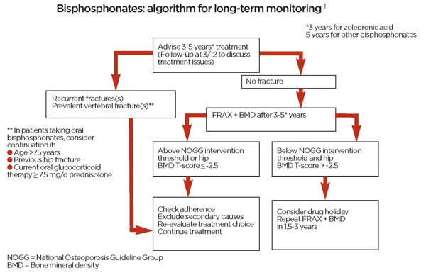
An algorithm for monitoring long-term bisphosphonate therapy in postmenopausal women is shown in figure 1.
Continuation of bisphosphonate treatment beyond three years (for zoledronic acid) and five years (for alendronate, risedronate and ibandronate) respectively, is recommended in the following situations:
• Age 75 years or more
• Previous hip or vertebral fracture
• Fragility fracture/s during treatment, after nonadherence and causes of secondary osteoporosis have been excluded
• Ongoing oral treatment with glucocorticoids at daily doses of ≥ 7.5 mg prednisolone (or equivalent)
Practical tip 7: After discontinuing treatment, reassess fracture risk after a new fracture (regardless of when this occurs), or after 18 months-3 years, if no fracture occurs.
There is currently no evidence base to guide decisions about treatment beyond ten years, and management should be considered on an individual basis.
Conclusion
Osteoporosis is one of the leading causes of morbidity and reduced quality of life, and carries an enormous health, emotional and financial burden worldwide. The bone loss occurs silently over years, and patients do not access healthcare services until complications such as fragility fractures occur. With the ageing population, it is estimated that the burden will become higher. The development of fracture assessment tools and national guidelines aims to improve patient care, and help avoid some of the long-term complications for patients.
REFERENCES
1 Compston J, Cooper A, Cooper C et al. Arch Osteoporos 2017;12:43. https://doi.org/10.1007/s11657-017-0324-5
2 Kanis JA, Melton LJ, Christiansen C et al. The diagnosis of osteoporosis. J Bone Miner Res 1994;9:1137-41
3 Johnell O, Kanis JA. An estimate of the worldwide prevalence and disability associated with osteoporotic fractures. Osteoporos Int 2006;17:1726-33
4 British Orthopaedic Association. The care of patients with fragility fracture. British Orthopaedic Association. 2007
5 Van Staa TP, Dennison EM, Leufkens HG, Cooper C. Epidemiology of fractures in England and Wales. Bone 2001;29(6):517-22
6 Kanis JA, Oden A, Johnell O et al. The burden of osteoporotic fractures: a method for setting intervention thresholds. Osteoporos Int 2001;12:417-27
7 Sernbo I, Johnell O. Consequences of a hip fracture: a prospective study over 1 year. Osteoporos Int 1993;3:148-53
8 Assessment of fracture risk and its application to screening for postmenopausal osteoporosis. Report of a WHO Study Group. World Health Organ Tech Rep Ser 1994;843:1-129
9 Lyles KW, Schenck AP, Colón-Emeric CS. Hip and other osteoporotic fractures increase the risk of subsequent fractures in nursing home residents. Osteoporos Int 2008;19(8):1225-33
10 Fink HA, Kuskowski MA, Taylor BC et al. Association of Parkinson's disease with accelerated bone loss, fractures and mortality in older men: the Osteoporotic Fractures in Men (MrOS) study. Osteoporos Int 2008;19:1277-82
11 Sinaki M. Exercise and osteoporosis. Arch Phys Med Rehabil 1989;70(3):220-29
12 National Institute for Health and Care Excellence. CG146. Osteoporosis: assessing the risk of fragility fracture. NICE. London. 2012 (Updated Feb 2017)
13 Kanis JA, McCloskey EV, Johansson H et al. A reference standard for the description of osteoporosis. Bone 2008; 42(3):467-75
14 Silverman SL. Selecting patients for osteoporosis therapy. Ann NY Acad Sc 2007;1117:264-72
15 Czerwinski E, Badurski JE, Marcinowska-Suchowierska E, Osieleniec J. Current understanding of osteoporosis according to the position of the World Health Organization (WHO) and International Osteoporosis Foundation. Orthop Traumatol Rehabil 2007;9(4):337-56
16 Kanis JA. Assessment of fracture risk and its application to screening for postmenopausal osteoporosis: synopsis of a WHO report. WHO Study Group. Osteoporos Int 1994;(6):368-81
17 Kanis JA, Johnell O, Oden A et al. FRAX and the assessment of fracture probability in men and women from the UK. Osteoporos Int 2008;19:385-397
18 Hippisley-Cox J, Coupland C. Derivation and validation of updated QFracture algorithm to predict risk of osteoporotic fracture in primary care in the United Kingdom: prospective open cohort study. BMJ 2012;344:e3427
19 Gillespie LD, Robertson MC, Gillespie WJ et al. Interventions for preventing falls in older people living in the community. Cochrane Database Syst Rev 2012 Sep 12;(9):CD007146
20 Blasco J, Martinez-Ferrer A, Macho J et al. Effect of vertebroplasty on pain relief, quality of life, and the incidence of new vertebral fractures: a 12-month randomized follow-up, controlled trial. J Bone Miner Res 2012;27(5):1159-66
21 National Osteoporosis Guideline Group 2017. www.sheffield.ac.uk/NOGG/NOGG%20Guideline%202017.pdf
 Download this article in PDF format