SEARCH

McCloskey O, Maxwell AP. Diagnosis and management of nephrotic syndrome. Practitioner 2017;261(1801):11-15
Diagnosis and management of nephrotic syndrome
22 Feb 2017
AUTHORS
Dr Oonagh McCloskey MBChB MRCP, Specialist Registrar in Nephrology
Professor Alexander P. Maxwell MD PhD FRCP, Consultant Nephrologist
Regional Nephrology Unit, Belfast City Hospital, Belfast, UK
Article
Nephrotic syndrome is defined by a triad of clinical features: oedema, substantial proteinuria (> 3.5 g/24 hours) and hypoalbuminaemia (< 30 g/L). It is often associated with hyperlipidaemia, thromboembolism and an increased risk of infection. Nephrotic syndrome develops following pathological injury to renal glomeruli. This may be a primary problem, with a disease specific to the kidneys, or secondary to a systemic disorder such as diabetes mellitus. The most common cause in children is minimal change glomerulonephritis. In white adults, nephrotic syndrome is most frequently due to membranous nephropathy whereas in populations of African ancestry the most common cause of nephrotic syndrome is focal segmental glomerulosclerosis. Diabetic nephropathy is the most common multisystem disease that can cause nephrotic syndrome. Patients typically present with periorbital oedema (most noticeable in the morning) or dependent pitting oedema (more common later in the day). Proteinuria should be documented by a quantitative measurement e.g. urine protein: creatinine ratio (PCR) or albumin: creatinine ratio (ACR). PCR > 300-350 mg/mmol indicates nephrotic range proteinuria. Urgent referral to a nephrologist (ideally within 2 weeks) is necessary and a renal biopsy is usually performed. This will establish what form of glomerular disease is responsible. Additional tests may be undertaken to assess if nephrotic syndrome is secondary to another disorder e.g. systemic lupus erythematosus or amyloidosis.
Nephrotic syndrome is a well known presentation of kidney disease with a triad of clinical features namely oedema, substantial proteinuria (> 3.5 g/24 hours) and hypoalbuminaemia (< 30 g/L). It is often associated with hyperlipidaemia, thromboembolism and an increased risk of infection.
Nephrotic syndrome is uncommon in general practice. A GP may only see two or three adult cases in their career.
Nephrotic syndrome develops following pathological injury to renal glomeruli. This may be a primary problem, with a disease specific to the kidneys, or secondary to a systemic disorder such as diabetes mellitus.1,2
The most common cause in children is minimal change glomerulonephritis.3
In white adults, nephrotic syndrome is most frequently due to membranous nephropathy whereas in populations of African ancestry the most common cause of nephrotic syndrome is focal segmental glomerulosclerosis (FSGS).1,2 Diabetic nephropathy is the most common multisystem disease that can cause nephrotic syndrome.
Presentation
Patients typically present with periorbital oedema (most noticeable in the morning) or dependent pitting oedema (more common later in the day). Facial oedema is rare in heart failure or liver cirrhosis as patients with these conditions are less able to lie flat. As the volume of peripheral oedema increases fluid accumulates in the limbs, trunk and genitals. Patients with nephrotic syndrome may also become breathless due to pleural effusion and ascites.
Individuals with substantial proteinuria may notice frothy or foamy urine, as protein in the urine acts like a detergent. Severe hypoalbuminaemia can result in white nails or if nephrotic syndrome has been transient the development of white bands in nails (Muerhrcke’s lines).
The coincidental finding of a very elevated serum cholesterol concentration on a lipid profile may trigger assessment for other features of nephrotic syndrome.4 If hyperlipidaemia has been present for a prolonged duration, then skin xanthomata may develop.
New onset of hypertension and renal impairment may also be secondary to the glomerular disease causing nephrotic syndrome. Occasionally the first presentation of nephrotic syndrome is a thromboembolic event (deep vein thrombosis or pulmonary embolus) associated with the hypercoagulable state found in this condition.5
Causes
Nephrotic syndrome is a direct consequence of glomerular pathology1,2 and the precise cause can only be established by histological assessment of a kidney biopsy (see table 1, for pathological causes).
Proteinuria
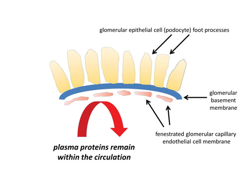
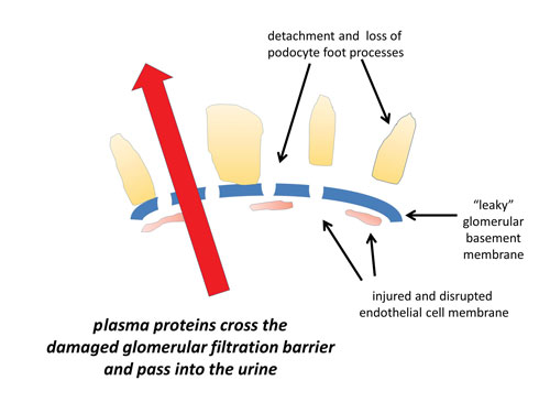
Proteinuria results from structural or functional failure of the glomerular barrier between the blood and urinary space. Proteinuria is associated with injury to podocytes (glomerular epithelial cells), capillary endothelial cells and disruption of the glomerular basement membrane function, see figure 1. Raised intraglomerular pressure may also contribute to proteinuria.
Hypoalbuminaemia
Massive urinary loss of protein leads to hypoalbuminaemia. Hepatic albumin synthesis increases but is insufficient to compensate for the urinary protein loss and a fall in serum albumin ensues. Hepatic synthesis of other proteins is also increased and accounts for the hyperlipidaemia and prothrombotic state associated with nephrotic syndrome.
Oedema
This may result from two mechanisms. The underfill hypothesis proposes that reduced oncotic pressure, due to hypoalbuminaemia, results in fluid shifts from the intravascular space to the interstitial compartment. This triggers salt and water retention, via the renin angiotensin aldosterone system and antidiuretic hormone release, and further promotion of fluid movement into the interstitial compartment.
The overfill hypothesis suggests that a defect in sodium excretion is the primary problem leading to fluid retention. This may be triggered by proteins lost in the urine that activate the epithelial sodium channel (ENaC) in the collecting duct promoting salt and water retention.6 Low oncotic pressure encourages movement of fluid into the interstitial compartment with resulting clinical signs of oedema.
Hyperlipidaemia
Hyperlipidaemia correlates inversely with the serum albumin level and extremely high serum cholesterol concentrations (> 10 mmol/L) may be seen in some patients.4 An atherogenic lipid profile (low HDL/high LDL) is triggered by increased hepatic synthesis of VLDL and urinary loss of HDL.
Hypercoagulability
Hypercoagulability is the net result of increased urinary loss of anticoagulant proteins, such as antithrombin III and protein C, increased hepatic synthesis of clotting factors, enhanced platelet aggregation and intravascular volume contraction.5
Diagnosis
Nephrotic syndrome is recognised by the presence of oedema, proteinuria and hypoalbuminaemia. The initial diagnostic step is a dipstick urinalysis (3-4+ protein suggests the nephrotic range). Proteinuria should be documented by a quantitative measurement e.g. urine protein: creatinine ratio (PCR) or albumin: creatinine ratio (ACR).
PCR > 300-350 mg/mmol indicates nephrotic range proteinuria.7 In practice, PCR and ACR measurements are as accurate as a 24-hour urine collection to quantify proteinuria.7
Hypoalbuminaemia is identified by measuring serum albumin (liver function tests or bone profile). It is essential to measure renal function (serum creatinine; eGFR) because nephrotic syndrome can be complicated by renal failure. A lipid profile will confirm the secondary hyperlipidaemia.
Urgent referral to a nephrologist (ideally within 2 weeks) is necessary and a renal biopsy is usually performed. This will establish what form of glomerular disease is responsible. Additional tests may be undertaken to assess if nephrotic syndrome is secondary to another disorder e.g. systemic lupus erythematosus (SLE) or amyloidosis, see table 2.
Potential complications
Deteriorating renal function
Deteriorating renal function may be an acute insult or represent kidney disease progression. Acute kidney injury (AKI) is commonly due to a prerenal insult from intravascular volume depletion secondary to aggressive diuresis and/or sepsis. The risk of AKI is also increased by concurrent use of angiotensin converting enzyme inhibitors (ACEi) or angiotensin II receptor blockers (ARB) as part of the general management of nephrotic syndrome. Pragmatic advice to reduce the risk of AKI would be to have ‘sick day rules’ for patients asking them to stop ACEi /ARB temporarily in the setting of acute illness, e.g. vomiting and diarrhoea.8 Other causes of AKI include drug-induced interstitial nephritis or renal vein thrombosis.1
Chronic kidney disease (CKD) may develop as most causes of nephrotic syndrome are associated with progressive loss of renal function (with the exception of minimal change disease). The risk of CKD is directly correlated with the severity of proteinuria and there is evidence that proteinuria itself is directly associated with chronic injury and fibrosis of the renal tubulointerstitium.
Thromboembolism
The annual incidence of venous thromboembolism is ten times higher in patients with nephrotic syndrome than the general population and up to 10% of adults with nephrotic syndrome have a clinical thrombotic event.5 The risk of thrombosis also correlates inversely with serum albumin concentration. There is no clear evidence on how this risk should be minimised. In clinical practice many would consider anticoagulation with low molecular weight heparin or warfarin if serum albumin was < 20 g/L.
Infection
Nephrotic patients are prone to bacterial infection. Loss of circulating immunoglobulins and complement factors in the urine impairs the immune response to encapsulated organisms.
In addition zinc and transferrin are required for lymphocyte function and these are lost in the urine.
There is no evidence to support the use of routine antibiotic prophylaxis in nephrotic syndrome,9 but a recent trial indicates that vaccinating against pneumococcus may be of benefit.10
Nutritional deficiency
Patients with nephrotic syndrome are in marked negative protein balance. Loss of muscle mass is often masked by oedema. Loss of binding proteins in the urine can lead to copper, zinc and iron deficiency. Dietetic input is invaluable.
Other metabolic effects
Thyroid binding globulin is lost in the urine and total circulating thyroxine is reduced but free thyroxine and thyroid stimulating hormone are normal.
Usually this causes no clinical change in thyroid status. Protein binding of drugs may be altered by low serum albumin necessitating dose modification.
Treatment
All patients with nephrotic syndrome should be referred to a nephrologist to help establish the underlying diagnosis and advise on further management.
General management principles
Oedema is managed by fluid and dietary salt restriction (< 80 mmol/day; < 2 grams salt/day) coupled with loop diuretics such as furosemide or bumetanide. Patients with nephrotic syndrome may be relatively resistant to oral loop diuretic treatment and stepwise addition of a thiazide diuretic may be needed to trigger an effective diuresis.
If these measures are insufficient to improve oedema, then hospital admission for daily intravenous loop diuretics may be necessary.
Close monitoring of the patient’s weight, electrolytes and renal function is essential since a rapid diuresis can result in hypokalaemia and AKI.
Daily measurement of the patient’s weight is a simple method of gauging response to diuretics with a maximum target weight loss of 0.5-1.0 kg/day.
Proteinuria may be partly reduced by altering glomerular haemodynamic pressure with prescription of an ACEi
(or ARB). Use of ACEi (or ARB) will also help control hypertension if present.
Statins are often coprescribed for the secondary hyperlipidaemia but there is limited evidence for efficacy of these agents in nephrotic syndrome.4
Disease specific treatments
Immunosuppressive medications may be used to target the underlying glomerular pathology causing the nephrotic syndrome. Steroids are used alone or in combination with a variety of other drugs including cyclophosphamide, calcineurin inhibitors, mycophenolate mofetil and rituximab.11,12 Treatment involves two phases; induction of remission (absence of proteinuria and resolution of oedema), followed by maintaining remission. The total duration of treatment varies, depending on the disease activity and clinical response.
Induction regimens are briefly described for the three most common renal diseases that cause nephrotic syndrome in adults.12,13,14
Minimal change disease
Induction: Prednisolone 1 mg/kg per day (maximum 80 mg daily).13 If unsuccessful by 16 weeks consider alternatives such as cyclophosphamide or calcineurin inhibitors. If frequently relapsing or steroid dependent, then consider cyclophosphamide or calcineurin inhibitors.
Focal segmental glomerulosclerosis
Induction: Prednisolone 1 mg/kg per day (maximum 80 mg daily). If unsuccessful at 16 weeks KDIGO guidelines recommend ciclosporin.12 If this fails to induce remission within 4-6 months, high-dose dexamethasone and mycophenolate mofetil are recommended.
Membranous nephropathy (idiopathic)
As this glomerular disease may spontaneously remit in approximately one third of patients, it is common practice to manage patients conservatively for six months (i.e. no immunosuppression, but the treatment defined under ‘general management principles’ above). Immunosuppression may be commenced earlier in the case of severe, life-threatening symptoms of nephrotic syndrome.14
Induction: Options include a regimen of monthly alternating steroids and cyclophosphamide for a total of six months, or calcineurin inhibitors.
Monitoring and follow-up
Patients with active nephrotic syndrome should be under regular nephrology clinic follow-up to ensure treatment regimens are adjusted and monitor progression of renal disease.
GPs may be asked to check renal function, blood count and liver function particularly for those patients receiving immunosuppression. Any new abnormalities or complications of nephrotic syndrome should be discussed with the nephrologist.
Long-term nephrology clinic follow-up is appropriate for most patients with nephrotic syndrome particularly if the cause is associated with progressive CKD.
REFERENCES
1 Swaminathan S, Leung N, Lager DJ et al. Changing incidence of glomerular disease in Olmsted County, Minnesota: A 30-year renal biopsy study. Clin J Am Soc Nephrol 2006;1(3):483-87
2 Hanko JB, Mullan RN, O'Rourke DM et al. The changing pattern of adult primary glomerular disease. Nephrol Dial Transplant 2009;24(10):3050-54
3 Wong W. Idiopathic nephrotic syndrome in New Zealand children, demographic, clinical features, initial management and outcome after twelve-month follow-up: results of a three-year national surveillance study. J Paediatr Child Health 2007;43(5):337-41
4 Kong X, Yuan H, Fan J et al. Lipid-lowering agents for nephrotic syndrome. Cochrane Database Syst Rev 2013, Issue 12. Art. No.: CD005425. DOI: 10.1002/14651858.CD005425.pub2
5 Mahmoodi BK, ten Kate MK, Waanders F et al. High absolute risks and predictors of venous and arterial thromboembolic events in patients with nephrotic syndrome: results from a large retrospective cohort study. Circulation 2008;117(2):224-30
6 Bockenhauer D. Over or underfill: not all nephrotic states are created equal. Pediatr Nephrol 2013;28(8):1153-56
7 Methven S, MacGregor MS, Traynor JP et al. Comparison of urinary albumin and urinary total protein as predictors of patient outcomes in CKD. Am J Kidney Dis 2011;57(1):21-8
8 National Institute for Health and Care Excellence. CG169. Acute kidney injury: detection, prevention and management. NICE. London. 2013. www.nice.org.uk/guidance/cg169
9 McCaffrey J, Lennon R, Webb NJ. The non-immunosuppressive management of childhood nephrotic syndrome. Pediatr Nephrol 2016;31(9):1383-1402
10 Pittet LF, Posfay-Barbe KM, Chehade H et al. Optimizing seroprotection against pneumococcus in children with nephrotic syndrome using the 13-valent pneumococcal conjugate vaccine. Vaccine 2016;34(41): 4948-54
11 Hahn D, Hodson EM, Willis NS, Craig JC. Corticosteroid therapy for nephrotic syndrome in children. Cochrane Database Syst Rev 2015, Issue 3. Art. No.: CD001533. DOI: 10.1002/14651858.CD001533.pub5
12 Kidney Disease: Improving Global Outcomes (KDIGO) Glomerulonephritis Work Group. KDIGO clinical practice guideline for glomerulonephritis. Kidney Int Suppl 2012;2:139-274
13 Palmer SC, Nand K, Strippoli GFM. Interventions for minimal change disease in adults with nephrotic syndrome. Cochrane Database Syst Rev 2008, Issue 1. Art. No.: CD001537. DOI: 10.1002/14651858.CD001537.pub4
14 Chen Y, Schieppati A, Chen X et al. Immunosuppressive treatment for idiopathic membranous nephropathy in adults with nephrotic syndrome. Cochrane Database Syst Rev 2014, Issue 10. Art. No.: CD004293. DOI:10.1002/14651858.CD004293.pub3
USEFUL INFORMATION
For patients
ARTICLE IN PDF
 Download this article in PDF format
CLICK ON IMAGES
PRESET SEARCHES
PubMed
NICE evidence search
Immediate CPD prompts
These handy templates*, associated with all articles, prompt drafting of personal learning, reflection and planning.
Save your record straight into your personal development folder on your device or cloud storage system.
*Adobe fillable PDFs. Adobe reader needed for iPad
Comment or question
If you, or your peer group, would like to comment on an article or have a question for the authors, write to:
Editor
Case report submissions
Do you have an interesting case that you would like to share with other readers?
Your article would need to be around 1,500 words long, include a brief review of the literature to put the case in context, and be fully referenced in the Vancouver style.
Submissions for consideration for publication should be sent to: [email protected]