SEARCH

Practitioner 2011;255(1737): 15-17
Which patients with renal colic should be referred?
23 Feb 2011
AUTHORS
Dr Anil Xavier MB BS MRCP
Specialist Registrar in Nephrology
Professor Alexander P Maxwell MD PhD FRCP
Consultant Nephrologist
Regional Nephrology Unit, Belfast City Hospital, Belfast
Article
Renal stone disease is a common, and potentially recurrent, condition that affects 5-15% of the population worldwide.1 Symptomatic renal stone disease is at least twice as common in men as women.2 The incidence has risen over two decades and 1-2 people per thousand present with an acute episode of pain caused by renal stones each year.2,3 Annually there are more than 12,000 renal stone-related hospital admissions1 and considerably more GP consultations for management of renal colic. Data from NHS Direct showed that 80% of patients with renal colic symptoms were advised to contact their GP as the first port of call.4 The challenge for GPs is to identify which cases may be treated safely at home and which need immediate referral to hospital.
Presentation
Renal colic is classically sudden in onset, unilateral, and radiates from loin to groin (see table 1). The site of pain may suggest the position of obstruction, with renal pelvic or upper ureteric stones usually causing more flank pain and tenderness while lower ureteric stones cause pain radiating towards the ipsilateral testicle or labia. The pain waxes and wanes, depending on the movement of the stone in the ureter, which causes ureteric spasm that can last from a few minutes to hours. Renal colic pain is usually severe and disabling. Indeed, women who have previously experienced both labour pains and renal colic usually state that kidney stone-related pain is more severe than the pain associated with childbirth.
Other common symptoms associated with renal colic include nausea and vomiting, haematuria and irritative lower urinary tract symptoms such as dysuria and frequency. Studies have shown that up to 90% of patients with ureteric lithiasis can have haematuria on presentation.5 Another important aid to diagnosis is that usually patients with renal colic are very restless compared with patients with peritonitis who tend to lie still.
Fever accompanying renal colic symptoms is suggestive of additional complications such as urinary tract infection or pyonephrosis. A febrile patient with renal colic requires immediate hospital admission.
Symptoms suggestive of renal colic along with a positive dipstick for haematuria have a reported sensitivity of 84% and specificity of 99% 6 but it is important to consider other differential diagnoses for this clinical presentation including bowel pathologies (appendicitis, diverticulitis, perforation), a major vascular event (leaking abdominal aortic aneurysm) or a gynaecological emergency (ectopic pregnancy).
Pain control
Once a preliminary diagnosis of renal colic has been made, speedy administration of analgesia becomes a priority with the options being NSAIDs and/or opioids depending on suitability (see table 2). If there is no improvement in symptoms within an hour with adequate analgesia consider urgent referral to hospital.
NSAIDs
An NSAID is preferred over an opiate drug as an initial analgesic choice as the NSAID can help reduce smooth muscle tone and thus ureteric spasm. Diclofenac has the best evidence base for this class of analgesic. It is most effective given as an intramuscular injection (usual adult dose 75 mg) and the injection can be repeated once more at 30 minutes. Diclofenac can also be given rectally or orally (75 to 150 mg daily in divided doses) when renal colic pain is less severe and for ongoing analgesia. Other drugs in this group that have been tried include ketorolac and indometacin.
NSAIDs should be avoided in patients with known contraindications such as a history of hypersensitivity to aspirin or other NSAIDs (including those in whom attacks of asthma, angioedema or urticaria have been precipitated by an NSAID) or persons with a coagulation defect. It is important to avoid repeated NSAID dosing in those with chronic kidney disease (eGFR2) or a solitary kidney. An NSAID should be used with caution in patients who are dehydrated or who have been prescribed drugs that block the renin-angiotensin axis such as ACE inhibitors (ACEi) or angiotensin receptor blockers (ARB). The risk of acute kidney injury is increased when an NSAID and ACEi or ARB are co-prescribed.
Opiates
Opiates are certainly as effective as NSAIDs but are more likely to be associated with nausea.7 Opiates are used primarily in those who have contraindications to NSAIDs but occasionally may need to be given in conjunction with NSAIDs to provide adequate analgesia.
Diamorphine or morphine are the most commonly used opiates for renal colic either intramuscularly or subcutaneously at doses varying from 2.5 mg to 10 mg, depending on age and response. The other commonly used, and effective, agent in this group is pethidine. It is important to administer an antiemetic as well, given that nausea is a frequent side effect of opiates.
If pain is less severe or NSAIDs are contraindicated, paracetamol in combination with a weaker opioid (codeine or tramadol) can be prescribed for ongoing pain relief.
Patients should also be advised to drink enough fluids to remain hydrated and pass dilute (colourless) urine.
Primary care
Patients with undiagnosed severe abdominal pain require urgent clinical assessment. A careful history and clinical examination including: temperature, pulse, blood pressure, abdominal findings and urine dipstick analysis (if possible) will help to exclude other emergencies such as peritonitis or septic shock.
If there is still diagnostic uncertainty it is safer to arrange immediate assessment in hospital.
About 90% of stones will pass spontaneously8 and thus it is often appropriate to manage renal colic at home particularly if the patient has experienced several episodes before and can recognise when to call for reassessment if the pain is not settling or returns suddenly.
The ideal patients who may be managed in the community are those:
- less than 60 years of age
- with adequate social support
- contactable by telephone for clinical review
- willing to stay at home.
And, most importantly:
- have significant improvement or resolution in symptoms within an hour of being given appropriate analgesia.9
These patients can be provided with adequate oral or rectal analgesia for ongoing relief but should be advised to call again as an emergency if: the renal colic pain worsens or returns suddenly; they have recurrent vomiting or they develop a fever.
Urgent referral
Patients with signs of peritonitis, systemic infection, septic shock as well as those whose diagnosis is unclear should be referred urgently to hospital for assessment. Patients who are systemically unwell with renal stones are more likely to have an infected and obstructed urinary tract system that needs urgent imaging and possible drainage. Patients who are at risk of sudden deterioration or who may need specialist input (see table 3, above) should also be considered for urgent referral to hospital.
Follow up
All patients who are managed at home should have renal tract imaging within a week by fast track referral to radiology or as an urgent urology outpatient referral, as per local guidelines, to rule out an obstructed urinary system. Radiological investigation usually includes a plain X-ray (KUB) followed by an intravenous urogram (IVU) or non-contrast helical computerised tomography (CT).10
Smaller stones (
Further screening is recommended to identify risks for recurrent stone formation e.g. hypercalcaemia, metabolic acidosis or where stones have formed in the presence of urinary tract infection. Further evaluation is also recommended for children with renal stones or young age of onset of stone formation as a metabolic problem is more likely to be present.
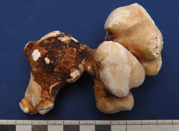
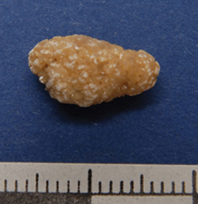
It is worthwhile identifying those patients who have non-calcium containing stones (urate or cystine), see figure 1, as specialist management can reduce recurrence.11
Patients with recurrent stones should be advised to maintain a copious fluid intake (>2 L/day) to reduce the concentration of the urine. A reduction in salt intake (ideally
A substantial decrease in dietary salt intake can reduce urinary calcium excretion and this measure coupled with increased fluid intake helps to prevent recurrence of calcium-containing renal calculi.
REFERENCES
1 Moe OW. Kidney stones: pathophysiology and medical management. Lancet 2006;367:33-44
2 Scott R. Epidemiology of stone disease. Br J Urol 1985;57:491-49
3 Romero V, Akpinar H, Assimos DG. Kidney Stones: A global picture of prevalence, incidence, and associated risk factors. Rev Urol 2010;12(2-3):e86-e96
4 Banks I. The NHS Direct Healthcare Guide 2000. Radcliffe Medical. Oxford. 2000
5 Elton TJ, Roth CS, Berquist TH. A clinical prediction rule for the diagnosis of ureteral calculi in emergency departments. J Gen Intern Med 1993;8(2):57-62
6 Eskelinen M, Ikonen J, Lipponen P. Usefulness of history-taking, physical examination and diagnostic scoring in acute renal colic. Eur Urol 1998; 34:467-73
7 Holdgate A, Pollock T. Systematic review of the relative efficacy of non-steroidal anti-inflammatory drugs and opioids in the treatment of acute renal colic. BMJ 2004; 328(7453):1401
8 Segura JW, Preminger GM, Assimos DG et al. Ureteral Stones Clinical Guidelines Panel summary report on the management of ureteral calculi. The American Urological Association. J Urol 1997; 158:1915-21
9 Wright PJ, English PJ, Hungin AP. Managing acute renal colic across the primary-secondary care interface: a pathway of care based on evidence and consensus. BMJ 2002; 325(7377):1408-1412
10 Ulahannan D, Blakeley CJ, Jeyadevan N. Benefits of CT urography in patients presenting to the emergency department with suspected ureteric colic. Emerg Med J 2008;25(9):569-71
11 Courtney AE, Maxwell AP. Renal stone recurrence can be prevented. Practitioner 2007:251(1691):18-33
12 Breslau NA, Brinkley L, Hill KD. Relationship of animal protein-rich diet to kidney stone formation and calcium metabolism. J Clin Endocrinol Metab 1988; 66:140-6
USEFUL INFORMATION
NHS Clinical Knowledge Summaries: Renal colic - acute management
www.cks.nhs.uk/renal_colic_acute
The Edinburgh Renal Unit website provides information for patients and clinicians on kidney stones
www.edren.org
British Association of Urological Surgeons guidelines for the acute management of first presentation of renal/ureteric lithiasis www.baus.org.uk
ARTICLE IN PDF
 Download this article in PDF format