SEARCH

Welch K, Thekkumpurath P. Diagnosis and management of vascular dementia. Practitioner June 2022;266(1859):11-14
Diagnosis and management of vascular dementia
24 Jun 2022
AUTHORS
Dr Killian Welch BSc MBChB MPhil MD MRCPsych, Consultant Neuropsychiatrist
Dr Parvez Thekkumpurath MB BS MRCPsych, Consultant Old Age Psychiatrist, Royal Edinburgh Hospital, Edinburgh, UK
Competing interests: None
Article
Vascular dementia describes chronic progressive impairment of cognitive function arising from cerebrovascular injury. Although overlap exists with Alzheimer’s disease (around 20% of dementia patients have a mixed form) vascular dementia is regarded as the second most common cause of dementia in the UK, a reasonably pure form constituting 15-20% of dementia cases in Europe and North America. Vascular cognitive impairment (VCI) refers to the full spectrum from mild cognitive impairment to dementia associated with, and presumed to be caused by, cerebrovascular disease. The most common cause of VCI is cerebral small vessel disease. The presentation of vascular dementia is often insidious, with disorganisation, loss of drive, irritability, mental rigidity, difficulties with planning and problem solving, emotional lability and other mood changes, and sometimes inappropriate or disinhibited social behaviour. Slowed gait is common in those with advanced disease. Physical examination may identify gait impairment, bradykinesia, rigidity, and focal neurological signs. Screening tools are insensitive in vascular dementia, in which memory can be relatively preserved. All patients should have their BP checked. Vascular risk factors are not essential for the diagnosis but should be identified, as should any reversible medical causes. Patients with impairment of sufficient severity to classify as dementia will require input from a dedicated dementia service or community mental health team. Objective evidence of cognitive impairment is required for diagnosis: the cognitive profile is dominated by executive dysfunction, slowed processing speed and impaired retrieval. Imaging is a prerequisite for diagnosis.
Vascular dementia (VaD) describes chronic progressive impairment of cognitive function arising from cerebrovascular brain injury. Although overlap exists with Alzheimer’s disease (AD) (around 20% of dementia patients have a mixed form1) VaD is regarded as the second most common cause of dementia in the UK. A reasonably pure form constitutes 15-20% of dementia cases in Europe and North America.2
As with AD, the incidence of VaD increases steeply with age, contributing to the growing epidemic of dementia in ageing populations worldwide.3 The estimated incidence in people aged 65-69 years is 0.3-2.2 per 1,000 per annum, rising to 9.3-15.9 per 1,000 per annum in those aged over 90.4
It has been argued that, owing to improvement in the management of vascular disease, the incidence of vascular dementia may have decreased in recent decades. Indeed, the age-specific risk of all-cause dementia in the USA and Europe has declined by about 20% per decade since the late 20th century, paralleled by reductions in the burden of cerebral small vessel disease.5
Aetiology
Dementia, renamed major neurocognitive disorder in DSM-5, is the loss of cognitive functioning (thinking, remembering, and reasoning) to such an extent that it interferes with a person's daily life and activities.6
Vascular cognitive impairment (VCI) is a broader concept. It covers the full spectrum of cognitive impairment from mild cognitive impairment to dementia associated with, and presumed to be caused by, cerebrovascular disease.
Diagnostic criteria for VaD have been refined in recent years and currently require establishing the presence of dementia syndrome with clinical features indicative of vascular aetiology. These include onset after stroke, decline in complex attention and frontal executive function, and evidence of cerebrovascular disease from history, examination or imaging.6
However, establishing a link between onset of dementia and the presence of cerebrovascular disease can be challenging. Vascular brain changes are common in people with other dementias such as AD as well as in older people without dementia.7 These considerations help explain why for many patients, especially older patients in their eighth or ninth decade, labelling their dementia as having a mixed aetiology is more appropriate.
The most common cause of VCI is cerebral small vessel disease, such as arteriolosclerosis and cerebral amyloid angiopathy. Other less common causes include CADASIL (cerebral autosomal dominant arteriopathy with subcortical infarcts and leukoencephalopathy) a type of subcortical small vessel disease with a genetic basis.8 Typical features of CADASIL are shown in table 1.8
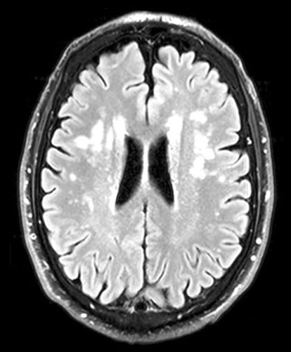
Arteriolosclerosis predominantly affects subcortical brain regions, including the corona radiata and basal ganglia, and patients typically have multiple lacunar infarcts (cystic lesions < 1.5 cm) or extensive, confluent white matter lesions,9 (see figure 1).
Cerebral amyloid angiopathy, the second most common cerebral small vessel disease, is caused by beta-amyloid deposition in small arteries. Vascular integrity is lost leading to haemorrhage, most commonly in lobar locations while sparing the basal ganglia and brainstem.8Table 2, lists the specific vascular dementia syndromes.10
Presentation
Though vascular dementia can present with the classical stepwise decline, insidious deterioration is more common, with disorganisation, loss of drive, irritability, mental rigidity, difficulties with planning and problem solving, emotional lability and other mood changes, and sometimes inappropriate or disinhibited social behaviour.11
The cognitive profile is dominated by executive dysfunction, slowed processing speed and impaired retrieval (memory difficulties that improve with cueing, e.g. being able to identify a word when given multiple choice options).
Slowed gait is common in those with advanced disease.11 Impaired balance, falls and incontinence can also occur.
The extent of vascular disease may be unrecognised until revealed by imaging. Imaging is a prerequisite for diagnosis. The presence of vascular damage is not sufficient for a vascular dementia diagnosis and, as mentioned above, the number of patients for whom vascular pathology is a contributor to their dementia likely exceeds those with a pure vascular dementia.12
If a single ischaemic or haemorrhagic stroke is large enough, or simply damages a crucial brain region such as the thalamus or language cortices, (i.e. a strategic infarct), it can be associated with substantial cognitive impairment. There may be stroke-related symptoms and signs, such as aphasia or motor weakness, meaning the association between vascular brain damage and cognitive impairment should be clear.
Though cognitive impairment may be static or improve if vascular risk factors are treated and there are no further strokes, stroke is a strong and independent risk factor for incident dementia.13 At any point following stroke, more than one in five people will have dementia; in around a quarter of these dementia will have predated stroke, but in three-quarters it will be a new diagnosis.14
This relationship between stroke and dementia is complex and variable. Stroke may lead to a clear picture of VaD, may trigger Alzheimer’s pathology or exacerbate prestroke cognitive decline of either vascular or Alzheimer’s pathology.
Assessment
Insidious or stepwise cognitive deterioration, potentially in association with overt stroke pathology, should prompt consideration of vascular dementia as a potential diagnosis. Equally, executive dysfunction, cognitive slowing, mood and behavioural changes along with gait and balance problems in the absence of overt memory problems should also prompt consideration of vascular dementia. Collateral history should be sought, including a timeline of cognitive and functional changes. Vascular risk factors should be identified but are not essential for the diagnosis (see table 3).
Potentially reversible medical causes which can masquerade as dementia should be considered. This could include depression, but neuropsychiatric disorders are particularly common in vascular dementia, so comorbidity is also likely.
Other possibilities are brain lesions (including subdural haematoma following recent head injury), nutrient or vitamin deficiencies (B1, B6, B12, folate), endocrine conditions (including hypothyroidism), chronic infection and inflammation, medication (including anticholinergics, benzodiazepines, opioids), alcohol, sensory impairment (loss of hearing or vision).
Physical examination is particularly important if vascular dementia is suspected. It may identify gait impairment, bradykinesia, rigidity, and focal neurological signs.
Objective evidence of cognitive impairment is required for diagnosis. Screening tools for dementia may be particularly insensitive in vascular dementia, in which memory can be relatively preserved. There is data to support the use of the Montreal Cognitive Assessment in separating vascular dementia from controls;15 its relative sensitivity to executive deficits makes it intuitively a good choice. As ever, normal performance on a screening tool does not exclude dementia.
All patients should have their blood pressure (BP) checked and a urine dipstick. Blood tests should include full blood count, urea & electrolytes, thyroid function tests, vitamin B12 and folate, liver function tests, calcium, lipid profile and glucose/HbA1c. Substance use should be enquired about and gamma-glutamyl transferase tested if alcohol excess is suspected.
Lupus anticoagulant, antiphospholipid, and antinuclear testing should be performed in selected patients with atypical presentations or at increased risk for these conditions (e.g. young people with recurrent strokes) and syphilis serology if indicated.
It is useful to distinguish patients with atherosclerotic ischaemic disease from those with cardioembolic disease (e.g. atrial fibrillation, vascular heart disease); ECG will obviously be helpful in identifying an arrhythmia. Imaging will likely be deferred until review by secondary care but is central to diagnosis. Magnetic resonance imaging (MRI) is more sensitive than computed tomography for signs of cerebral small vessel disease, including microbleeds.
Management of vascular risk factors
Since risk factors for VCI are modifiable, targeting these would be expected to reduce the incidence and progression of VCI/VaD. In patients who have experienced a stroke there is clear guidance on the use of antithrombotics, antihypertensives, lipid lowering agents and consideration of endarterectomy (depending on patient characteristics).16
Hypertension
The situation with VaD is more complex. Hypertension, especially high BP during midlife, is associated with a higher risk of developing cognitive impairment and dementia.17 This has implications beyond pure VaD and may help to explain why so many patients have a mixed aetiology dementia.
It is thought, for example, that one of the main mechanisms underlying cognitive dysfunction in hypertension involves cerebral vessel remodelling, leading to changes in cerebral autoregulation and perfusion, and thus affecting the ability to clear beta-amyloid;18 this has obvious implications for the development of AD pathology as well as VCI.
Studies examining the impact of tight BP control on the development of dementia have reported mixed results. The SPRINT-MIND study, for example, reported reduction in the risk of MCI in intensive compared with standard BP control (i.e. BP target < 120 mmHg vs < 140 mg mmHg) by 20%, with a 16% risk reduction in both MCI and probable dementia (though no statistically significant reduction was seen for probable dementia alone).
While midlife hypertension undoubtedly has a deleterious effect on later life cognitive function,19 intervention for high BP in those aged 80 years or older has not been shown to reduce incidence of dementia and orthostatic hypotension is associated with an increased risk of dementia and cognitive decline.20
Studies examining the impact of hypertension control on progression of vascular dementia are lacking. Nonetheless, given the strong evidence that lowering BP with antihypertensive drugs was associated with a reduced risk of stroke,21 and the likelihood that it reduces incidence of mild cognitive impairment and dementia,22 effectively treating hypertension in primary care is a public health priority.
While vigilance for orthostatic hypotension is important (i.e. check sitting and standing BP in these patients; if there is a postural drop ≥ 10 mmHg, record the standing BP), expert consensus suggests treating hypertension in overt VCI.23 Guidance in this specific population is lacking, but NICE advises a target BP of 140/90 mmHg for those younger than 80 years and 150/90 mmHg for people 80 or older.24 Any advantages of aggressive treatment that allows BP to reach below 120 mmHg are still unclear and may be associated with harm in some.25
Other risk factors
The main role of statins in the treatment of vascular dementia is in preventing further ischaemic events. Despite good evidence for secondary stroke prevention, they have not been shown to prevent cognitive decline or dementia in people at risk of vascular disease.26
Blood glucose control in patients with diabetes should be optimised given it can reduce macrovascular and microvascular disease risk. Assistance should be given to stop smoking, alcohol intake minimised and weight loss advised and supported if indicated.
Referral
The appropriate secondary care referral route will depend on local services. Young patients (under the age of 65), atypical or rapidly progressive cases will likely be referred to neurology. Cases arising after a clear stroke will likely be under the care of stroke services.
Patients with impairment of sufficient severity to classify as dementia will require input from a psychiatric team, either a dedicated dementia service or community mental health team.
Specific treatments
General issues in the management of dementia were discussed in our previous article on AD in this journal.27 The focus is on ensuring the safety of the patient and others, giving reassurance and support to the patient and carers, and identifying and treating comorbidity. A multidisciplinary team is often required, including nurses, physiotherapists, occupational therapists, psychologists, and social workers.
The effects of cholinesterase inhibitors and memantine are small and of uncertain clinical benefit in patients with dementia with a solely vascular aetiology.28 Many patients have a mixed dementia however, with coexisting AD, and are more likely to benefit.
Comorbidity
Neuropsychiatric comorbidity is common in vascular dementia. Apathy is reported in more than 50%,29 agitation in 40%,30 and depression in 25% (compared with 15% in AD).31 Though evidence is weak, antidepressant treatment should be considered if depression is suspected. SSRIs, with their lower side effect burden are favoured. These generally well tolerated drugs are, however, still associated with risks, including postural hypotension, falls, hyponatraemia and bleeds, so treatment must be individualised. They may also, with careful patient selection and open discussion of risks, be trialled in agitation.
Though there is little data in vascular dementia, post-stroke psychosis (delusions and/or hallucinations with poor insight) has been reported to occur in around 5% of stroke patients.32 Given the concerns about metabolic syndrome, adverse cardiac effects and association with stroke, antipsychotics are used with caution in this population. In the presence of frank psychosis use will likely be justified, but this would necessitate psychiatric input, and unfortunately a Cochrane review suggests effects can be disappointing.33
REFERENCES
Further reading
For healthcare professionals
Zanon Zotin MC, Sveikata L, Viswanathan A, Yilmaz P. Cerebral small vessel disease and vascular cognitive impairment: from diagnosis to management. Curr Opin Neurol 2021;34:246-57
This paper provides a good overview of the aetiology and clinical presentation of vascular dementia as well as a helpful discussion on the complex area of managing risk factors to try and slow progression.
References
1 Custodio N, Montesinos R, Lira D et al. Mixed dementia: a review of the evidence. Dement Neuropsychol 2017;11(4):364-70
2 Wolters FJ, Ikram MA. Epidemiology of vascular dementia: nosology in a time of epiomics. Arterioscler Thromb Vasc Biol 2019;39:1542-49
3 Wolters FJ, Ikram MA. Epidemiology of dementia: the burden on society, the challenges for research. Methods Mol Biol 2018;1750:3-14
4 Bowler JV. Modern concept of vascular cognitive impairment. Br Med Bull 2007;83:291-305
5 Schrijvers EMC, Verhaaren BFJ, Koudstaal PJ et al. Is dementia incidence declining?: trends in dementia incidence since 1990 in the Rotterdam study. Neurology 2012;78:1456-63
6 American Psychiatric Association. Diagnostic and Statistical Manual of Mental Disorders. Fifth Edition. American Psychiatric Association. Arlington, VA. 2013
7 Fernando MS, Ince PG. Vascular pathologies and cognition in a population-based cohort of elderly people. J Neurol Sci 2004;226:13-17
8 Alhusaini S, Smith EE. Vascular cognitive impairment. Pract Neurol 2019; https://practicalneurology.com/articles/2019-jan/asymptomatic-carotid-artery-stenosis/pdf
9 Roman GC, Erkinjuntti T, Wallin A et al. Subcortical ischaemic vascular dementia. Lancet Neurol 2002;1:426-36
10 Stewart R. Vascular and Mixed Dementia in Oxford Textbook of Old Age Psychiatry, 3rd edition, 2021
11 Johnson JCS, McWhister L, Hardy CJD et al. Suspecting dementia: canaries, chameleons and zebras. Pract Neurol 2021;21:300-12
12 de Reuck J, Deramecourt V, Cordonnier C et al. The incidence of post-mortem neurodegenerative and cerebrovascular pathology in mixed dementia J Neurol Sci 2016;366:164-66
13 Kuźma E, Lourida I, Moore SF et al. Stroke and dementia risk: a systematic review and meta-analysis. Alzheimers Dement 2018;14:1416-26
14 Craig L, Hoo ZL, Yan TZ et al. Prevalence of dementia in ischaemic or mixed stroke populations: systematic review and meta-analysis. J Neurol Neurosurg Psychiatry 2022;93:180-87
15 Ghafar M, Miptah HN, O'Caoimh R. Cognitive screening instruments to identify vascular cognitive impairment: a systematic review. Int J Geriatr Psychiatry 2019;34:1114-27
16 National Institute for Health and Care Excellence. Clinical knowledge summaries. Stroke and TIA https://cks.nice.org.uk/topics/stroke-tia/management
17 McGrath ER, Beiser AS, DeCarli C et al. Blood pressure from mid- to late life and risk of incident dementia. Neurology 2017;89:2447-54
18 Muller M, van der Graaf Y, Visseren FL et al. Hypertension and longitudinal changes in cerebral blood flow: the SMARTMR study. Ann Neurol 2012;71:825-33
19 Iadecola C, Yaffe K, Biller J et al. Impact of hypertension on cognitive function: a scientific statement from the American Heart Association. Hypertension 2016;68:e67-94
20 Sierra C. Hypertension and the risk of dementia. Front Cardiovasc Med 2020;7:5
21 Gaciong Z, Siński M, Lewandowski J. Blood pressure control and primary prevention of stroke: summary of the recent clinical trial data and meta-analyses Curr Hypertens Rep 2013;15:559-74
22 Ding J, Davis-Plourde KL, Sedaghat S et al. Antihypertensive medications and risk for incident dementia and Alzheimer's disease: a meta-analysis of individual participant data from prospective cohort studies. Lancet Neurol 2020;19:61-70
23 Zanon Zotin MC, Sveikata L, Viswanathan A, Yilmaz P. Cerebral small vessel disease and vascular cognitive impairment: from diagnosis to management. Curr Opin Neurol 2021;34:246-57
24 National Institute for Health and Care Excellence. NG136. Hypertension in adults: diagnosis and management. NICE. London. 2019 www.nice.org.uk/guidance/ng136
25 Turana Y, Tengkawan J, Chia YC et al. High blood pressure in dementia: How low can we go? J Clin Hypertens 2020;22:415-22
26 McGuinness B, Craig D, Bullock R et al. Statins for the prevention of dementia. Cochrane Database Syst Rev 2016 Jan 4;(1):CD003160
27 Szalek A, Thekkumpurath P, Welch K. Diagnosing and managing Alzheimer’s disease. Practitioner 2022;266:21-25
28 Battle CE, Abdul-Rahim AH, Shenkin SD et al. Cholinesterase inhibitors for vascular dementia and other vascular cognitive impairments: a network meta-analysis. Cochrane Database Syst Rev 2021;2:CD013306
29 Santos MAO, Bezerra LS, Correia CDC, Bruscky IS. Neuropsychiatric symptoms in vascular dementia: epidemiologic and clinical aspects. Dement Neuropsychol 2018;12(1):40-44
30 Anor CJ, O’Connor SB, Saund AB et al. Neuropsychiatric symptoms in Alzheimer disease, vascular dementia, and mixed dementia. Neurodegener Dis 2017;17(4-5):127-34
31 Asmer MS, Kirkham J, Newton H et al. Meta-analysis of the prevalence of major depressive disorder among older adults with dementia. J Clin Psychiatry 2018;79(5)
32 Stangeland H, Orgeta V, Bell V. Poststroke psychosis: a systematic review. J Neurol Neurosurg Psychiatry 2018;89:879-85
33 Mühlbauer V, Möhler R, Dichter MN et al. Antipsychotics for agitation and psychosis in people with Alzheimer's disease and vascular dementia. Cochrane Database Syst Rev 2021 Dec 17;12(12):CD013304
34 Di Donato I, Bianchi S, De Stefano N et al. Cerebral Autosomal Dominant Arteriopathy with Subcortical Infarcts and Leukoencephalopathy (CADASIL) as a model of small vessel disease: update on clinical, diagnostic, and management aspects. BMC Med 2017;15:41
ARTICLE IN PDF
 Download this article in PDF format