SEARCH

Armon K, Bale P. Identifying heritable connective tissue disorders in childhood. Practitioner 2012; 256 (1752):19-23
Identifying heritable connective tissue disorders in childhood
20 Jun 2012
AUTHORS
Dr Kate Armon BMedSc BMBS DCH DRCOG DM FRCPCH, Paediatric Rheumatology Consultant, Norfolk and Norwich University Hospital NHS Foundation Trust, Norwich, UK. Honorary Senior Lecturer, University of East Anglia, Norwich, UK
Dr Peter Bale MBChB MRCPCH, Specialty Registrar level 5 in Paediatrics, Ipswich Hospital, Ipswich, UK
Article
Heritable connective tissue disease is rare, each disorder estimated at 1-10 per 100,000. However, as a group they are prevalent enough to constitute an important diagnostic challenge. Connective tissue disorders most significantly affect three systems: musculoskeletal, ocular and cardiovascular.
The cardinal feature of the majority of these disorders is ligamentous laxity, or joint hypermobility. The joints show an increased range of movement, and the child may present with arthralgias, effusions and an increased risk of joint or soft tissue injury. By contrast, joint pain associated with a decreased range of movement or joint contractures suggests other aetiology, most commonly juvenile idiopathic arthritis, and more rarely arthrogryposis or storage disorders.
Marfan syndrome
Case history
Chantelle is a 10-year-old girl complaining of intermittent lower back and knee pain. The pains are mostly in the evenings after a particularly active day. There is no night waking or limping. Chantelle is tall and thin, with long fingers. You find a mild scoliosis on forward flexion of her back. Her knees hyperextend but are otherwise normal. You plot her on the centile chart, noting that height has always been above the 98th centile. Her mother does not look very tall but, on enquiry, Chantelle’s father is 6’4” and had a heart valve replacement in his early forties.
Marfan syndrome is the most common heritable connective tissue disorder with an incidence of 2-3 per 10,000, affecting all ethnic groups. It is primarily caused by a mutation on the long arm of chromosome 15 (15q21.1), which results in an abnormality of fibrillin.1 There are, however, more recently described genetic mutations on chromosome 3, affecting the expression of transforming growth factor-ß (TGF-ß ) receptors causing the Marfan phenotype, and improving the molecular understanding of the condition.1
Fibrillin is a glycoprotein that forms the main structural component of microfibrils. Microfibrils provide a supporting scaffold for the deposition of elastin throughout the body, including skin, lung, dura mater, tendons, ciliary zonules of the lens, myocardium, heart valves and periosteum.
Marfan syndrome is an autosomal dominant condition with high penetrance but with striking pleiomorphism (varied phenotypes for the same single gene abnormality).2 In 25% of individuals there is no family history, suggesting the condition has presented de novo.
Clinical features
Chantelle’s appearance and her family history suggest Marfan syndrome, but you prescribe ibuprofen, or topical NSAIDs PRN, and ask her to return in two weeks while you look for further information.
Marfan syndrome may be suspected on antenatal ultrasound, but this is rare and the diagnosis is often not made until late childhood. A high index of clinical suspicion is required. Long, thin extremities with proportionately more lengthening of the distal extremities (arachnodactyly) result in a tall individual with a low upper:lower segment ratio and an arm span greater than height.
Other skeletal characteristics include pectus deformity (carinatum or excavatum) and scoliosis.2 Scoliosis is normally convex to the right, it is equal in both sexes and children may also have increased lumbar lordosis. A narrow and high-arched palate with crowded dentition is common and may result in referral via the dentist. These patients have mild to moderate joint laxity and may present in their teenage years with general arthralgia. See boxes 1 and 2, above, for assessing joint hypermobility in adults and children respectively.
Myopia and astigmatism are common early problems and the patient will need glasses. In severe cases ectopia lentis, with characteristic upward dislocation of the lens, and detachment of the retina are found.2
Cardiac abnormalities include mitral valve prolapse, mitral regurgitation and arrhythmias.3 The cardiac problems related to Marfan syndrome can occur at any age and thus should be considered as soon as the diagnosis is suspected. They are more often diagnosed as the child gets older, especially in the teenage years and twenties but there are rare cases of children as young as eight months showing evidence of mitral valve prolapse.4 Weakness of the tunica media of the aorta results in aortic route dilatation, aortic insufficiency and ultimately dissection.
Additional problems include spontaneous pneumothorax, presenting primarily with pleuritic chest pain and dural ectasia (or spinal arachnoid cyst) caused by weakening of the connective tissue around the dural sac. The latter is a specific diagnostic marker of Marfan syndrome. It is usually asymptomatic and diagnosed by MRI, but may cause lower back and leg pain.
Assessment in primary care
When Chantelle returns, the NSAIDs have not helped and her symptoms are much the same. In the light of your reading you ask about glasses and find she is short sighted. You check the palate which does seem high arched. You listen to heart and lungs which are normal.
Upper:lower segment ratio is difficult to measure (lower segment measured from upper border of symphysis pubis to the floor), but you do measure arm span (middle finger to middle finger) and find that the span:height ratio is >1.05.
You also check the thumb sign (entire nail of the thumb extends beyond the ulnar border of the hand when the hand is clenched) and the wrist sign (the thumb overlaps the distal phalanx of the fifth finger when grasping the other wrist). Both are positive. You therefore decide to refer her to a paediatrician.
Diagnostic confirmation
Chantelle is seen by a paediatrician who examines her referring to the Ghent nosology 2010 (see box 3, above and box 4, opposite).5 The paediatrician decides there are sufficient systemic features to investigate her further, and requests an MRI spine, scoliosis plain films, plain film of hips, echocardiogram and an ophthalmology opinion. Blood is taken for plasma amino acids to screen for homocysteinuria which can clinically mimic Marfan syndrome. She tells the family that she suspects Marfan syndrome, and suggests a referral to a clinical geneticist for Chantelle’s whole family, pending the results of her investigations.
Referral and management
Any child in whom you have a clinical suspicion of Marfan syndrome should be referred to a paediatrician or paediatric rheumatologist. The likelihood of the diagnosis based on clinical characteristics will determine the requirement for further investigation.
Marfan syndrome is incurable. With advances in the understanding of the genetics, in particular the role of TGF-ß receptors in the regulation of fibrillin, this may change in the future.
Cardiovascular disease accounts for more than 90% of premature deaths in patients with the condition.5 Early diagnosis, meticulous echocardiographic follow-up and multidisciplinary assessment are essential.6
Children and young people should be encouraged to remain active, but avoid isometric exercise, such as weight lifting, and competitive or contact sports because of the small risk of aortic dissection on exercise. They need paediatric follow-up to detect and manage any emergent health issues.6 They will need annual echocardiography to measure and treat hypertension and increasing aortic root diameter.
There are no large randomised controlled trials (RCTs) of medical treatment, but beta blockade appears to be successful in slowing aortic root growth and improving survival.7 Cardiothoracic surgery may be needed if the diameter of the aortic root is > 5cm.8 The severity, and therefore the prognosis, is extremely variable and improvements in cardiac management have led to improved life expectancy.
Ehlers-Danlos syndromes
Case history
Sam’s health visitor refers him to you because she agrees with his nursery that his bruising seems excessive and there may be safeguarding issues.
When you see this three year old, his mother tells you that he falls over a lot and appears clumsy. When he cuts himself, it seems to take a while to heal and he is always bruised.
You note that he has marked pes planus, a blue tinge to his sclera, rather obvious scarring of his forehead and a knee, and more than average bruising, but in the usual places (extensor surfaces/bony prominences) and of different ages.
Sam was adopted and his mother is not aware of any family history. You do not feel there are any safeguarding concerns at this stage; rather you are worried about a possible connective tissue disorder. You tell his mother that you would like him to be seen in secondary care, and prior to making the referral you undertake some further reading.
The Ehlers-Danlos syndromes (EDS) share a triad of features:
• skin hyperextensibility
• articular hypermobility
• tissue fragility9
The abnormalities are caused by genetic defects resulting in the faulty synthesis or structure of collagen (type 3 and type 5). There is a wide variety of phenotypes and mode of inheritance. Eleven different types were described in the literature but a revised classification was agreed in 1997 comprising six groups.10 The condition has an estimated prevalence of 1 in 5,000 and ranges from mild to life threatening in its clinical course.
The diagnosis should be considered when the triad of features are present. Consider the patient’s full medical history along with that of their relatives, and familial appearance.
Classic EDS
This condition is characterised by the classical skin hyperextensibility and joint hypermobility. Abnormalities of the COL5A1 (long arm of chromosome 9) and COL5A2 (long arm of chromosome 2) are usually found. The mode of inheritance is autosomal dominant but as new mutations can arise the diagnosis should be considered even in those children where there does not appear to be a family history. Importance is therefore shifted to clinical suspicion by correctly considering the condition and identifying the features which are consistent with the diagnosis before then referring to a specialist and a geneticist to make the final diagnosis. With new mutations it is also important to consider that the phenotypic presentation can be more or less severe and thus it is important to consider the diagnosis and refer for further evaluation even if only a reduced number of features are present or the presentation is atypical.
The usual presentation is with poorly healing scars, joint aches and pains, clumsiness and bruising. Patients will be able to bend with amazing flexibility but pain may be experienced when performing their ‘party trick’. The prevalence is between 1 in 10,000 and 1 in 20,000.8
Hypermobility EDS
Joint hypermobility, joint dislocations and frequently musculoskeletal pain in the absence of radiographic abnormality, largely unresponsive to analgesia characterise this form of EDS. Skin manifestations are inconspicuous.
There is emerging evidence of a mutation in the gene encoding tenascin XB.8
Chronic pain in some adults may lead to a downward spiral of immobility, deconditioning, dependency and despair,11 and may result in patients being chair-bound and unable to work. Recurrent dislocations/subluxations (shoulders, elbows, patellae and fingers) may result in secondary osteoarthritis.11
If their child experiences joint aches and pains they may fear the child faces a similar future.
There is significant controversy surrounding this diagnosis as there is a wide range of severity and it is sometimes difficult to differentiate from benign joint hypermobility syndrome (BJHS), indeed some leading experts would argue that it is the same condition.11 BJHS of all the connective tissue disorders will be the most commonly seen in general practice, if recognised. It appears to be dominantly inherited, but the genetic defect has
not been identified. It is characterised by joint hypermobility (see boxes 1 and 2) and subluxations, but lacks skin involvement.
Typically children present with arthralgia. They may walk late, are considered clumsy, have difficulty with handwriting12 and their joints ‘click’ (ligamentous). There are no evidence-based interventions, but there is a current NIHR-funded RCT on the efficacy and cost-effectiveness of a multidisciplinary team intervention strategy for the treatment of BJHS in childhood.13
Vascular EDS
This form of EDS is characterised by vascular, intestinal and uterine rupture. This rarely presents in childhood. The condition is associated with a defect in type 3 collagen due to a mutation of the COL3A gene (on the long arm of chromosome 2).9 It predisposes young adults to aortic dissection and rupture.
It has an autosomal dominant inheritance pattern, however a third are new mutations. The prevalence is between 1 per 100,000 and 1 per 200,000 of the population.
Kyphoscoliosis, arthrocalasia and dermatosparaxis EDS are rarer still.
Having read these descriptions, you think that Sam may have the classic type of EDS. You refer him to a paediatrician.
Confirming diagnosis
Diagnosis is based on typical clinical findings, and the exclusion of other conditions such as homocysteinuria, Stickler syndrome and osteochondrodysplasia, with biochemistry, radiology and genetic testing. Joint hypermobility can be assessed in adults using the Beighton score14 (see box 1) but the Bulbena score15 (see box 2) appears better for the child with joint laxity.
Sam scores 8/9 on the Beighton scale and 7/10 on the Bulbena scale. His skin is elastic and he has classical tissue paper scarring.
The paediatrician explains that she suspects EDS classical type. She organises blood testing (including coagulation, amino acids and genetics), and X-rays of the feet and knees which are normal.
She arranges genetic referral, dental review, echocardiogram and ophthalmology review and liaises with the health visitor/school nurse to ensure that the nursery and subsequently the school are aware of the implications.
Management
It is important that Sam stays physically active, but he must avoid contact sports. If his skin is injured, he needs open access to the plastic surgery team for expert repair.
This genetic condition is lifelong. Symptom management and joint protection are important to improve quality of life and prevent secondary complications. The child and family must be encouraged to lead as normal a life as possible, mindful of the complications and implications of the condition.
Osteogenesis imperfecta
Case history
Sarah is a five-year-old girl who is brought to you by her mother, who is worried that she is shorter than her classmates. The child complains of pains after walking for just 5 minutes on her way to school. When looking through her records, you note she broke her wrist last year and femur when she was 20 months old following a simple fall when she had just learnt to walk.
Clinical features
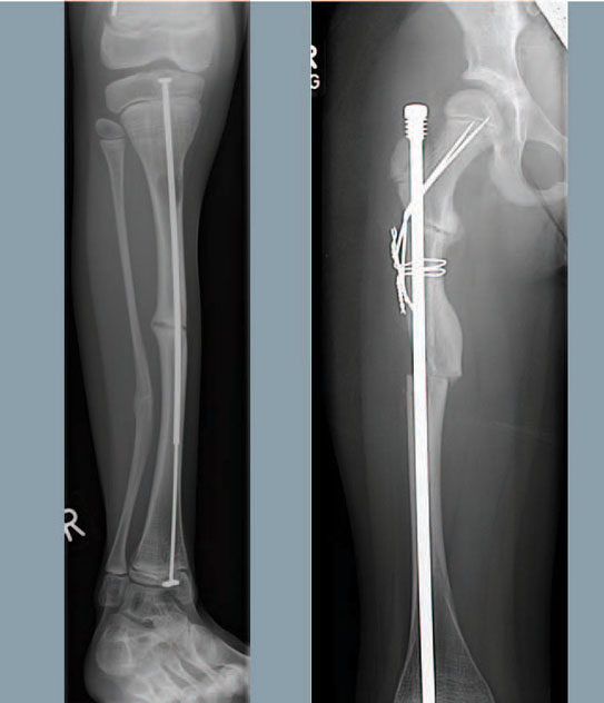
Osteogenesis imperfecta (OI) encompasses a group of rare heritable disorders associated with low bone mass and increased susceptibility to fractures. Around 90% of cases are the result of disturbances in type 1 collagen production, secondary to mutations in the genes COL1A1 and COL1A2, that code for the precursor of type 1 collagen. OI has an estimated prevalence of 1 per 15,000 to 1 per 20,000.
Classification is on the basis of severity, and nine types are currently described, see table 1.16 Functional collagen is required to form the organic matrix of bone within which the mineral component is deposited.
Increased bone fractures after minimal trauma is the cardinal feature. Additional features include blue sclera, hearing loss, scoliosis, deafness, and hypermobility.
Assessment in primary care
You enquire further about the fractures and note that they occurred after simple falls. There is no family history of OI. Sarah plots to the third centile for height. Her sclera are light blue tinged, teeth are normal. She has flat feet, ligamentous laxity, a slightly barrel shaped chest when viewed from the side and her sitting height seems reduced. You decide to refer her to a paediatrician.
Confirming diagnosis
Diagnosis is made on clinical features. Enquire about development and delayed motor milestones, detail activity levels and capacity for exercise (often reduced). Enquire about dairy product intake, vitamin D and sun exposure. Detail the fractures and dislocations, and other past history such as hernias, premature delivery.
Also record the family history (short stature, fractures, hernias, hearing loss (present in 50% of middle-aged patients with OI), early onset osteoporosis, teeth which chip or crack.
Examine the head shape carefully (for plagiocephaly, frontal bossing) eyes (blue sclera are normal up to six months old), teeth (for variable colour, translucent appearance), skin, musculoskeletal system (for bowing, pes planus, hypermobility), spine and growth.
Investigations will include a skeletal survey in any child with bony deformity. An AP and lateral spine is key to looking for vertebral crush fractures (commonly asymptomatic). Dual energy X-ray absorptiometry (DXA) is usually lower than predicted for height). Bone biochemistry (calcium, phosphate, alkaline phosphatase), parathyroid hormone and vitamin D levels should be checked. However, investigations alone cannot reliably identify all cases of OI, which may cause difficulty when assessing an infant with unexplained fractures and possible non-accidental injuries.16
Referral
Any child in whom OI is suspected should be referred. Accurate and prompt diagnosis is important because specialist multidisciplinary input, often
in conjunction with specific medical therapy, can dramatically improve outcomes. The importance of bone mass acquisition and spinal growth in childhood highlights the need for early diagnosis.
Sarah is found to have features of type I OI. She scores 6 on the Bulbena scale. Her spinal films show vertebral crush fractures at T8/9 and L1. Her DXA Z score is reported as -1.9 from age-related mean in the femoral neck.
Management
Sarah is referred to physiotherapy for core muscle strengthening. She is fitted with insoles. She is commenced on disodium pamidronate infusions for three days every four months.
There are recently designated nationally commissioned services
for severe, complex and atypical OI.
Less severely affected children may be looked after by a local multidisciplinary team with a paediatrician with a specialist interest in OI co-ordinating input.
The use of bisphosphonate therapy is now well established for improving bone mineralisation and reduction in fracture frequency. It is reported to improve back pain from vertebral crush fractures significantly. Most commonly IV pamidronate is used, but oral bisphosphonates are reported. Improvements in bone mass may be lower when given orally compared with IV but fracture risk is similar.17
Sarah’s walking distance and stamina improve and she reports less pain after six months of treatment with physiotherapy, insoles and pamidronate. Her spinal films show no further deterioration at one year, and her DXA Z score is still -1.9, but shows improvement in accordance with expected age-related gains. Genetic testing shows that her father is also affected, but has a milder phenotype. He has been referred to ENT because of deterioration in his hearing.
Conclusion
Connective tissue disorders are rare, but important, conditions because of their potential severity and the effect on the lives of those who suffer from them. Awareness of these conditions in children and early suspicion in primary care provides the opportunity to initiate early and appropriate specialist management and surveillance, which can dramatically improve the outcome for your patients.
The conditions often run in families and thus a thorough family history is important. Making a diagnosis in the child can have a positive outcome for other family members, as yet undiagnosed, who may benefit from intervention themselves. In children these conditions seldom require emergency admission at the time of first presentation and therefore you have time to consider the symptoms and signs and read around them before initiating appropriate referrals.
REFERENCES
1 Robinson PN, Arteaga-Solis E, Baldock C et al. The molecular genetics of Marfan syndrome and related disorders. J Med Genet 2006;43:769–87
2 Marfan syndrome. Online Mendelian Inheritance in Man. www.omim.org/entry/154700
3 Stuart AG, Williams A. Marfan’s syndrome and the heart. Arch Dis Child 2007;92:351-356
4 Rozendaal L, Blom NA, Hilhorst-Hofstee Y, Ten Harkel ADJ. Dilatation of the great arteries in an infant with Marfan syndrome and ventricular septal defect. Case Reports in Medicine vol 2011; Article ID 172109, 5 pages 2011.doi:10.1155/2011/172109
5 Loeys BL, Dietz HC, Braverman AC et al. The revised Ghent nosology for the Marfan syndrome. J Med Genet 2010;47:476-485
6 Anonymous. Health supervision for children with Marfan Syndrome. American Academy of Pediatrics, Committee on Genetics. Pediatrics 1996;98(5):978-82
7 Gao L, Mao Q, Wen D et al. The effect of beta-blocker therapy on progressive aortic dilatation in children and adolescents with Marfan’s syndrome: a meta-analysis. Acta Paediatrica 2011;100(9):101-5
8 National Institute for Health and Clinical Excellence: IPG394. External aortic root support in Marfan’s syndrome. NICE. London. 2011
9 Ehlers-Danlos syndrome type 1. Online Mendelian Inheritance in Man. www.omim.org/entry/130000
10 Beighton P, DePaepe A, Steinmann B et al. Ehlers-Danlos Syndromes: Revised Nosology, Villefranche 1997. Am J Med Gen 1998;77:31-37
11 Ross J, Grahame R. Joint hypermobility syndrome. BMJ 2011;342:c7167
12 Adib N, Davies K, Grahame R et al. Joint hypermobility syndrome in childhood. A not so benign multisystem disorder? Rheumatology 2005;44:744-750
13 Armon K, Macgregor A. Personal communication. Details at www.ukpmc.ac.uk/ GrantLookup/grants
14 Beighton PH, Solomon L, Soskolne C. Articular mobility in an African population. Ann Rheum Dis 1973;32:413-418
15 Bulbena A, Duro JC, Porta M et al. Clinical assessment of hypermobility of joints: assembling criteria. J Rheumatol 1992;19:115-22
16 Arundel P, Bishop N. Diagnosing osteogenesis imperfecta. Paed Child Health 2010;20:225-231
17 Bishop N. Management of osteogenesis imperfecta (OI). Paed Child Health 2011;21:569-570
ARTICLE IN PDF
 Download this article in PDF format
PRESET SEARCHES
Immediate CPD prompts
These handy templates* prompt immediate drafting of personal learning, reflection and planning. Save your record straight into your personal development folder on your device or Cloud storage system.
*Adobe reader needed for iPad
PubMed
Heritable connective tissue disorders
NICE Evidence