SEARCH

Hlaing Htwe S, Tidman M J. Prompt treatment of impetigo reduces risk of spread. Practitioner June 2020;264(1838):17-20
Prompt treatment of impetigo reduces risk of spread
22 Jun 2020
AUTHORS
Dr Su Hlaing Htwe MBBS MSc MRCP, Locum Registrar
Dr Michael J. Tidman MD FRCP(Edin) FRCP(Lond), Consultant Dermatologist
Department of Dermatology, Royal Infirmary of Edinburgh, Edinburgh, UK
Competing interests: None
Article
Impetigo is a common contagious bacterial infection of the skin. The causative organisms are either Staphylococcus aureus or, less commonly, Streptococcus pyogenes. The infection can occur at any age, but it is particularly common in children, especially the pre-school and early school age years, and tends to be more frequent during the summer months. It may arise on previously normal skin or complicate a pre-existing dermatosis, in particular eczema, herpes simplex infection, insect bites and scabies. Impetigo is usually transmitted by interpersonal contact or via fomites, but an individual may harbour the bacteria on the skin, particularly in the anterior nares or on the perineum. The non-bullous form typically presents as a moist or exudative area of skin, progressing to the formation of a golden or honey-coloured crust. The crust eventually dries up and separates leaving erythema which gradually fades but scarring tends not to occur. Bullous impetigo presents as one or more flaccid blisters containing a turbid liquid. The blisters usually rupture quickly to leave an erosion with a rim of scale. The diagnosis is essentially a clinical one, but if in doubt a swab should be taken for bacteriological culture. Good hygiene measures help to prevent the spread of impetigo to other areas of the body and other people. Affected children should be kept away from school until the impetigo has healed or until 48 hours after initiation of antibiotics. Patients with symptoms and signs of a complicating condition, such as cellulitis or sepsis, immunocompromised patients with widespread impetigo, and those failing to respond to treatment should be referred. Referral should also be considered for bullous impetigo, particularly in babies, frequently recurring impetigo, patients who are systemically unwell, and those at high risk of complications.
Impetigo is a common contagious superficial bacterial infection of the skin. The causative organisms are either Staphylococcus aureus or, less commonly, Streptococcus pyogenes (Group A beta-haemolytic streptococcus).1 The infection can occur at any age, but it is particularly common in children especially the pre-school and early school age years,1 and tends to be more frequent during the summer months. It may arise on previously normal skin or complicate a pre-existing dermatosis, in particular eczema, herpes simplex infection, insect bites and scabies.1,2
Impetigo is usually transmitted by interpersonal contact or via fomites (such as towels, utensils or toys) but an individual may harbour the bacteria on their skin, particularly in the anterior nares or on the perineum, and asymptomatic colonisation in these regions is not uncommon. Other predisposing factors for impetigo include: warm temperatures, high humidity, poor hygiene, overcrowded living conditions and skin abrasions.1
There are two clinical variants of impetigo; non-bullous and bullous.1 The more common non-bullous form is the result of either staphylococcal or streptococcal infection, or both.
The bullous form is caused by exfoliative (epidermolytic) toxin-producing strains of Staph. aureus. The toxins are serine proteases that target desmoglein 1, an important epidermal cell adhesion protein, and their interaction induces a cleavage plane very high in the epidermis, resulting in a fragile, thin-roofed blister.
Presentation
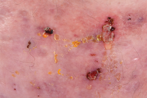
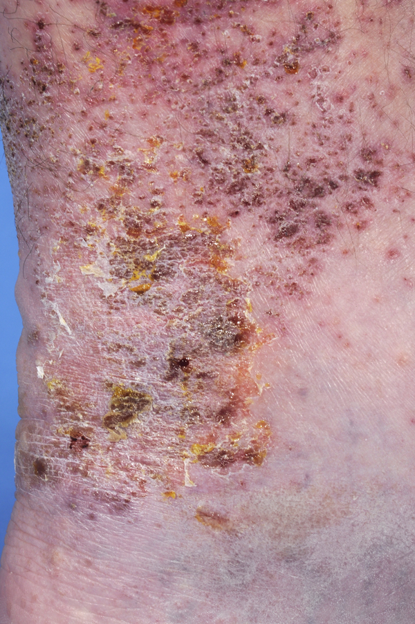
The non-bullous form of impetigo typically presents as a moist or exudative area of skin, progressing to the formation of a golden or honey-coloured crust (see figure 1 and figure 2).
It is usually uncomfortable and itchy. The crust eventually dries up and separates initially leaving erythema which gradually fades. Scarring tends not to occur unless the lesions have been heavily infected or excoriated. Pigmentary change (hyper- or hypo-pigmentation) may occur in heavily pigmented skin.
The face is a common site for impetigo, but any area can be affected. In severe cases, it can be associated with regional lymphadenopathy and constitutional symptoms.
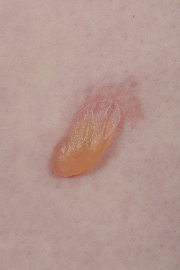
Bullous impetigo presents as one or more flaccid blisters containing a turbid liquid (see figure 3). The blisters usually rupture quickly to leave an erosion with a rim of scale. Healing follows the same course as in non-bullous impetigo.
Assessment
Enquiries should be made about the general health of the patient, the temperature should be taken and the regional lymph nodes palpated. It is also useful to establish whether impetigo is recurrent, and whether there are other affected individuals in the family or among social contacts.
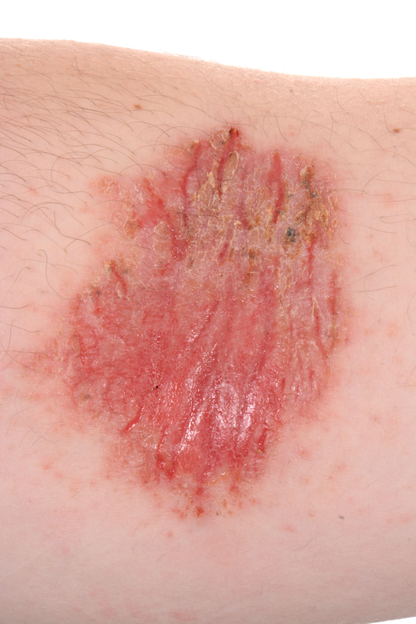
It is usually evident whether impetigo is primary or secondary to an underlying skin problem, such as atopic eczema (see figure 4), venous or discoid eczema, tinea capitis, insect bites or scabies.1,2 Recurrent impetigo localising to the same area of the body, especially the face, should raise the possibility of secondary bacterial infection of recrudescent herpes simplex.
Diagnosis
The diagnosis of impetigo is essentially a clinical one, but in cases of doubt a swab should be taken for bacteriological culture. A skin biopsy is rarely necessary, except in cases of putative impetigo that does not respond to standard therapy, in order to exclude underlying rare dermatological conditions such as pemphigus or Darier’s disease.
In cases of recurrent impetigo, carriage of potentially pathogenic bacteria should be excluded, and consideration should also be given to excluding carriage in family members or close contacts.
If preceding cold sores are thought to be a possibility, swabs for virology and bacteriology should be taken at the outset of an outbreak of impetigo.
Management
Although primary impetigo usually resolves spontaneously within 2-3 weeks, secondary impetigo tends to be more persistent. Healing occurs more quickly and the risk of spread to others is reduced if it is actively treated.
NICE has recently set out an antimicrobial prescribing strategy for impetigo, with the primary aims of optimising antibiotic use and reducing the development of antibiotic resistance.3,4
General measures
Good hygiene measures help to prevent the spread of impetigo to other areas of the body and other people.1 These include washing the affected area with soap and water, rigorous hand hygiene, the avoidance of scratching, and minimal sharing of fomites.
Parents should be advised to keep affected children away from school and childcare facilities until the impetigo has healed or until 48 hours after initiation of antibiotics. Likewise, adolescents and adults should stay away from work. (Food handlers are legally required to inform their employers immediately should they develop impetigo.)4
It is advisable to provide patients or carers with a detailed information leaflet such as that available from the British Association of Dermatologists, see External links.
When prescribing for impetigo the preferences of individuals and carers, including the practicalities of administration and the potential adverse effects, should be considered.1 Treating any pre-existing skin condition is crucial in the management of impetigo.
Localised non-bullous impetigo
The first line of treatment for localised non-bullous impetigo in an otherwise well patient is a topical antiseptic; hydrogen peroxide 1% cream is recommended. This is available over the counter (OTC) and should be applied two to three times a day for five days.3,5
Hydrogen peroxide 3% or 6% solution is also obtainable OTC. Patients and carers should be warned that hydrogen peroxide has a mild bleaching action.
Although other topical antiseptics are available, there is a lack of evidence to support their use in impetigo.3
If hydrogen peroxide is unsuitable (for instance if the impetigo is close to the eyes) or ineffective, a short course of topical antibiotics should be offered: fusidic acid 2% cream or mupirocin 2% cream/ointment can be applied thrice daily for 5-7 days (see tables 1 and 2).3,4
Widespread non-bullous impetigo
Depending on the severity and the presence or absence of systemic symptoms, the initial treatment for widespread non-bullous impetigo is a course of either a topical antibiotic (fusidic acid or mupirocin) or an oral antibiotic.3
Flucloxacillin is the recommended first choice oral antibiotic, but clarithromycin and erythromycin are alternatives if an allergy to penicillin or unpalatability are concerns (see tables 1 and 2).3,4
Bullous impetigo and systemically unwell patients
Patients with bullous impetigo, and those who are systemically unwell or at high risk of complications, should be offered a short course of oral antibiotics (flucloxacillin, clarithromycin or erythromycin) as initial treatment (see tables 1 and 2, right).3,4
If these regimens are ineffective, swabbing for bacteriological culture and antibiotic sensitivities should be undertaken, and the antibiotic changed according to the results, taking into consideration local antimicrobial resistance data.3
There is an increased risk of the development of antibiotic resistance from the repeated use of topical antibiotics.3 If methicillin-resistant Staphylococcus aureus (MRSA) is isolated it is recommended that the local microbiologist is contacted for advice on appropriate antibiotic treatment.3
In cases of frequently recurring impetigo, skin (perineum, groin, axillae) and nasal swabs should be sent for microbiological testing. If carriage is confirmed, a decolonisation regimen is recommended.4
Nasal carriage should be eliminated with chlorhexidine and neomycin cream four times daily for ten days as first choice, or mupirocin three times daily for five days if this is unsuitable or ineffective. The chlorhexidine and neomycin cream formulation contains arachis oil and should therefore be avoided in those who are allergic to peanut or soya.
For the skin an antiseptic such as chlorhexidine 4% body wash or triclosan 2% should be applied daily for five days in the bath or shower, paying particular attention to the perineum, axillae, groin, sub-mammary region and hands. A benzalkonium chloride-containing product is an alternative. General hygiene measures should also be employed.4
Periodic checking for recolonisation is advisable if impetigo continues to recur.
Potential complications
Serious complications with impetigo are unusual. Occasionally, deeper infection such as cellulitis, erysipelas or ecthyma can occur, particularly with streptococcal infection, and sepsis is a possibility in immunocompromised individuals.
Nephritogenic strains of Strep. pyogenes may, rarely, cause post-streptococcal glomerulonephritis, which poses a risk of chronic renal disease.1 Routine urinalysis two to three weeks after an episode of impetigo should be considered.
Streptococcal impetigo may be associated with subsequent erythema multiforme, scarlet fever and urticaria.1
In neonates, infants and young children, the haematogenous spread of staphylococcal exfoliative toxins from a small focus of impetigo may result in staphylococcal scalded skin syndrome (SSSS), characterised by large areas of painful superficial blister formation associated with fever and irritability.
The diagnosis is easily and quickly made by removing a small portion of blister roof with sterile scissors (which is painless) and examining a frozen section. In SSSS the blister roof comprises just the stratum corneum and stratum granulosum, in contrast to toxic epidermal necrolysis (which SSSS can clinically resemble) where the roof is formed by the entire thickness of necrotic epidermis. With appropriate antibiotic therapy and nursing care the prognosis of SSSS is good.
Referral
Although only a minority of cases of impetigo require specialist care, it is recommended that patients with symptoms and signs of a complicating condition, such as cellulitis or sepsis, immunocompromised patients with widespread impetigo, and those failing to respond to treatment despite adequate compliance should be referred.3,4
Referral should also be considered for those with bullous impetigo, particularly babies, cases of frequently recurring impetigo, patients who are systemically unwell, and those at high risk of complications.3,4
Additionally, specialist advice should be sought when optimisation of treatment is required for a skin disorder predisposing to impetigo, and when there is doubt concerning the diagnosis of impetigo. If there is a significant local outbreak, for instance in a school (or residential home), referral to a consultant in communicable disease control should be made.4
Conclusion
GPs will commonly encounter impetigo, and NICE guidance has provided a rational strategy for its management. Optimisation of treatment for any predisposing skin conditions is an important part of the management of impetigo.
REFERENCES
1 Hay RJ, Morris-Jones R. Bacterial infections in Rook’s Textbook of Dermatology, Chapter 26. 9th Edition. Eds. Griffiths C, Barker J, Bleaker T, Chalmers R, Creamer D. John Wiley & Sons Ltd. Oxford. 2016 ISBN 9781118441190
2 Elliot AJ, Cross KW, Smith GE et al. The association between impetigo, insect bites and air temperature: a retrospective 5-year study (1999-2003) using morbidity data collected from a sentinel general practice network database. Fam Pract 2006;23:490-96
3 National Institute for Health and Care Excellence. NG153. Impetigo: antimicrobial prescribing. NICE. London. 2020 www.nice.org.uk/guidance/ng153
4 National Institute for Health and Care Excellence. Clinical knowledge summaries. Impetigo cks.nice.org.uk/impetigo
5 Leitch CS, Leitch AE, Tidman MJ. Quantitative evaluation of dermatological antiseptics. Clin Exp Dermatol 2015;40:912-15
ARTICLE IN PDF
 Download this article in PDF format
CLICK ON IMAGES
EXTERNAL WEBLINKS
British Association of Dermatologists
NHS conditions
DermNetNZ