SEARCH

Sedgwick C, Sharma S. Diagnosing heart disease in children and adolescents. Practitioner 2017;261(1805):11-15
Diagnosing heart disease in children and adolescents
22 Jun 2017
AUTHORS
Dr Catherine Sedgwick BSc (Hons) MBBS, Honorary Clinical Fellow, Cardiac Research Centre, St George’s University of London, London, UK
Professor Sanjay Sharma MD FRCP (UK) FESC (UK), Professor of Cardiology and Head of Cardiac Research Centre, Cardiology Clinical and Academic Group, St George’s University of London, London, UK
Article
Abstract
Heart disease in children and adolescents can be congenital, in which structural defects of the heart and major blood vessels are present from birth, acquired, whereby disease develops during life, or genetic, including diseases affecting the heart muscle, electrical system or the aorta. The incidence of congenital heart disease has decreased over the past 30 years, with approximately 1 in 180 babies born with congenital heart disease in the UK each year. Congenital heart disease is typically classified as cyanotic or non-cyanotic. Tetralogy of Fallot is the most common form of cyanotic congenital heart disease. Non-cyanotic congenital heart diseases include ventricular septal defect (VSD), atrial septal defect and bicuspid aortic valve. VSD are seen in 50% of all children with congenital heart disease and are the most common congenital heart defects. Several cardiac diseases are genetic and can manifest in childhood. Most are primary cardiomyopathies, ion channel diseases, coronary artery disease from familial hypercholesterolaemia or aortopathies. Hypertrophic cardiomyopathy is the most common cardiomyopathy, affecting 1 in 500 people. Acquired heart disease in young people is often precipitated by infection. A self-limiting systemic vasculitis, Kawasaki disease typically affects children aged six months to five years. Myocarditis, acute inflammation of the heart muscle, is implicated in 8.6% of cases of sudden cardiac death. Hypercholesterolaemia, hypertension, diabetes, and obesity are major risk factors for cardiovascular disease and, as levels increase in young people, the cardiac health of future generations is at risk.
Heart disease in children and adolescents can be congenital, in which structural defects of the heart and major blood vessels are present from birth, acquired, whereby disease develops during life, or genetic, including diseases affecting the heart muscle, electrical system or the aorta.
Congenital heart disease can vary in severity, from small asymptomatic heart defects to conditions requiring immediate postnatal surgical intervention. The incidence of congenital heart disease has fallen over the past 30 years, with approximately 1 in 180 babies born with congenital heart disease in the UK each year.1
As treatment options have improved, and survival rates from surgical interventions have increased, there are now twice as many adults with congenital heart disease as children.2
Acquired heart disease, often precipitated by infection, is becoming less common in high income countries.3,4 However, as rates of obesity and type 2 diabetes increase in the younger population, evidence shows that children with the highest cholesterol and blood pressure levels will continue to express these traits into adolescence, threatening their cardiovascular health in adulthood.5
Congenital heart disease
Structural heart defects are caused by failure of the heart to develop fully in utero, resulting in disturbed blood flow to, from or around the heart. If a significant amount of blood shunts from the right to the left side of the heart, without passing through the respiratory system, cyanosis and cardiovascular compromise ensues.
Investigations
Most congenital heart defects are detected antenatally or in the immediate postnatal period. However, some conditions will not be identified until the six week check. Table 1, shows symptoms of congenital heart disease by age. Auscultation may reveal abnormal heart sounds or a murmur, as outlined in table 2.
An electrocardiogram (ECG) is often normal in mild congenital heart disease, although there may be evidence of right axis deviation and right ventricular hypertrophy in cases of pulmonary hypertension and left ventricular hypertrophy in septal defects.
Chest x-rays may be normal or feature cardiomegaly, increased pulmonary vascular markings or signs of cardiac failure. A transthoracic 2D echocardiogram is the gold standard for investigating suspected congenital heart defects as it provides both the anatomical information and haemodynamic evaluation required for diagnosis.
A 3D echocardiogram may provide more detailed information in preparation for surgery.
Congenital heart disease is typically classified as either cyanotic or non-cyanotic. The most common diseases are listed in table 3.
Cyanotic
Tetralogy of Fallot: This is the most common form of cyanotic congenital heart disease. The tetralogy of abnormalities includes: a large ventricular septal defect (VSD); right ventricular outflow tract (RVOT) obstruction; an overriding aorta and right ventricular hypertrophy.6
Usually diagnosed antenatally, and often in patients with Down’s syndrome, it is commonly detected while the neonate is still ‘pink’. Full surgical repair takes place in the first year of life and prognosis depends on the degree of RVOT obstruction. If left untreated mortality is 50%.7
Transposition of the great arteries: As the name suggests, the aorta and pulmonary arteries are transposed in this disease, with the aorta rising from the right ventricle and the pulmonary artery from the left. Survival depends on the existence of a shunt, allowing oxygenated blood into the systemic circulation.
Prostaglandins are given immediately to preserve ductal patency and an urgent atrial septostomy is performed until more definitive corrective surgery can take place. Postoperatively, 5-7% of neonates develop myocardial ischaemia or infarction, but 20-year survival stands at 90%.8
Non-cyanotic
Ventricular septal defect: VSDs are observed in 50% of all children with congenital heart disease9 and are the most common congenital heart defects. They occur when any part of the ventricular septum fails to develop in utero. VSDs are often observed in chromosomal disorders10 and are sometimes seen in fetal alcohol syndrome,11 gestational diabetes12 and with maternal cannabis use.13
A small VSD may be asymptomatic and usually closes spontaneously. However, a moderate to large VSD can cause respiratory distress during feeding and diuretics may be given to relieve heart failure symptoms. Large VSDs increase pulmonary vascular resistance and cause pulmonary hypertension, and surgical intervention is usually indicated. Prognosis is excellent, although female patients with pulmonary hypertension are advised against pregnancy as maternal mortality is 50%.14
Atrial septal defect (ASD): An ASD is an abnormal communication between the left and right atria of the heart, classified according to its location within the septum. The aetiology is usually unknown. Most infants will be asymptomatic, although haemodynamically significant ASDs will require surgical closure. Prognosis is excellent if surgery is performed before the age of 12.15
Patent ductus arteriosus (PDA): A PDA is a normal feature of the fetal heart and it usually closes spontaneously in the hours after birth.
Risk factors for ongoing patency include genetic predisposition, asphyxia during delivery and exposure to viral infections such as rubella.16 A small PDA is usually asymptomatic but will require endovascular closure if patent at one year. A large PDA may cause feeding difficulties, poor growth and heart failure, for which surgical closure is indicated.
Prognosis for full-term infants is very good.17 However, PDA in preterm babies is often associated with other serious comorbidities so prognosis is difficult to establish.
Bicuspid aortic valve: Seen in 1-2% of the population and twice as common in males,18 a bicuspid aortic valve only has two leaflets instead of three. It is usually asymptomatic until later life when the valve may become calcified and require repair or replacement. There is an increased risk of aortic dissection and thoracic aortic aneurysm as the tensile strength of the aorta is reduced in these patients.
Once diagnosed, most patients with congenital heart disease will be managed in specialist congenital heart disease centres. Advances in treatment have led to a dramatic improvement in outcomes, with more than 85% of children expected to survive into adulthood.19
Genetic heart conditions
Several cardiac diseases are genetic and can manifest in childhood. The vast majority are primary cardiomyopathies, ion channel diseases, coronary artery disease from familial hypercholesterolaemia and aortopathies.
Cardiomyopathies
Cardiomyopathies are primary myocardial diseases and are mostly feared for their association with sudden cardiac death.
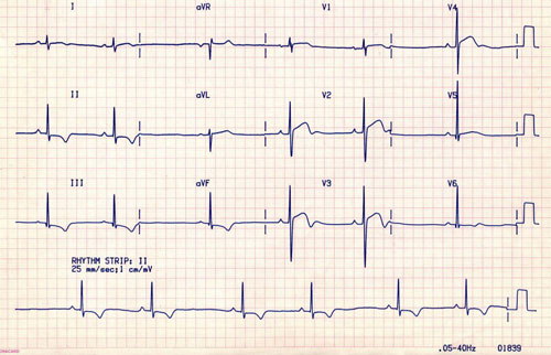
Hypertrophic cardiomyopathy is the most common cardiomyopathy, affecting 1 in 500 people. It is characterised by left ventricular hypertrophy (LVH) in the absence of a cardiac or systemic cause. Often asymptomatic, the first ‘symptom’ may be sudden cardiac death. ECG changes include LVH, ST segment changes and T wave inversion (see figure 1). Transthoracic echocardiogram shows asymmetrical septal hypertrophy and is diagnostic. Cardiac MRI is a useful modality to identify areas of segmental hypertrophy.
Medical management includes rate and rhythm control and a surgical myectomy may be performed to relieve left ventricular outflow tract (LVOT) obstruction. Many patients wear an implantable cardioverter defibrillator to offer protection against sudden death.
Dilated cardiomyopathy is inherited in up to 40% of cases and presents with heart failure and serious arrhythmias. Management is with conventional heart failure therapy.
Ion channel disease
Mutations within genes encoding sodium and potassium ion channels within the heart may cause a variety of sequelae including: Long QT syndrome, catecholaminergic polymorphic ventricular tachycardia (CPVT) and Brugada syndrome. Some of these diseases have been implicated in sudden infant death syndrome.
Long QT syndrome usually causes polymorphic ventricular tachycardia (VT) during adrenergic surges such as exercise, sudden loud auditory stimuli, heightened emotion or fear. Certain commonly prescribed drugs, such as the macrolide and quinolone antibiotics or antihistamines, may precipitate arrhythmias, which might manifest as transient syncope or sudden cardiac death. In some cases, cerebral hypoperfusion due to VT may cause transient epileptiform movements, leading to a misdiagnosis of epilepsy.
Risks are highest in prepubertal males and girls in menarche, and management is with beta blockers.
Acquired heart disease
Acquired heart disease in young people is often precipitated by infection. Common examples are described below.
Kawasaki disease
A self-limiting systemic vasculitis, Kawasaki disease typically affects children aged six months to five years. It is the most common childhood acquired heart disease in high income countries and the incidence in the UK is 8.39 per 100,000 children.3 Typical features include: fever, rash, bilateral conjunctivitis and inflammation of the mouth and extremities. The aetiology is thought to have both an infective and genetic component.
Diagnosis is made clinically, with echocardiography used to identify associated coronary artery aneurysms. Medical treatment is with aspirin and intravenous immunoglobulins. Prognosis is dependent on the extent of coronary artery involvement.
Myocarditis
Acute inflammation of the heart muscle is implicated in 8.6% of cases of sudden cardiac death.21 Signs and symptoms range from fatigue, chest pain and fever, to LV dysfunction and severe cardiac failure. Most commonly precipitated by Coxsackie virus, myocarditis may also be caused by toxins, drug hypersensitivity and immune-mediated reactions.
An endomyocardial biopsy is the gold standard for diagnosis. Prognosis is dependent on the level of cardiac dysfunction with dilated cardiomyopathy often persisting into adulthood.
Pericarditis
Occasionally seen in younger patients, pericarditis is inflammation of the pericardium, the membranous sac surrounding the heart. Common aetiologies include viral infection, systemic lupus erythematosus (SLE) and myocardial infarction.
Symptoms include substernal chest pain, classically relieved on leaning forwards, and abdominal pain in children. A pericardial rub is heard on auscultation in 60-85% of cases. Serial ECGs may reveal concave ST elevation, PR depression and T wave inversion. In complex, persistent cases, echo-guided pericardiocentesis and biopsy are required for diagnosis. Medical management of the underlying cause is usually sufficient in this self-limiting illness.
Rheumatic heart disease
Rheumatic fever was a major cause of chronic structural heart disease and death in children in the UK until 1960. With an incidence of 1 in 100,000 in high income countries,4 prevalence today is associated with areas of low socioeconomic status. Carditis occurs in 40% of patients, where Lancefield group A beta-haemolytic streptococci typically attack the mitral valve. Clinical features include fever, tachycardia and a pericardial rub. Management includes bed rest, penicillin and supportive treatment of cardiac complications. Long-term follow-up is usually required.
Modifiable risk factors in children for CVD
Hypercholesterolaemia, hypertension, diabetes, and obesity are major risk factors for cardiovascular disease and, as levels increase in young people, the cardiac health of future generations is at risk.
Hypertension
The Global Burden of Disease 2010 report ranked hypertension as the second highest risk factor contributing to the global burden of disease22 and Din-Dzietham and colleagues (2007) reported that hypertension in children is increasing in prevalence as a result of rising levels of childhood obesity.23
Diabetes
In 2009, The Royal College of Paediatrics and Child Health estimated that 22,783 children under 18 were living with diabetes, with a prevalence of type 2 diabetes of 3 per 100,000 of the population in England.24
Hypercholesterolaemia
Data submitted by GP practices in 2011 to the Information Services Division in Scotland estimated that high cholesterol levels were observed in 0.3 boys per 1,000 registered children and less than 0.1 girls per 1,000 registered children.25
Obesity
In 2011, 17% of boys and 16% of girls in England were obese, compared with 11% of boys and 12% of girls in 1995.26 Obesity is an independent risk factor for cardiovascular disease but it also increases the likelihood of hypertension, diabetes and hypercholesterolaemia,27 and usually persists into adulthood.28
Conclusion
As diagnosis, treatment options and survival rates of congenital heart disease have improved in children and adolescents over the past 30 years, we have also witnessed a reduction in those acquired heart diseases precipitated by infection.
In cases of parents diagnosed with a cardiomyopathy or ion channel disease, it would be prudent to screen the children of these patients to identify whether they are also at risk of an inherited cardiac disease.
Perhaps the most concerning trend is the increase in obesity in young people and its associated effects on hypertension, diabetes and hypercholesterolaemia in this population. The Royal College of Paediatrics and Child Health recently published its strategies to combat childhood obesity and recommends further training for healthcare professionals.29
REFERENCES
1 British and Irish Network of Congenital Anomaly Researchers (BINOCAR) www.binocar.org
2 Avila P, Mercier LA, Dore A et al. Adult congenital heart disease: a growing epidemic. Can J Cardiol 2014;(12 Suppl): S410-9.
3 Harnden A, Mayon-White R, Perera R et al. Kawasaki disease in England: ethnicity, deprivation, and respiratory pathogens. Pediatr Infect Dis J 2009;28(1):21-24
4 Cilliers AM. Rheumatic fever and its management. BMJ 2006;333(7579):1153-56
5 British Heart Foundation Health Promotion Research Group, Department of Public Health, University of Oxford; Children and Young People Statistics 2013 www.bhf.org.uk/publications/statistics/children-and-young-people-statistics-2013
6 Monaco M, Williams I. Tetralogy of Fallot: fetal diagnosis to surgical correction. Minerva Pediatr 2012;64(5):461-70
7 Piazza L, Chessa M, Giamberti A et al. Timing of pulmonary valve replacement after tetralogy of Fallot repair. Expert Rev Cardiovasc Ther 2012;10(7):917-23
8 Villafane J, Lantin-Hermoso MR, Bhatt AB et al. D-transposition of the great arteries: the current era of the arterial switch operation. J Am Coll Cardiol 2014;64(5):498-511
9 Minette MS, Sahn DJ. Ventricular septal defects. Circulation 2006;114(20):2190-97
10 Beke A, Papp C, Toth-Pal E et al. Trisomies and other chromosome abnormalities detected after positive sonographic findings. J Reprod Med 2005;50(9):675-91
11 Steeg CN, Woolf P. Cardiovascular malformations in the fetal alcohol syndrome. Am Heart J 1979;98(5):635-37
12 Loffredo CA, Hirata J, Wilson PD et al. Atrioventricular septal defects: possible etiologic differences between complete and partial defects. Teratology 2001;63(2):87-93
13 Williams LJ, Correa A, Rasmussen S. Maternal lifestyle factors and risk for ventricular septal defects. Birth Defects Res A Clin Mol Teratol 2004;70(2):59-64
14 Thorne S, MacGregor A, Nelson-Piercy C. Risks of contraception and pregnancy in heart disease. Heart 2006;92(10):1520-25
15 Murphy JG, Gersh BJ, McGoon MD et al. Long-term outcome after surgical repair of isolated atrial septal defect. Follow-up at 27 to 32 years. N Engl J Med 1990;323(24):1645-50
16 Anilkumar M. Patent ductus arteriosus. Cardiol Clin 2013;31(3):417-30
17 Schneider DJ. The patent ductus arteriosus in term infants, children, and adults. Semin Perinatol 2012;36(2):146-53
18 Congenital Heart Defects UK www.chduk.co.uk/types-of-chd-and-operations/bicuspid-aortic-valve/
19 Ramrakha P, Hill J. Oxford Handbook of Cardiology. 2nd edition. Oxford University Press. 2012
20 Steriotis AK, Sharma S. Eur Cardiol Rev 2015;10(1):31-6
21 Schultz JC, Hilliard AA, Cooper LT Jr et al. Diagnosis and treatment of viral myocarditis. Mayo Clin Proc 2009;84(11):1001-09
22 Institute for Health Metrics and Evaluation (2012). Global Burden of Diseases, Injuries, and Risk Factors Study 2010. Institute for Health Metrics and Evaluation. USA
23 Din-Dzietham R, Lie Y, Bielo MV, Shamsa F. High blood pressure trends in children and adolescents in national surveys, 1963 to 2002. Circulation 2007;116:1488-96
24 Royal College of Paediatrics and Child Health. Growing up with diabetes: children and young people with diabetes in England. RCPCH. London. 2009
25 Information Services Division Scotland (2012). Practice team Information (PTI). www.isdscotland.org/pti#
26 Joint Health Surveys Unit. National Child Measurement Programme 2011/12. The Information Centre. Leeds. 2012
27 World Health Organization (2000). Obesity – preventing and managing the global epidemic. Report of a WHO Consultation on Obesity. WHO. Geneva. 2000
28 Serdula M, Ivery D, Coates R et al. Do obese children become obese adults? A review of the literature. Prev Med 1993;22:167-77
29 Royal College of Paediatrics and Child Health. Position statement: Childhood obesity. RCPCH. London. 2012
ARTICLE IN PDF
 Download this article in PDF format
PRESET SEARCHES
PubMed
Diagnosing heart disease in children and adolescents review
NICE evidence search