SEARCH

Davidson L, Derry C. Seizure classification key to epilepsy management. Practitioner 2015; 259 (1785):13-19
Seizure classification key to epilepsy management
24 Sep 2015
AUTHORS
Dr Louise Davidson MBChB MRCP, Neurology Registrar
Dr Chris Derry MRCP PhD, Consultant Neurologist
Western General Hospital, Edinburgh, UK
Article
Abstract
The diagnosis of epilepsy carries significant implications for physical, psychosocial and financial wellbeing as well as a small but significant increased risk of mortality. The diagnosis is often incorrect, potentially in up to 20% of cases, so should be revisited if seizures are not responding to treatment. Evidence indicates that misdiagnosis is significantly more common among nonspecialists. SIGN recommends that the diagnosis of epilepsy should be made by an epilepsy specialist, ideally in the setting of a dedicated first seizure or epilepsy clinic. An incorrect diagnosis of epilepsy can be harmful. There is an exhaustive list of epilepsy mimics that can result in misdiagnosis and expose patients to unnecessary treatment with antiepileptic drugs. Diagnosis relies primarily on the history. Investigations can support the diagnosis but cannot make it in isolation, and negative investigation findings are common in epilepsy. Brain imaging will be undertaken in most patients with epilepsy, but is not routinely required in those with a definite diagnosis of genetic generalised epilepsy. The EEG has limitations and can sometimes cloud rather than clarify the diagnostic picture. Distinguishing between a genetic generalised epilepsy and a focal epilepsy is vital as this influences investigation, treatment and prognosis. Generally medication should not be started following a single seizure except in specific circumstances or in cases where the risk of recurrence is high.
Epilepsy is a common condition, with an annual incidence of 5 per 10,000 per year and prevalence of 7.5 per 1,000.1
The management of epilepsy often involves a range of medical professionals, including neurologists, epilepsy specialist nurses, GPs, learning disability teams, and emergency, general and geriatric medicine physicians.1 Hence, an up to date working knowledge of epilepsy among all healthcare professionals is essential in order to deliver optimal patient care.
The diagnosis of epilepsy carries significant implications for physical, psychosocial and financial wellbeing as well as a small but significant increased risk of mortality.3 An incorrect diagnosis of epilepsy can be harmful, and it is, therefore, vital that every effort is made to reach a correct diagnosis. Evidence indicates that misdiagnosis is significantly more common among nonspecialists.2,4,5
It is well recognised that there is an exhaustive list of epilepsy mimics that can result in misdiagnosis and expose patients to unnecessary antiepileptic drugs (AEDs).2 Moreover, epilepsy itself is not a single entity, but comprises a range of seizure types and epilepsy syndromes.
Making an accurate assessment of seizure type and classification is important, both for treatment and prognostication.1,3
There have been a number of recent advances in the field of epilepsy and SIGN has recently published updated guidance (SIGN 143).3
The terminology, see box 1, and classification of seizures and epilepsy has changed over the years, see below.6
Classification
The most widely used classification system for epilepsy is that published by the International League Against Epilepsy (ILAE).3,7,8 The most recent ILAE classification update was in 2010.9
Classification largely depends on two characteristics:1,3,9
• Site of seizure onset determines whether a seizure is:
— Generalised (i.e. simultaneous involvement of both hemispheres)
— Focal (i.e. starting from one area
of the brain)
• Presumed aetiology
— Structural-metabolic (an underlying cause is present, previously referred to as symptomatic, see box 1
— Epilepsy of unknown origin (presumed underlying cause but not identified, previously referred to as cryptogenic)
— Genetic (previously idiopathic)
Classification is frequently overlooked but is key in epilepsy management,1 and the importance of this is highlighted in the updated SIGN guideline.3
Distinguishing between a genetic generalised epilepsy and a focal epilepsy is vital, as this influences investigation, treatment and prognosis. In focal epilepsies, identifying the underlying cause is important.3
Focal epilepsies
The focal epilepsies are so called because seizures start in a small area of the brain; the location of this abnormal area dictates the clinical features of the attack.
Focal seizures are usually described as simple partial (seizures in which awareness is retained) or complex partial (awareness is impaired).
The new ILAE 2010 classification has replaced these descriptions by the single term focal seizure; if impairment of consciousness clearly occurs, the adjective dyscognitive (i.e. focal dyscognitive seizure) can be applied.
Initial symptoms: Some patients with focal epilepsy lose awareness early in a seizure, and may report no warning or initial symptoms. However, some initial symptoms are common, and depend on the site of seizure onset.
Common clinical symptoms include:6
• Motor – spasms, jerking (frontal onset)
• Sensory – tingling or burning (parietal onset)
• Autonomic – rising abdominal sensation, palpitations (temporal onset)
• Psychic – altered perception of size or distance, dysphasia, altered memory (temporal onset)
Observed behavioural changes: If consciousness is lost or impaired, automatic behaviours (automatisms) may be seen and described by witnesses.
Simple automatisms include lip smacking, chewing, fidgeting or plucking at clothes; occasionally more complex behaviours including undressing or wandering.6
Automatisms are most common in seizures arising from the temporal lobes, although may occur in focal seizures arising from other areas.
Focal seizures can spread to involve both hemispheres, and develop into a generalised tonic-clonic seizure (i.e. secondary generalisation).
A summary of features of focal seizures, subdivided by the site of seizure onset, is given in table 1.1,2,6
Genetic (idiopathic) generalised epilepsies
Prior to the ILAE 2010 classification, these epilepsies (characterised by onset in diffuse, bilateral networks of neurones) were called idiopathic generalised epilepsies. This term remains in widespread clinical use, although the 2010 system has changed this to genetic generalised epilepsies.
There are several clues in the history that point towards a genetic generalised epilepsy. Individuals typically report no warning at all of seizures, and have usually one or more of three seizure types:
tonic-clonic seizures; myoclonic jerks (brief electric shock like jerks of the limbs); or absences (brief staring spells, usually lasting a few seconds only). There are subtypes of genetic generalised epilepsy (syndromes), the most common being juvenile myoclonic epilepsy and childhood absence epilepsy.
Specific features suggesting a genetic generalised epilepsy:3,10
• Childhood or teenage onset
• Seizures triggered by sleep deprivation and alcohol
• Early morning tonic-clonic seizures or myoclonic jerks
• Brief absence seizures
• Photostimulation response on EEG
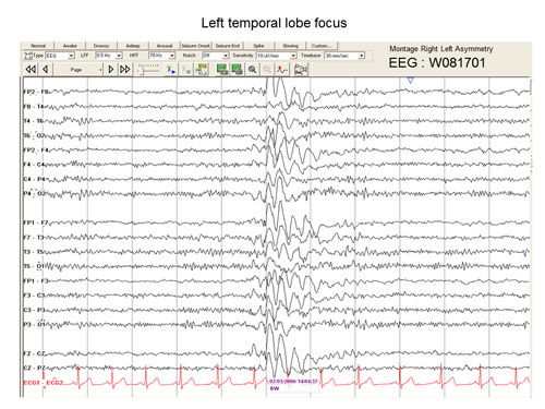
• Generalised three per second spike and wave or polyspike and wave on the EEG, see figure 1.
Diagnosis
SIGN states that the diagnosis of epilepsy should be made by an epilepsy specialist, ideally in the setting of a dedicated first seizure or epilepsy clinic.3
The differential diagnosis of patients presenting with episodic altered states of consciousness is exhaustive and full discussion is beyond the scope of this article. A list of epilepsy mimics is shown in table 2.2 In many cases, the key challenge is differentiating between seizure, syncope and psychogenic attack.1,2,3 Distinguishing features of each are highlighted in table 3.1,2,3,6
The diagnosis of epilepsy relies primarily on the history. Investigations can support the diagnosis but cannot make it in isolation,1,3 and negative investigation findings are common in epilepsy. Given that awareness is often impaired in attacks, a detailed witness account is crucial.2,3,6 It is important to establish what happened before, during and after the attack, from both the patient and the witness.3,6 Table 4, highlights some of the important questions to cover.1,2,6
In cases where there is an established history of epilepsy, it is good practice to go over the history again asking questions as for a new patient, especially if the patient has treatment-resistant seizures.
The diagnosis of epilepsy is often incorrect, potentially in up to 20% of cases,2 so should be revisited if seizures are not responding to treatment.
Additional questions to be considered in such situations include:6
• When was the diagnosis of epilepsy made?
• Who made the diagnosis?
• What investigations were performed and what were the results?
• What treatments have been tried and at what dose?
• How did they affect the seizures?
• Were there any side effects?
Diagnostic investigations
Investigations cannot replace an adequate history in the diagnosis of epilepsy, but can aid in clarifying the diagnosis and refining the epilepsy syndrome. Several modalities can be used, and the key techniques are discussed here.
EEG
The EEG has its place in the diagnosis and classification of epilepsy.1,3,11 However, it has limitations and can sometimes cloud, rather than clarify, the diagnostic picture.
Many patients with epilepsy have a normal EEG between attacks, and so a normal test neither confirms nor refutes the diagnosis. In contrast, non-specific EEG abnormalities are common in people without epilepsy (especially the elderly, patients with migraine and those on psychotropic medication).3
Epileptiform abnormalities (changes associated strongly with epilepsy) occur in approximately 0.5% of healthy young adults and so, although suggestive, are not unequivocal evidence of epilepsy.3
The EEG is, therefore, best seen as a supportive tool in diagnosis and classification, but one that is not routinely indicated, and best requested and interpreted by an epilepsy specialist.
In difficult cases, ambulatory EEG or video telemetry (combined EEG and video monitoring) can be used with the aim of obtaining an ictal EEG (i.e. EEG changes during an attack itself).
An ictal EEG is far more likely than an interictal EEG to give definitive diagnostic information, but is only really practically possible to obtain in individuals having frequent attacks.1,3,6
Hand-held video
In difficult cases (for example when distinguishing epileptic from non-epileptic attacks), video recording of attacks can provide useful information to clinicians experienced in diagnosing epilepsy. Based on anecdotal evidence, SIGN has suggested the use of hand-held videos such as smart phones to record attacks. Patient consent must be obtained before taping attacks.3
Brain imaging
Neuroimaging is an important part of the evaluation of patients with epilepsy. Advances in imaging techniques have resulted in structural lesions being identified in some people previously deemed to have epilepsy of unknown cause.6 Imaging identifies lesions in 21-37% of patients with epilepsy.3
The sensitivity and specificity of MRI is much higher than CT, and MRI is the recommended modality of structural imaging in epilepsy.1,3
Brain imaging will be undertaken in most people with epilepsy, but is not routinely required in those with a definite diagnosis of genetic generalised epilepsy (as this is not associated with an increased prevalence of brain lesions.3)
ECG
An ECG should be carried out in all patients presenting with altered consciousness, particularly in the elderly where disorders of cardiac rhythm can mimic epilepsy. If necessary, prolonged ECG recording may be required.1,3
Management
The ultimate goal of epilepsy management is to achieve seizure freedom without adverse drug side effects. This is achievable in most cases.1 In addition to pharmacological therapy and surgical intervention, it is also important to consider a variety of lifestyle influences on seizure control and how the diagnosis impacts on quality of life.3
The following recommendations have been made by SIGN.3
Starting AED therapy
The decision to prescribe AEDs is significant, as it commits the patient to prolonged treatment with a significant risk of adverse effects. It is therefore essential that every effort is made to ensure the diagnosis is correct before starting treatment. If there is uncertainty, a period of observation is reasonable as this will often clarify the diagnosis and guide the need for treatment.1,3
The decision to start treatment should be shared between the epilepsy specialist and the patient.3
Generally, medication should not be started following a single seizure except in specific circumstances or in cases where the risk of recurrence is high:1,3
• Individuals wishing to avoid recurrent seizures e.g. drivers
• The patient has a structural cerebral disorder
• The EEG shows unequivocal epileptic discharges
• History of previous myoclonus, absence or focal seizure prior to the presenting tonic-clonic seizure.
Choice of AED monotherapy
Monotherapy is the preferred treatment strategy in newly diagnosed epilepsy.1 The drug of choice will be strongly influenced by the epilepsy classification, side-effect profiles, medication history, gender and the patient’s wishes.1,3 All AEDs have an effect on focal epilepsies but evidence-based treatment of generalised epilepsies is restricted to sodium valproate, lamotrigine and topiramate.1
Dosing for each AED should start low and be increased slowly to the maximum tolerated or recommended dose as set out by the BNF.3 The first-line recommended drug for focal epilepsies is lamotrigine which is as effective and better tolerated than carbamazepine, topiramate and oxcarbazepine.3
Options for genetic generalised epilepsy include valproate, lamotrigine or topiramate with valproate remaining the recommended first-line choice.1,3 However, in certain circumstances
(e.g. women of childbearing age) an alternative such as lamotrigine or levetiracetam is often used in order to reduce the risk of teratogenicity.3
Epilepsy resistant to AED therapy
This is defined as a patient who has failed to achieve sustained seizure freedom despite two trials of AED treatment, given at adequate doses, either as monotherapy or combination therapy.3,13 However, before this conclusion is reached, it is important to ask the following questions:3
• Is the diagnosis of epilepsy correct?
• Was the choice of AED appropriate for the epilepsy syndrome?
• Is the patient compliant with their medication?
• Is there concurrent drug or alcohol misuse?
• Is there an underlying structural or metabolic cause?
Combination therapy is often needed in treatment-resistant epilepsy. The goal is to minimise seizures (ideally achieving seizure freedom), using the minimum number of drugs, usually two or three.3
Carbamazepine, gabapentin, lacosamide, lamotrigine, levetiracetam, perampanel, pregabalin, topiramate, sodium valproate and zonisamide are all suitable adjuvant therapies for the treatment of focal epilepsy.3
Lamotrigine, levetiracetam, ethosuximide, sodium valproate and topiramate can be used in the adjunctive treatment of generalised epilepsy.3
Before commencing any AED, patients need to be warned of the potential side effects both in the acute and chronic setting. They need to be provided with clear instructions about seeking urgent medical attention should they develop symptoms such as rash, sedation, bruising or vomiting, especially within the first few weeks of treatment.3
Longer-term side effects such as osteoporosis and increased risk of clinical fracture should also be explained and the patient given dietary and lifestyle advice in relation to this.3
Blood monitoring
Routine blood monitoring of AED serum concentrations is not necessary although it can be useful in the following circumstances:1,3
• Adjustment of phenytoin dose
• To establish drug compliance
• Assessment of suspected toxicity
• If drug metabolism could potentially change such as during pregnancy
• Unexplained loss of seizure control
Surgery
In true cases of drug resistant epilepsy, despite a variety of trials of combination therapies appropriate for the epilepsy syndrome, neurosurgical intervention needs to be considered as soon as possible as younger patients have better outcomes.3
Surgery is divided into curative and palliative. The aim of curative surgery (e.g. anterior and medial temporal lobe resection) is complete seizure freedom and approximately 70% of patients will achieve this.3,14 Palliative surgery applies to those who are not suitable for resective surgery (e.g. vagus nerve stimulation and callostomy) and the aim is to reduce seizure activity.
In both groups, there is potential for improved seizure control, reduction in AED treatment and enhanced quality of life.3
Women with epilepsy
One key area of concern is the teratogenicity of AEDs. Most AEDs are potentially teratogenic, although sodium valproate is associated with a higher rate of fetal malformations, and should not be prescribed in women of childbearing potential unless other alternatives are ineffective.
Women of childbearing age who have epilepsy need careful counselling on contraception and pregnancy as well as general epilepsy management. Patients who are better informed on this topic tend to have safer pregnancies in relation to their epilepsy and to use more reliable contraception.3
Some of the key recommendations from SIGN include:3
• To reduce the chance of contraception failure, those who take any combined hormonal contraception or progesterone-only pill should not be prescribed an AED that induces hepatic enzymes
• Patients should receive pre-pregnancy counselling at diagnosis and at regular intervals throughout their management
• Women should be informed that sodium valproate is associated with a higher rate of teratogenicity compared with other AEDs
• Patients should be well informed about pregnancy- and epilepsy-related issues such as the importance of smoking cessation
• Women should have their epilepsy reviewed by a specialist team before conception and seizure control optimised
This topic is extensive and we recommend reading the section ‘Epilepsy and women’s health’ in the SIGN guideline for further details.
Mortality in epilepsy
Premature death in those with epilepsy compared with the general population has been widely reported in the literature,3,15 including drowning, road traffic accidents, and suicide.3
Studies have reported a three-fold increase in suicide risk compared with the general population, and vigilance for signs of depression is important.3,16
Sudden unexplained death in epilepsy (SUDEP) has attracted widespread attention in recent years. It is defined as ‘sudden, unexpected, unwitnessed,
non-traumatic, non-drowning death of a person with epilepsy, with or without a seizure, excluding documented status epilepticus and in whom post mortem examination does not reveal a structural or toxicological cause of death.’ 3,17
Fortunately it is uncommon, affecting 1 in 1,000 people with epilepsy per year, but the predominant risk factor is poor seizure control, especially generalised tonic-clonic seizures at night.3 It is now explicitly recommended that the small risk of SUDEP be discussed with the patient, at an appropriate time and by an appropriate healthcare professional.
Drug adherence should be strongly encouraged.3
Conclusion
Epilepsy is a complex group of disorders, and presents a range of diagnostic and management challenges. Patients with epilepsy present to a broad spectrum of professionals, from GPs to general physicians, epilepsy nurses and neurologists, and can experience a range of comorbidities.
Up to date evidence-based guidelines have recently been published by SIGN to assist these professionals in management decisions. Good communication, between doctor and patient, and between healthcare professionals involved in the care of patients with epilepsy, is essential.
REFERENCES
1 Smith PE. The Bare Essentials: Epilepsy. Pract Neurol 2008;8:195-202
2 Smith PE. Epilepsy: mimics, borderland and chameleons. Pract Neurol 2012;12:299-307
3 Scottish Intercollegiate Guidelines Network. SIGN 143. Diagnosis and management of epilepsy in Adults. SIGN. Edinburgh. 2015 (www.sign.ac.uk)
4 Smith D, Defalla BA, Chadwick DW. The misdiagnosis of epilepsy and the management of refractory epilepsy in a specialist clinic. QJM 1999;92(1):15-23
5 Scheepers B, Clough P, Pickles C. The misdiagnosis of epilepsy: findings of a population study. Seizure 1998;7(5):403-6
6 Leach JP, O’Dwyer R. Epilepsy Simplified, 1st edition. Tfm publishing limited. January 2011
7 Proposal for revised clinical and electroencephalographic classification of epileptic seizures. From the Commission on Classification and Terminology of the International League Against Epilepsy. Epilepsia 1981;22:489-501
8 Proposal for classification of epilepsies and epileptic syndromes. Commision on Classification and Terminology of the International League Against Epilepsy. Epilepsia 1989;30(4):389-99
9 Berg AT, Berkovic SF, Brodie MJ et al. Revised terminology and concepts for organization of seizures and epilepsies: report of the ILAE Commission on Classification and Terminology, 2005-2009. Epilepsia 2010;51(4):676-85
10 Cutting S, Lauchheimer A, Barr W, Devinsky O. Adult-onset idiopathic generalized epilepsy: clinical and behavioural features. Epilepsia 2001;42(11):1395-8
11 King MA, Newton MR, Jackson GD et al. Epileptology of the first-seizure presentation: a clinical, electroencephalographic and magnetic resonance imaging study of 300 consecutive patients. Lancet 1998;352(9133):1007-11
12 Zinn L, Marsan CA. Incidence and prognostic significance of 'epileptiform' activity in the EEG of non epileptic subjects. Brain 1968;91(4):751-78
13 Kwan P, Arzimanoglou A, Berg AT et al. Definition of drug resistant epilepsy: consensus proposal by the adhoc Task Force of the ILAE commission on therapeutic strategies. Epilepsia 2010;51(6):1069-77
14 McIntosh AM, Wilson SJ, Berkovic SF. Seizure outcome after temporal lobectomy: current research practice and findings. Epilepsia 2001;42(10):1288-1307
15 Nilsson L, Tomson T, Farahmand BY et al. Cause specific mortality in epilepsy: a cohort study of more than 9000 patients once hospitalized for epilepsy. Epilepsia 1997;38(10):1062-8
16 Christensen J, Vestergaard M, Mortensen PB et al. Epilepsy and risk of suicide: a population based case-control study. Lancet Neurol 2007;(8):693-8
17 Nashef L. Sudden unexpected death in epilepsy: terminology and definitions. Epilepsia 1997;38(11 suppl):S6-8
ARTICLE IN PDF
 Download this article in PDF format
PRESET SEARCHES
PubMed
Epilepsy in primary care
EXTERNAL WEBLINKS
SIGN 143. Diagnosis and management of epilepsy in adults