SEARCH

Practitioner 2011; 255 (1743):25-28
Evaluating first seizures in adults in primary care
20 Sep 2011
AUTHORS
Dr Laura Mantoan MD MRCP, Clinical Research Associate
Professor Dimitri M Kullmann MA DPhil FRCP FMedSci, Professor of Neurology, UCL Institute of Neurology, Queen Square, London
Article
Many patients experiencing a first seizure will present to their GP. A seizure is defined as the clinical manifestation resulting from an excessive and abnormal discharge of a population of neurones.1 The individual lifetime risk of developing a non-febrile seizure is around 5%2 and approximately a third of patients will go on to experience further seizures and hence be diagnosed as having epilepsy.
Assessment
When assessing a possible first seizure, assuming that the patient has recovered and is not in an emergency situation, the first question to be answered is whether the paroxysmal event really was a seizure.
It is important to have an accurate seizure diagnosis for several reasons. First, there are many seizure imitators, some of which can be fatal if missed. Examples include cardiac syncope due to arrhythmias, valvular abnormalities or ischaemia, hypotension, hypoglycaemia and transient ischaemic attacks (TIAs). More benign disorders such as hyperventilation, panic attacks, migraine, parasomnias and non-epileptic (or psychogenic) seizures can also mimic epileptic seizures. Second, a seizure diagnosis has implications for preconception counselling, recreational activities and driving. Ultimately, the diagnosis of a seizure is clinical and is best established by taking an accurate history.
A review evaluating three studies on misdiagnosis of epilepsy in the adult UK population found misdiagnosis rates of 5.6-26%,3 while an evidence review by the RCGP states an overall misdiagnosis rate of 25% for adults and children.4 A misdiagnosis of epilepsy carries social stigma, a driving restriction of at least one year, the risk of teratogenic and other harmful side effects of antiepileptic medication, and potential disadvantages for employment and insurance, while the underlying disease may remain untreated.
History
Table 1, above, summarises a series of clinical features in the patient narrative that were found to be suggestive of a seizure as opposed to simple syncope in one study.5 However, none of the clinical markers alone is reliable for an accurate diagnosis, and in case series 15-41% of patients diagnosed with seizures were actually found to have syncopal events. Among common pitfalls are positive motor manifestations in the form of myoclonic jerks, which can occur in up to 90% of individuals with syncope.6 Conversely, patients can have abnormal cardiac events such as asystole or bradycardia during seizures,7, 8 while some inherited arrhythmogenic cardiac syndromes such as the Brugada syndrome or long QT syndrome can initially present with seizures.9 Because an accurate description of the paroxysmal event is paramount, the patient should ideally be asked to bring a witness to the consultation, allow the clinician to contact such a witness by telephone, or provide a written description of the paroxysm. The witness should be advised to try to video the event if it recurs.
Further important features to be established from the history include the type of seizure experienced (generalised or partial) and the timing of the event (early morning or nocturnal), as this may provide important clues to the diagnosis. For instance, early morning myoclonic jerks followed by a generalised tonic clonic seizure (GTCS) in a young person are suggestive of juvenile myoclonic epilepsy, while violent nocturnal movements may need further investigations to distinguish between a parasomnia and a frontal lobe seizure.
The past medical history should be probed for previous syncopal events or possible seizures (suggested by a history of absences, early morning myoclonus, epigastric aura, olfactory hallucinations or feelings of déjà vu). In one study up to three quarters of patients presenting with a first seizure (most commonly a GTCS) had experienced multiple previous seizures before medical contact.10
Risk factors
Risk factors for seizure development should be identified, including:
• head injury
• complex febrile convulsions in childhood
• central nervous system infections
• stroke
The family history should also be established, looking not only for relatives with epilepsy but also sudden unexplained deaths and cardiac disease: although Mendelian (dominant or recessive) forms of epilepsy or cardiac conduction abnormality are rare, common idiopathic epilepsies are increasingly recognised as a group of genetic disorders with complex inheritance.
Once a putative diagnosis of a seizure has been established, the second question to answer is whether the seizure was provoked (acute symptomatic)11 or unprovoked (spontaneous).
An acute symptomatic seizure is caused by an insult to a previously healthy brain. Leading causes are:
• head injury
• ischaemic or haemorrhagic stroke
• alcohol withdrawal
• drugs
• underlying infection
• haemorrhage
• metabolic derangements such as hypoglycaemia or hyponatraemia
A spontaneous seizure, in contrast, occurs without an insult to the brain. Nevertheless, this distinction is not absolute because spontaneous seizures are more likely to occur in susceptible individuals following exposure to alcohol, recreational drugs, sleep deprivation and photic stimulation. Depending on the clinical situation, the potential causes need to be identified, investigated and managed accordingly, as the different causes carry different prognoses and risks of recurrence.
Examination
The third important element of the consultation is the physical examination. Importantly, has the patient fully recovered from the paroxysmal event? The examination should include assessment of the level of consciousness and orientation, cardiovascular system (determine if there are heart murmurs or arrhythmias) and nervous system. Is there any focal neurology, meningism or evidence of raised intracranial pressure suggested by headaches, nausea and vomiting, visual disturbances, papilloedema and a depressed level of consciousness, alone or in combination?
The patient’s skin can provide valuable additional information (for instance, a petechial rash, stigmata of liver disease, neurofibromatosis or tuberous sclerosis). Blood pressure, pulse rate and temperature should also be measured as part of the clinical assessment.
There is no evidence supporting extensive laboratory testing in the patient who has fully recovered from a first seizure, but the NICE guidelines recommend measuring plasma glucose, and electrolytes including calcium, because these can potentially identify easily reversible seizure precipitants.12 Pregnant women should have their blood pressure measured and urine screened for proteinuria.
A 12-lead electrocardiogram (ECG) is recommended both by NICE and SIGN. This may reveal a long QT interval, or ST elevation with T-wave inversion suggestive of Brugada syndrome.13
Referral
The timing and urgency of the referral are both determined by the patient’s degree of recovery after the seizure, see figure 1, opposite. Referral to an emergency department for urgent neuroimaging and lumbar puncture should be considered if the patient has not fully recovered, and/or if the following risk factors are present:
• an underlying acute illness
• residual focal neurological signs
• headache
• fever
• decreased level of consciousness
• pregnancy
• head trauma
• immunocompromise (including known positive HIV status)
• a history of malignancy
• therapeutic levels of anticoagulation
• bleeding diathesis
Computed tomography (CT) is the preferred imaging modality in the acute setting; it is more readily available out of hours and it reveals the presence of blood earlier than magnetic resonance imaging (MRI).12, 14 If the patient has fully recovered, both NICE and SIGN guidelines recommend referral to a first seizure clinic, where the patient should be reviewed by an epilepsy specialist.
Current evidence suggests that brain imaging is not necessary if the clinical diagnosis is a form of idiopathic generalised epilepsy, because this is not associated with intracranial abnormalities.15 In all other forms of epilepsy, MRI is the imaging modality of choice because of its higher sensitivity than CT.
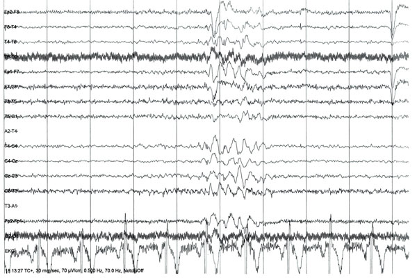
An electroencephalogram (EEG) will also be requested by the specialist,12, 14 although a normal interictal EEG does not exclude epilepsy. Conversely, the EEG may show ictal abnormalities in 0.5% of individuals who do not have epilepsy. Its value in the patient presenting with a first seizure lies in the identification and classification of epilepsy syndromes with characteristic EEG signature patterns. It has prognostic value if performed within the first few weeks after a seizure, as patients with abnormalities on EEG have been found to have a higher risk of recurrence when showing a pattern typical of idiopathic epilepsy.16
Treatment and lifestyle advice
Current guidelines state that the decision to start antiepileptic drug (AED) treatment should be taken by the patient in conjunction with an epilepsy specialist once the diagnosis has been made.12,14 The majority of patients will, in practice, not take up the option of starting an AED after a first symptomatic seizure. The MESS (Multicentre Study of Early Epilepsy and Single Seizure) trial, which compared immediate versus delayed AED treatment in more than 1,000 patients, failed to detect an effect of drug treatment on the course of the disease or the risk of seizure recurrence at five years.17
AEDs are however generally started if there is a history of previous seizures, a congenital neurological disorder, or if the patient finds the risk of recurrence unacceptable. This is influenced by the patient’s occupation and neuroimaging and EEG results.12, 14 Patients may also take into consideration the risk of sudden unexpected death in epilepsy in deciding whether to start an AED, although this risk rises with severity of epilepsy and is, in several studies, only appreciable in patients taking multiple AEDs.18
Every first seizure consultation should also include a discussion of the risks of seizure recurrence,18 fitness to drive, and sensible advice about managing lifestyle risk factors (sleep deprivation, alcohol, recreational and therapeutic drugs) and avoidance of hazardous sports activities (solitary sports, climbing, diving and swimming), and taking a bath while at home alone. For sources of further advice regarding safety at home, see Useful information box, below.
Patients should be offered a follow-up appointment to ensure continuity of epilepsy care. A recent audit showed that 20% of patients referred to a first seizure clinic from the emergency department were lost to follow-up19 and the national sentinel audit on epilepsy-related deaths revealed that only 31% of patients were seen by a specialist within one month, 54% had to wait between one and six months and 15% waited for longer than six months.18
In the meantime, while patients are waiting for their appointment with the specialist, GPs should offer them a follow up consultation, and give appropriate lifestyle advice as mentioned above. If seizures recur, the patient should be reviewed and reassessed. Helpful telephone advice and information on how to refer the patient to a first seizure clinic can usually be obtained from the on-call neurology specialist registrar or the epilepsy specialist nurse at the local neurology unit.
Conclusion
When evaluating a patient with a first seizure an accurate history is key and other potentially life-threatening diagnoses, especially cardiac disease, must be ruled out. Patients who have fully recovered should be directly referred to a first seizure clinic, where further tests and AEDs may be considered. Doctors should provide support and advice to the first seizure patient and be aware of the medical and social consequences of a diagnosis of seizures or epilepsy.
Patients who have not recovered from their seizure, have an abnormal examination, or about whom there is diagnostic doubt, should be referred to the emergency department or specialist services for further investigations including brain imaging and lumbar puncture.
Table 1 Criteria suggestive of seizure or syncope with high specificity and sensitivity5
Figure 1
Management algorithm for the evaluation of adult first seizures
REFERENCES
1 ILAE Commission Report. The epidemiology of the epilepsies: future directions. International League Against Epilepsy. Epilepsia 1997;38(5):614–618
2 Forsgren L, Bucht G, Eriksson S et al. Incidence and clinical characterization of unprovoked seizures in adults: a prospective population-based study. Epilepsia 1996;37(3):224–229
3 Chowdhury FA, Nashef L and Elwes RDC (2008). Misdiagnosis in epilepsy: a review and recognition of diagnostic uncertainty. Eur J Neurology 2008;15:1034–1042
4 Stokes T, Shaw EJ, Juarez-Garcia A et al. Clinical Guidelines and Evidence Review for the Epilepsies: diagnosis and management in adults and children in primary and secondary care. Royal College of General Practitioners. London. 2004
5 Sheldon R, Rose S, Ritchie D et al. Historical criteria that distinguish syncope from seizures. J Am Coll Cardiol 2002;40(1):142–148
6 Bergfeldt L. Differential diagnosis of cardiogenic syncope and seizure disorders. Heart 2003;89(3):353–358
7 Strzelczyk A, Cenusa M, Bauer S et al. Management and long-term outcome in patients presenting with ictal asystole or bradycardia. Epilepsia 2011;52(6):1160–1167
8 Surges R, Taggart P, Sander JW et al. Too long or too short? New insights into abnormal cardiac repolarization in people with chronic epilepsy and its potential role in sudden unexpected death. Epilepsia 2010;51(5):738–744
9 Johnson JN, Hofman N, Haglund CM et al. Identification of a possible pathogenic link between congenital long QT syndrome and epilepsy. Neurology 2009;72(3):224–231
10 Hauser WA, Rich SS, Annegers JF et al. Seizure recurrence after a 1st unprovoked seizure: an extended follow-up. Neurology 1990;40(8):1163–1170
11 Guidelines for epidemiologic studies on epilepsy. Commission on Epidemiology and Prognosis, International League Against Epilepsy. Epilepsia 1993;34(4):592–596
12 National Institute for Clinical Excellence. CG20. The diagnosis and management of the epilepsies in adults and children in primary and secondary care. NICE. London. 2004
13 Benito B, Brugada J, Brugada R et al. Brugada syndrome. Rev Esp Cardiol 2009;62(11):1297–1315
14 Scottish Intercollegiate Guidelines Network. Guideline 70. Diagnosis and Management of Epilepsy in Adults. SIGN. Edinburgh. 2003
15 Guidelines for neuroimaging evaluation of patients with uncontrolled epilepsy considered for surgery. Commission on Neuroimaging of the International League Against Epilepsy. Epilepsia 1998;39(12):1375–1376
16 van Donselaar CA, Schimsheimer RJ, Geerts AT et al. Value of the electroencephalogram in adult patients with untreated idiopathic first seizures. Arch Neurol 1992;49(3):231–237
17 Marson A, Jacoby A, Johnson A et al. Immediate versus deferred antiepileptic drug treatment for early epilepsy and single seizures: a randomised controlled trial. Lancet 2005;365(9476):2007–2013
18 Hanna NJ, Black M, Sander JWS et al. National sentinel clinical audit of epilepsy-related death: epilepsy – death in the shadows. The Stationery Office. London. 2002
19 Bhatt H, Matharu MS, Henderson K et al. An audit of first seizures presenting to an Accident and Emergency department. Seizure 2005;14 (1):58–61
USEFUL INFORMATION
For healthcare professionals
NICE CG20. The epilepsies: the diagnosis and management of the epilepsies in adults and children in primary and secondary care
SIGN Guideline 70. Diagnosis and management of epilepsy in adults
For patients
NICE CG20. Epilepsy in adults and children: information for the public
Epilepsy Action
Epilepsy Society
ARTICLE IN PDF
 Download this article in PDF format