SEARCH

Jabbari E, Orrell R. GPs should be vigilant for Parkinson’s disease. Practitioner September 2021;265(1851):11-15
GPs should be vigilant for Parkinson’s disease
25 Sep 2021
AUTHORS
Dr Edwin Jabbari BSc MBBS MRCP PhD, Specialist Registrar in Neurology and Postdoctoral Clinical Researcher
Dr Richard Orrell MD FRCP FAAN, Associate Professor and Consultant Neurologist, Royal Free Hospital, National Hospital for Neurology and Neurosurgery and UCL Queen Square Institute of Neurology, London, UK
Competing interests: None
Article
Parkinson’s disease is the second most common neurodegenerative disease with an estimated 137,000 people living with the condition in the UK in 2015. Parkinsonism is defined as bradykinesia, in combination with at least one of rest tremor or rigidity. Patients with Parkinson’s disease may present with motor or non-motor symptoms. The prodromal features of Parkinson’s disease are often non-motor and can occur decades before the onset of motor symptoms. The motor signs of Parkinson’s disease typically start after the age of 50 years and are related to the core clinical features of parkinsonism: tremor, bradykinesia, rigidity and postural instability. The tremor of Parkinson’s disease is typically asymmetrical, seen at rest, low frequency (4-6 Hz) and classically described as pill rolling. Parkinson’s disease is a clinical diagnosis. The history should establish the onset, pattern (symmetry, axial vs limb predominant) and progression of symptoms. It should include a family history of dementia and/or parkinsonism, a past medical history of vascular risk factors and a current or previous drug history of antidopaminergic medications. Examination should include gait and posture, decreased blink rate and a lack of spontaneous facial movements. Speech will be hypophonic. Evidence of a resting tremor should be sought and the patient assessed for joint rigidity and bradykinesia. After initial assessment, prompt referral to a neurologist is important to confirm the diagnosis and discuss management. Early involvement of the multidisciplinary team is key.
Parkinson’s disease is the second most common neurodegenerative disease with an estimated 137,000 people living with the condition in the UK in 2015.1 The prevalence increases with age and is approximately 1.5 times higher in men compared with age-matched women.
A GP with an average list size of around 1,500 patients will only see one new case of Parkinson’s disease every 3.3 years.2 However, the global prevalence of the disease is projected to double by 2040, in part due to an ageing population.3
The clinical manifestations of Parkinson’s disease occur after dopaminergic cell loss in the substantia nigra due to the accumulation of aggregated alpha-synuclein into pathological intraneuronal inclusions (Lewy bodies). Other, non-dopaminergic pathologies e.g. vascular, also contribute to the clinical picture.
Disease progression is linked to ongoing deposition of Lewy body pathology and cell loss in the substantia nigra and brainstem, along with the spread of pathology to cortical brain regions.
Presentation
Patients with Parkinson’s disease may present with motor or non-motor symptoms. The prodromal features of Parkinson’s disease are often non-motor and can occur decades before the onset of motor symptoms. The majority of these non-motor symptoms are non-specific, such as depression, constipation and anosmia. However, more specific features such as REM sleep behaviour disorder, characterised by physically acting out vivid and often unpleasant dreams, should raise concern regarding the possibility of underlying Parkinson’s disease.
The motor features of Parkinson’s disease typically start after the age of 50 years and are related to the core clinical features of parkinsonism: tremor, bradykinesia, rigidity and postural instability. The reporting of these symptoms, in combination with their associated signs on examination, often leads to a clinical diagnosis of Parkinson’s disease being made.
The tremor of Parkinson’s disease is typically asymmetrical, seen at rest, low frequency (4-6 Hz) and classically described as pill rolling. In contrast, the presentation of bradykinesia and rigidity tend to be more subtle and non-specific such that patients may complain of stiffness, a frozen shoulder or generalised slowing down. Freezing of gait and falls tend to occur in more advanced Parkinson’s disease.
Examination and assessment
The history from a patient with parkinsonism aims to establish the onset, pattern (symmetry, axial vs limb predominant) and progression of symptoms. It is also important to establish if there is a family history of dementia and/or parkinsonism, a past medical history of vascular risk factors (smoking, diabetes, hypertension etc), or a current/previous drug history of antidopaminergic medications (antipsychotics, metoclopramide, sodium valproate etc).
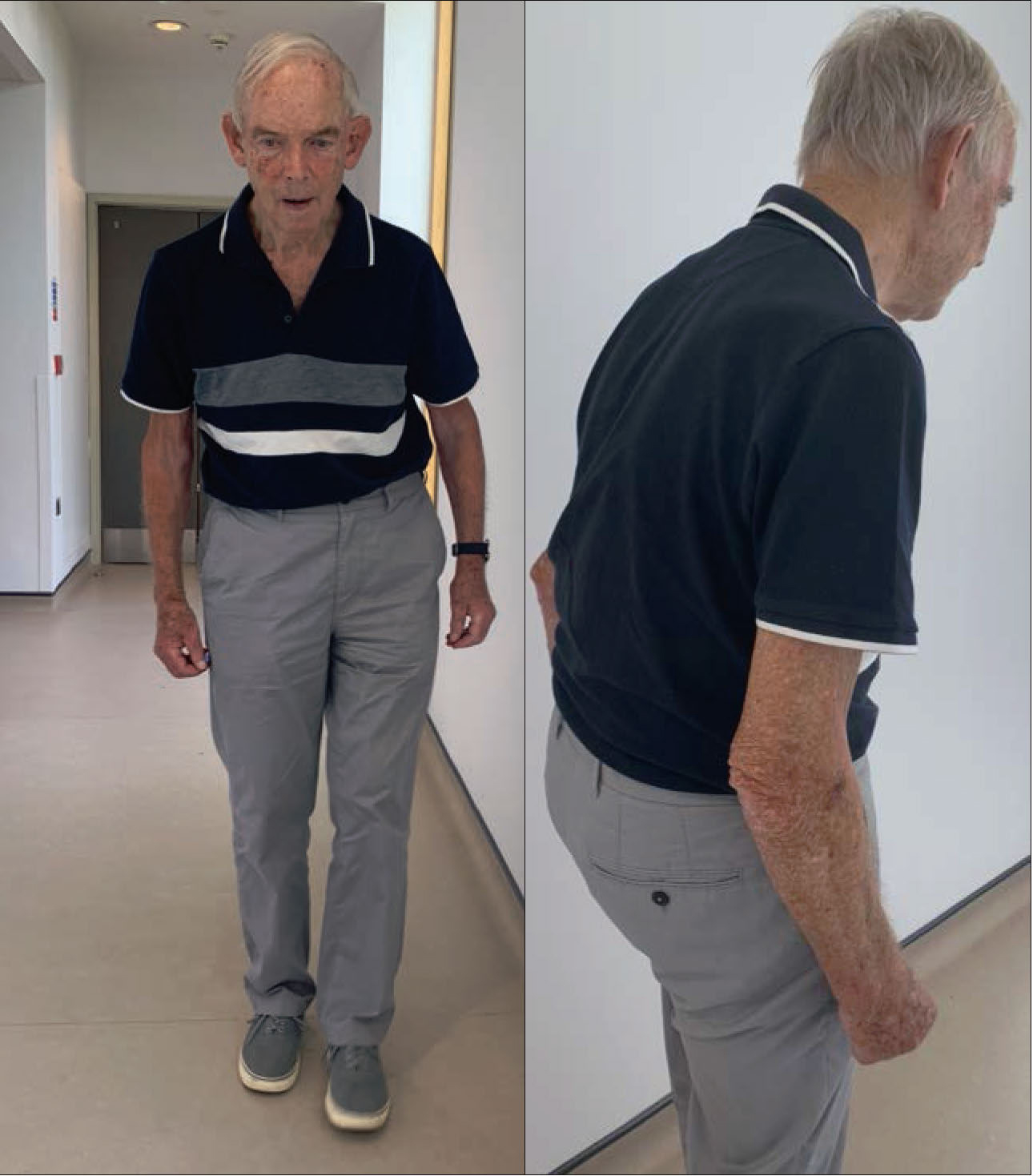
• The examination of a patient with parkinsonism begins when the patient walks into the assessment room by observing the gait. In Parkinson’s disease, a stooped posture and shuffling gait may be seen in established disease, (see figure 1, p11) but this may be more subtle, especially in patients at an early stage.
• Gait freezing may be elicited by asking the patient to walk through a narrow space, such as a doorway.
• Observation of the patient at rest will often reveal facial hypomimia with a decreased blink rate and a lack of spontaneous movements. Speech will be hypophonic.
• Rest tremor may be brought out by distracting the patient by asking them to close their eyes and count backwards.
In Parkinson’s disease, there may be a re-emergent tremor when the patient holds their arms out in front of them.
• Rigidity should be assessed for in all joints but is most easily detected at the wrist and elbow.
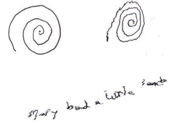
• Bradykinesia, defined as a decrease in amplitude and speed of repetitive movements, may be assessed using finger and foot tapping or pronation-supination hand movements. Asking the patient to write a sentence and draw a spiral is a useful way to elicit micrographia as a manifestation of bradykinesia (see figure 2).
The presence and severity of cognitive dysfunction should be assessed using simple screening tools such as the Montreal Cognitive Assessment (MoCA).
The history and examination should also assess the patient for the presence of any red flag features (see the section below) which may suggest an alternative diagnosis.
Confirming diagnosis
Parkinson’s disease is a clinical diagnosis and therefore relies on early and thorough assessment of patients with suspected parkinsonism. After initial assessment, prompt referral to a neurologist in a general neurology clinic is important to confirm the diagnosis and discuss management.
Onward referral to a movement disorders specialist at a tertiary centre may be indicated for patients who do not respond to initial symptomatic therapies or in cases where there is diagnostic doubt.
Using the International Parkinson and Movement Disorder Society (MDS) diagnostic criteria,4 a diagnosis of ‘clinically established Parkinson’s disease’ requires at least two supportive criteria and no red flag features (see table 1). Post-mortem studies have estimated that the accuracy of an ante-mortem diagnosis of Parkinson’s disease is around 80%.5
Baseline blood tests should be carried out to rule out electrolyte and metabolic causes of tremor, such as hypocalcaemia, hypomagnesaemia and hyperthyroidism. The neurologist may arrange for further blood tests to be carried out in the hospital setting, including copper studies and genetic testing for patients with young onset and/or familial parkinsonism.
Magnetic resonance imaging (MRI) should not be used to diagnose Parkinson’s disease. MRI is indicated if a structural cause of parkinsonism is suspected, if the patient has early onset parkinsonism, or if the patient has rapidly progressive parkinsonism with red flag features of alternative causes of parkinsonism (see below).
Similarly, dopamine transporter (DAT) scans should not be routinely used to diagnose Parkinson’s disease.
As the scan detects dopaminergic degeneration in the basal ganglia, it is most useful when there is diagnostic doubt about whether a patient has Parkinson’s disease or one of the non-degenerative benign causes of parkinsonism such as drug-induced parkinsonism or essential tremor.
Metaiodobenzylguanidine-single photon emission computed tomography (MIBG-SPECT), reflecting postganglionic cardiac autonomic denervation, has emerged as a reliable imaging tool to differentiate Parkinson’s disease (abnormal findings) from multiple system atrophy (normal findings),6 although it is not widely available in clinical practice.
Relatively benign differential diagnoses to consider include vascular parkinsonism, characterised by a predominant lower limb parkinsonism affecting gait in a patient with vascular risk factors; essential or dystonic tremor, characterised by a predominant postural tremor; and drug-induced parkinsonism.
In patients with young onset parkinsonism (age at onset < 50 years), monogenic and treatable causes should be considered including Wilson’s disease.
Important differentials to consider are progressive supranuclear palsy, corticobasal syndrome and multiple system atrophy which tend to have a more rapid rate of disease progression, are less responsive to levodopa and have distinct red flag features in association with parkinsonism, see table 1.
Potential complications and disease progression
Non-motor complications occur secondary to levodopa therapy (postural hypotension) or due to progression of the disease itself and manifest as autonomic dysfunction (urinary urgency/incontinence), pain and cognitive impairment/psychosis.
Motor complications are very common as the disease progresses with motor fluctuations (changes in the ability to move, commonly referred to as ‘on-off’ times) and dyskinesias (involuntary movements) occurring secondary to a less predictable response to levodopa therapy. In the latter stages of disease, parkinsonism tends to become symmetrical, the patient develops dysphagia (putting them at risk of aspiration pneumonia), and postural instability (manifesting as falls) becomes more prominent.
The rate of Parkinson’s disease progression is heterogeneous but is generally considered to be a slower progressing neurodegenerative disease with an estimated 45% survival at 10 years post-diagnosis.7
Certain clinical factors may predict a more rapid rate of decline including early autonomic dysfunction, older age and more severe motor impairment at onset.8
In contrast, Parkinson’s disease dementia occurs in up to 80% of patients with Parkinson’s disease after 20 years of symptomatic disease.9
Management
There are currently no proven disease-modifying therapies for Parkinson’s disease, although there are a number of clinical trials in progress.10 Therefore, the main focus of management is on supportive and symptomatic therapies. The NICE guidelines for the management of Parkinson’s disease were updated in 2017.11
Management can be divided into: information and support; supportive therapies; treatment of motor symptoms (including advanced Parkinson’s disease therapies); treatment of non-motor symptoms.
Information and support
Communication with Parkinson’s disease patients, both for the initial delivery of diagnosis and on subsequent follow-up, should be clear, honest and realistic. Patients should be empowered to participate in shared decision-making about their patient-centred care.
Patients should have an accessible point of contact via a hospital or community-based Parkinson’s disease nurse specialist, and this is usually arranged via the general neurology clinic. This is especially important with regard to future discussions about the progression of disease, potential medication side-effects and available support services (personal care, financial support etc). For further information for patients, including patient participation in research, see Useful information box.
Support should be provided for patients who are still working, including the provision of a letter to the employer if the patient’s job role needs to be modified. Patients should also be told about the need to inform the DVLA and insurers about their diagnosis.
Supportive therapies
Early involvement of the hospital and/or community based multidisciplinary team (MDT) is an important aspect of management. This includes:
• A Parkinson’s disease nurse specialist as an accessible point of contact
• Physiotherapy for balance problems and the provision of walking aids
• Occupational therapy for patients requiring home modifications because of difficulties with activities of daily living
• Speech and language therapy for patients who are experiencing difficulties with communication, swallowing or saliva
• Dietitians for advice on a protein redistribution diet, especially for Parkinson’s disease patients on levodopa who experience motor fluctuations
• Palliative care for advance care planning, including discussions about DNACPR, lasting power of attorney and advance decisions to refuse treatment such as artificial feeding.
Treatment of motor symptoms
NICE guidelines suggest the use of oral levodopa in patients in the early stages of Parkinson’s disease whose motor symptoms impact on their quality of life.11 This is on the basis that there is no evidence to suggest any benefit in delaying levodopa therapy,12 and levodopa has a more favourable risk-benefit profile in comparison with oral levodopa-sparing therapies such as dopamine agonists (DA), monoamine oxidase B (MAO-B) inhibitors and catechol-O-methyltransferase (COMT) inhibitors.13
The two most used immediate release formulations of levodopa are Sinemet and Madopar. The starting dose of both medications is 62.5 mg three or four times a day, with each dose consisting of 50 mg levodopa and 12.5 mg of a dopa-decarboxylase inhibitor to reduce peripheral dopaminergic side-effects such as nausea and vomiting. The dose is usually increased by 62.5 mg every 3-4 days until a good clinical response has been achieved (usual total levodopa maintenance dose of 400-800 mg/day). Domperidone may be coprescribed to reduce the risk of GI side effects occurring.
Adjunctive therapies that aim to boost dopaminergic activity (DA) and reduce the metabolism of dopamine (MAO-B inhibitors and COMT inhibitors) are used to treat motor fluctuations such as wearing-off and delayed on-periods. These fluctuations tend to occur after the initial honeymoon period when patients have an excellent response to levodopa.
If swallowing becomes impaired while on oral treatment, then a neurologist can oversee the conversion to non-oral treatments such as topical rotigotine (alongside considering advanced Parkinson’s disease therapies – see below).
Amantadine is the main oral treatment for levodopa-induced dyskinesias. Recently licensed adjunctive oral therapies such as opicapone and safinamide may have local prescribing restrictions.
Although these adjunctive therapies are usually initiated at secondary/tertiary care level, GPs have an active role in screening for associated complications, in particular dyskinesias, hallucinations and impulse control disorders. The identification of these complications should prompt urgent referral to the patient’s assigned consultant for appropriate management. Established treatments should never be stopped abruptly as this can cause severe morbidity or even death.
Advanced Parkinson’s disease therapies are reserved for patients who experience debilitating off-time or dyskinesias despite optimised oral adjunctive therapies, and include subcutaneous apomorphine, levodopa-carbidopa intestinal gel via jejunostomy, and deep brain stimulation.14 The implementation of such therapies, after an MDT discussion of potential benefits and inclusion/exclusion criteria, is usually coordinated by a movement disorders specialist at a tertiary centre.
Treatment of non-motor symptoms
Non-motor symptoms are a significant source of morbidity, and the early identification and treatment of these symptoms is important.
Many of the non-motor features are non-dopaminergic in origin and so the drug classes used are diverse. These treatments based on current NICE guidelines are summarised in table 2.
Conclusion
Parkinson’s disease is a common neurodegenerative disorder that GPs will encounter, and the prevalence of this condition is increasing. The initial presentation is heterogeneous, and a detailed assessment to establish the progression of motor and non-motor features is key to establishing an early and accurate diagnosis. Initial and subsequent clinical assessments must also include the exclusion of alternative causes of parkinsonism.
Parkinson’s disease is a clinical diagnosis, therefore MRI and DAT scans should be reserved for when there is diagnostic doubt.
As Parkinson’s disease progresses, the identification and management of late-stage features must be considered including cognitive impairment and falls.
Although clinical trials are ongoing, there are no proven disease-modifying therapies. Therefore, management focuses on supportive and symptomatic treatments, delivered through a good working relationship between the patient, GP, nurse specialist and neurologist.
Symptomatic therapies should be considered for both motor and non-motor features which are impacting on quality of life. GPs should be aware of the potential side-effects of these medications and refer for specialist input when indicated. Most importantly, involvement of the MDT, including physiotherapy and speech and language therapy, is vital in optimising the independence of Parkinson’s disease patients.
REFERENCES
1 Parkinson's UK. The incidence and prevalence of Parkinson's in the UK report. Parkinson's UK. 2017
2 Scottish Intercollegiate Guidelines Network. SIGN 113. Diagnosis and pharmacological management of Parkinson’s disease. SIGN. Edinburgh. 2010 www.sign.ac.uk
3 Dorsey ER, Sherer T, Okun MS, Bloem BR. The emerging evidence of the parkinson pandemic. J Parkinsons Dis 2018;8:S3-8
4 Postuma RB, Berg D, Stern M et al. MDS clinical diagnostic criteria for Parkinson's disease. Mov Disord 2015;30:1591-1601
5 Rizzo G, Copetti M, Arcuti S et al. Accuracy of clinical diagnosis of Parkinson disease: a systematic review and meta-analysis. Neurology 2016;86:566-76
6 Braune S, Reinhardt M, Schnitzer R et al. Cardiac uptake of [123I]MIBG separates Parkinson's disease from multiple system atrophy. Neurology 1999;53:1020-25
7 Williams-Gray CH, Mason SL, Evans JR et al. The CamPaIGN study of Parkinson's disease: 10-year outlook in an incident population-based cohort. J Neurol Neurosurg Psychiatry 2013;84:1258-64
8 De Pablo-Fernandez E, Lees AJ, Holton JL, Warner TT. Prognosis and neuropathologic correlation of clinical subtypes of Parkinson disease. JAMA Neurol 2019;76:470-79
9 Hely MA, Reid WG, Adena MA et al. The Sydney multicenter study of Parkinson's disease: the inevitability of dementia at 20 years. Mov Disord 2008;23:837-44
10 McFarthing K, Buff S, Rafaloff G et al. Parkinson’s disease drug therapies in the clinical trial pipeline: 2020. J Parkinsons Dis 2020;10:757-74
11 National Institute for Health and Care Excellence. NG71. Parkinson's disease in adults: diagnosis and management. NICE. London. 2017
12 Verschuur CVM, Suwijn SR, Boel JA et al. Randomized delayed-start trial of levodopa in Parkinson's disease. N Engl J Med 2019;380:315-24
13 PD MED Collaborative Group, Gray R, Ives N et al. Long-term effectiveness of dopamine agonists and monoamine oxidase B inhibitors compared with levodopa as initial treatment for Parkinson's disease (PD MED): a large, open-label, pragmatic randomised trial. Lancet 2014;384:1196-1205
14 Worth PF. When the going gets tough: how to select patients with Parkinson's disease for advanced therapies. Pract Neurol 2013;13:140-52
ARTICLE IN PDF
 Download this article in PDF format
EXTERNAL WEBLINKS
For healthcare professionals - Parkinsons.org.uk
A free one-hour course for GPs and nurses which gives guidance on how to examine, assess and refer patients where Parkinson’s disease is suspected