SEARCH

Mbizvo GK, Derry C. Diagnosis and management of sleep-related epilepsy in adults. Practitioner Sept 2018;262(1818):11-16
Diagnosis and management of sleep-related epilepsy in adults
24 Sep 2018
AUTHORS
Dr Gashirai K Mbizvo MBChB (Hons) MRes (Dis) MRCP, Clinical Research Fellow, Centre for Clinical Brain Sciences and Muir Maxwell Epilepsy Centre, University of Edinburgh, Edinburgh, UK
Dr Chris Derry BSc MB BS MRCP PhD, Consultant Neurologist, Department of Clinical Neurosciences and Sleep Medicine, Centre for Clinical Brain Sciences, University of Edinburgh, Edinburgh, UK
Competing interests: None
Article
Abstract
Epilepsy usually presents with seizures occurring during both wakefulness and sleep. However, there are some epilepsies in which seizures appear mainly or exclusively during sleep. Nocturnal epilepsies account for 10-15% of all epilepsies, and 80% of nocturnal epilepsies in adults are focal. They present a diagnostic challenge as they can be difficult to differentiate from normal movements and behaviour during sleep and also from several non-epileptic, sleep-related, motor and behavioural disorders. Sleep-related hypermotor epilepsy (SHE) is often idiopathic, although there can be a positive family history in up to 40% of cases, and the genetic form shows an autosomal dominant inheritance pattern. Although the median age of onset is 14 years, this type of epilepsy can occur at any age. More than 90% of the seizures in SHE occur during sleep. Seizures occur during the N2 stage of NREM sleep so are particularly likely to occur within 30 min of falling asleep (even during daytime naps), and shortly before waking. Seizures in SHE are simple partial seizures which easily wake the patient so that they will usually be able to recall the seizures, often describing auras of somatic sensations or feeling unable to breathe. SHE seizures have a rapid onset and offset, a short duration (usually < 2 min), and a stereotyped motor pattern for that individual. Several semi-purposeful hyperkinetic movements may be seen, including bipedal or bimanual movements (running, cycling, or kicking), and axial movements (rocking, sitting or twisting). SHE is characterised by a high frequency of attacks (3-8 events or more per night), and occurrence in clusters.
Epilepsy usually presents with seizures occurring during both wakefulness and sleep. When seizures occur exclusively or predominantly (more than 90%) during sleep, this is termed nocturnal epilepsy.1
Nocturnal epilepsies account for 10-15% of all epilepsies,2 and 80% of nocturnal epilepsies in adults are focal.3 They present a diagnostic challenge as they can be difficult to differentiate from normal movements and behaviour during sleep and also from several non-epileptic, sleep-related, motor and behavioural disorders.
This article largely focuses on sleep-related hypermotor epilepsy (SHE), previously called nocturnal frontal lobe epilepsy (NFLE), as this is not an uncommon presentation of nocturnal epilepsy yet it is often misdiagnosed as a non-epileptic sleep disorder, such as a parasomnia.4,5 Nocturnal temporal, parietal, and occipital lobe epilepsies (NTLE, NPLE, and NOLE respectively) are extremely rare and tend to overlap with SHE.2 Generalised nocturnal epilepsies are likely to cause fewer diagnostic difficulties because there is greater recognition of the clinical characteristics of generalised seizures.4
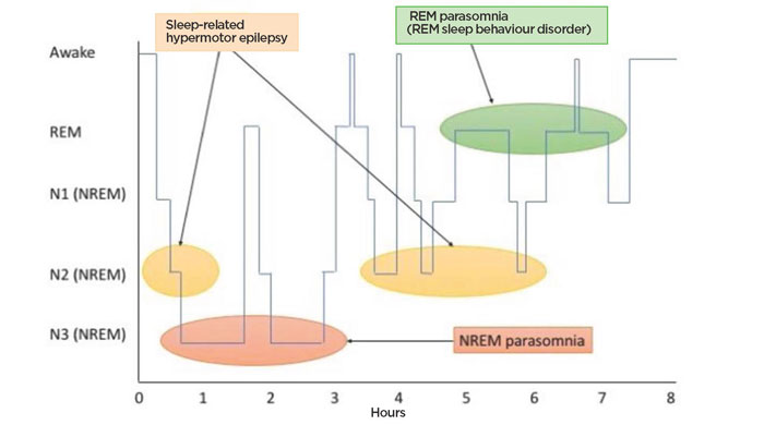
Figure 1 provides an overview of normal sleep physiology, which helps contextualise nocturnal epilepsy and its differential diagnoses.4
Epidemiology
More than 90% of the seizures in SHE occur during sleep. The term NFLE was replaced by SHE as these seizures occur during sleep regardless of the time of day, they are often extrafrontal originating in regions such as the temporal lobe (epileptogenic zone) and subsequently activate frontal lobe networks (symptomatic zone). Moreover, their motor activity is more excessive than in other types of frontal lobe epilepsy.2,6 SHE seizures occur during N2 of NREM sleep. As shown in figure 1, above, not only does N2 take up the majority of total sleep time, but it is also particularly prevalent within the first 30 minutes of sleep or shortly before waking. SHE seizures follow these timelines, meaning that patients present with seizures throughout the night, but particularly within 30 minutes of going to sleep (even during daytime naps), and shortly before waking.4
Seizures in SHE are simple partial seizures, and therefore they involve no loss of consciousness. They may be preceded by an aura. As they occur at an N2 depth of sleep, they wake the patient up. Patients usually recall the seizures, describing auras of somatic sensations or feeling unable to breathe.4
The estimated prevalence of SHE is 1.8 per 100,000.7 The exact prevalence of SHE is difficult to determine because there are few epidemiological studies and the condition presents diagnostic difficulty.4 However, there is a consensus among specialists that SHE is probably not that rare, but often underestimated as a result of misdiagnosis.4,5
Although the median age of onset is 14 years, SHE can occur at any age.
It is often idiopathic, although there can be a positive family history in up to 40% of cases, and the genetic form shows an autosomal dominant inheritance pattern.4
Clinical history
When faced with a patient presenting with sleep-related motor and behavioural complaints, it is important to take a detailed history from the patient and also from a witness (normally a partner or family member). Assessment should address the following key questions:
1 Diagnosis: Is this epilepsy? If it is not epilepsy, are the movements and behaviour abnormal? As a general principle, epileptic seizures are brief, generally lasting no more than 2-3 minutes, and stereotyped. These principles hold true for nocturnal seizures. Certain seizures will be easier to recognise from sleep.
2 Safety: Does the patient pose a risk to self or others as a result of the sleep-related motor or behavioural complaint?
3 Triggers: Are there modifiable trigger factors? Is the presentation indicative of serious or treatable underlying pathology?
Differential diagnoses
Generalised epilepsies
Patients can have generalised tonic-clonic seizures (GTCS) while awake or asleep. In GTCS normally the muscles in the arms and legs become stiff, the patient becomes cyanosed, and there is rhythmic jerking and twitching. There may also be associated seizure markers including lateral biting of the tongue, incontinence, and myalgia. Juvenile myoclonic epilepsy is a genetic generalised epilepsy characterised by frequent myoclonic jerks in the morning, usually within the first hour of waking up, and GTCS. It generally begins in early adolescence3,5 and is a lifelong condition.8
Temporal lobe epilepsy
Temporal lobe epilepsy (TLE) is common and patients may experience these seizures during both wakefulness and sleep. The characteristics of temporal lobe seizures are a rising epigastric feeling, déjà vu or jamais vu, olfactory hallucinations (e.g. metallic taste), auditory hallucinations, and automatisms (e.g. hand fidgeting, chewing, or lip smacking).2 The presence of events while the patient is awake usually enables accurate clinical diagnosis of the sleep-related attacks in TLE.4
Sleep-related hypermotor epilepsy
The semiology of frontal lobe seizures in SHE is extremely variable between patients, although stereotyped for the individual. Common features are a rapid onset and offset, a short duration for each seizure (usually < 2 minutes), a high frequency of attacks throughout the night (3-8), and occurrence in clusters (e.g. several nights of seizures followed by weeks of remission). We have already described the aura and preserved consciousness.
Additionally, several semi-purposeful hyperkinetic movements may be seen, including bipedal or bimanual movements (running, cycling, or kicking), and axial movements (rocking, sitting or twisting). There can be prominent repetitive vocalisations that include unintelligible shouting or swearing. Tonic posturing is seen and includes head turning to the side of the seizure activity and fencing postures (contralateral shoulder abduction, external rotation, and elbow flexion). Additionally, patients tend to experience brief fragments of epileptic events that look like periods of arousal without dramatic motor features throughout NREM sleep.4 This is not an exhaustive list of the possible movements and behaviour seen in SHE. These events are often so bizarre and dramatic that they are frequently misdiagnosed as parasomnias or as non-epileptic attacks.
Parasomnias
In parasomnias complex motor or behavioural events occur during sleep in association with abnormal intrusions of one stage of sleep or wakefulness into another.6 They are divided into NREM parasomnias and REM parasomnias.
Table 1 summarises the NREM parasomnias, of which sleepwalking, confusional arousals, and sleep terrors are the most common. However, these conditions present as a spectrum and they often result in patients injuring themselves. They arise from a failure to make the transition between wakefulness and N3 of NREM sleep appropriately, meaning that certain parts of the brain are ‘awake’ while other parts are ‘asleep’.4 This may help explain the profound complexity of the behaviours seen, and the patient’s ability to interact strongly with the environment while retaining a near total lack of recall.
The association with N3 NREM sleep means that parasomnias tend to occur during the first third of sleep, normally around one to two hours after falling asleep. While they are very common in children, they occur in only 4% of adults.6
REM sleep behaviour disorder (RBD) is the most common REM parasomnia.6Figure 1 highlights atonia of the voluntary muscles during REM sleep, the stage in which vivid dreaming occurs.
In the absence of that atonia, patients act out their dreams, normally in violent ways such as punching, kicking, or falling out of bed. Standing and walking is uncommon, but injury is not.
Patients recall the dreams if awoken by them, and the content is usually unpleasant. The events last seconds or minutes, usually in the final third of sleep. Age of onset for RBD is normally over 50 years and 80% of sufferers are men.
RBD is a strong prodromal marker for neurodegenerative disease, particularly the synucleinopathies (Parkinson’s disease, dementia with Lewy bodies, and multiple system atrophy). More than 80% of patients with RBD develop degenerative disease when followed up for more than 15 years.4,6
Trying to differentiate between SHE and parasomnias can be very difficult.4Table 2 summarises seven key questions that should help focus the history to try to determine whether the presentation is SHE or a parasomnia.
The Frontal Lobe Epilepsy and Parasomnias (FLEP) Scale should also be used to help guide the history and differentiate between SHE and parasomnias. This is shown in table 3, p14.9 A score below zero favours parasomnias, 0-3 is inconclusive, and >3 favours SHE. The tool has positive and negative predictive values of 91-100% but should always be used in conjunction with a detailed clinical history.4
A complete history should aim to identify trigger factors and conditions that might be associated with SHE or parasomnias. Patients with SHE or parasomnias may complain of excessive daytime sleepiness resulting from interrupted sleep. Sleep deprivation is a trigger factor for seizures and parasomnias as is alcohol and/or drug abuse.
It is important to enquire about recent febrile illness and stress, which can trigger parasomnias. There is a high prevalence of intellectual disabilities and psychiatric or behavioural problems in families with SHE.5
Nocturnal dissociative seizures
Dissociative seizures (pseudoseizures) can occur when patients are awake and during pseudosleep, where they appear asleep to the observer but the EEG demonstrates an awake pattern of electrical activity.
More than half (58%) of patients with non-epileptic attack disorder (NEAD) report some events during sleep in addition to those when awake. Isolated nocturnal dissociative seizures are rare.4 A history of events exclusively during sleep is more suggestive of epilepsy.10
Periodic limb movements of sleep
These movements involve recurrent, stereotyped, extension of the hallux and flexion of the ankle plus or minus the knee and hip. They last 0.5-5 seconds and occur at predictable intervals, every 5-90 seconds for a duration of minutes to hours. They are often a feature of restless legs syndrome.4,6
Obstructive sleep apnoea
Obstructive sleep apnoea (OSA) presents with excessive daytime sleepiness as a result of disordered sleep due to prolonged and recurrent apnoeic events throughout sleep. It generally occurs in patients who are overweight.
It can be a trigger factor for parasomnias, therefore it is important to enquire about night-time snoring and daytime sleepiness. OSA itself can also result in abnormal arousals that look similar to those from the fragmented epileptic events in SHE.4
Nocturnal panic attacks
Patients with panic disorders are at risk of nocturnal panic attacks. These present with awakening with intense fear. The patient is often unable to return to sleep for prolonged periods. There is retained awareness of events. Isolated nocturnal panic attacks are rare, so the presence of daytime panic disorder gives a clue to the diagnosis. There is also a close association with NEAD.4
Hypnic jerks (sleep starts)
These are a common part of normal sleep and consist of brief jerks of a limb or the whole body during the transition between wakefulness and sleep.
There can be a hallucinatory sensation of falling. Trigger factors include sleep deprivation, excessive caffeine, and stimulant intake.2,4
Investigations
Most patients with sleep-related motor and behavioural complaints will have normal clinical examination findings.
The examination should include looking for signs of injury, trigger factors (e.g. OSA), and underlying disease (such as focal neurology or Parkinsonism). Many of the sleep-related motor and behavioural disorders are diagnoses that can be made on clinical grounds, and often investigation results will be normal. It is therefore important to rationalise the use of these investigations.
EEG
Half of patients with SHE have a normal EEG when awake and asleep.4 This is because seizures often originate in deeper parts of the brain not easily traced by surface EEG electrodes, and because epileptiform changes are often masked by the prominent movement and muscle artefact resulting from complex frontal seizure behaviours.
The EEG in parasomnia is also normal. Therefore, routine EEG does not normally advance the diagnosis of sleep-related motor and behavioural complaints. It is, however, worth ordering because in the 50% of cases where the trace does show epileptiform activity, further investigation is not required and the patient can commence treatment.
If a routine EEG is non-diagnostic, patients can undergo video telemetry.
There is likely to be a long waiting list for this investigation as even large specialist centres in the UK normally only have a handful of telemetry beds and there are only around 30-40 telemetry units.11
The investigation also involves an inpatient stay of one to two weeks, during which the EEG may still fail to detect epileptiform activity.4
Figure 2 shows a diagnostic algorithm that can be used as an aid in capturing SHE or parasomnias using video observation. This is accurate in 94% of cases.4
MRI
An MRI should be ordered because there is a small chance (14%) that it may identify a treatable local epileptogenic abnormality in SHE. However, the majority of MRI scan results in SHE and parasomnias will be normal.4
Polysomnography
Polysomnography is the gold standard investigation, but it is not easily accessible in the UK. There are fewer than 90 centres offering the investigation, and long waiting times.12
It combines EEG with various other investigations including eye movement, respiratory airflow, electromyography, and video.2 It is particularly helpful when trying to identify the co-occurrence of trigger factors such as OSA. The EEG may still fail to identify epileptiform discharges, for the reasons mentioned earlier.
Home video
Patients and partners should be encouraged to try and record events at home if possible, e.g. using a smart phone.
Management
Nocturnal epilepsy syndromes tend to respond well to antiepileptic drug therapy and the avoidance of seizure triggers. All patients should receive counselling about seizure precautions, complications, and driving safety.
Drugs
There is a paucity of evidence on specific treatments for nocturnal epilepsies. As a general principle, they are treated using the same range of drugs as focal and generalised awake seizures.13 Lamotrigine and carbamazepine are usually good options for focal epilepsy.
Lamotrigine tends to be better tolerated, although patients should be warned that they should stop the drug immediately if they develop a rash.
This tends to occur less frequently if dosing is gradually increased. Lamotrigine is normally started at a dose of 25 mg once daily for 14 days, increased to 50 mg once daily for a further 14 days, then increased in steps of up to 100 mg every 7-14 days. The usual maintenance dose is 100-200 mg daily, in 1-2 divided doses.14
Levetiracetam also works well for focal epilepsies, although there is some evidence that its efficacy is less than that of lamotrigine (when both are compared with carbamazepine).15 However, there is a lack of head-to-head trials.
Other options include oxcarbazepine and topiramate.13 There is emerging evidence that lacosamide may be effective in nocturnal focal epilepsy, with up to 65% of patients with drug-resistant seizures achieving ≥ 50% reduction in seizure frequency.16
Chronopharmacology describes the use of differential drug dosing times to reduce seizures at peak times, while keeping the total daily dose the same. For nocturnal seizures, this involves increasing the evening dose and reducing the morning dose. It may provide some benefit, although the evidence is limited.13
Clonazepam is a broad spectrum benzodiazepine that may work well for parasomnias and the other non-epileptic disorders of movement and behaviour. The typical dose is 0.5-1 mg at night.2
Surgery
The use of epilepsy surgery in SHE is rare, as surgery is reserved for drug-resistant cases with lesional aetiology. A recent study demonstrated that in 80% of patients with lesional nocturnal epilepsy, 76% of patients achieved freedom from seizures.17
Trigger management
Several studies have shown that managing OSA with continuous positive airway pressure helps reduce seizures and parasomnias. Insomnias can be managed with melatonin, although the effect of melatonin on seizure frequency is unclear.13
Safety
Patients and families should be advised to take seizure precautions including moving the mattress onto the floor, removing obstructing furniture, timing seizures and learning to use buccal midazolam or call 999 after 3-5 minutes during a seizure, and training in first aid and cardiopulmonary resuscitation.13
A patient must not drive and must notify the DVLA following a seizure during sleep. Relicensing may be granted if, over the course of at least one year from the date of the sleep seizure, the patient establishes a history or pattern of seizures occurring only ever while asleep. Alternatively, regardless of preceding seizure history, if a person establishes a pattern of asleep seizures only (all seizures had onset during sleep), starting at least three years prior to licence application and there have been no other unprovoked seizures during those three years, a licence may be issued.18
Complications
As a whole, epilepsy remains a major concern for premature death.19,20 Individuals with epilepsy are 20-40 times more likely to die a sudden death than those without epilepsy.13 This risk of sudden unexpected death in epilepsy (SUDEP) remains the same for SHE as for other epilepsies.21 One might have expected SUDEP risk to be even higher in SHE than the other epilepsies given the daily occurrence of seizures during sleep in this condition and the fact that nocturnal seizures independently increase the risk of SUDEP. However, GTCS also independently increase the risk of SUDEP, and SHE is associated with a low occurrence of these seizures.22 Other SUDEP risk factors include sleeping in the prone position, and a chronic history of epilepsy. Factors that can reduce risk of SUDEP include adequate seizure control, nocturnal supervision (e.g. sharing a bedroom), and epilepsy alarms.13
Conclusion
The diagnosis of nocturnal epilepsy, parasomnias and other non-epileptic abnormal movement and behaviour disorders in sleep largely remains clinical and a careful history is the most important diagnostic step. Observation of events by video may also be helpful.
Drug management of the nocturnal epilepsies is similar to that for awake epilepsy, and there should also be a focus on dealing with trigger factors such as OSA and other triggers of poor sleep. Nocturnal epilepsy can be a risk factor for SUDEP.
REFERENCES
1 Thomas RH, King WH, Johnston JA et al. Awake seizures after pure sleep-related epilepsy: a systematic review and implications for driving law. J Neurol Neurosurg Psychiatry 2010;81(2):130-35
2 St Louis EK, Foldvary-Schaefer N. Sleep-related epilepsy syndromes. UpToDate 2018. www.uptodate.com/contents/sleep-related-epilepsy-syndromes [Last accessed Sep 3 2018)
3 Yaqub BA, Waheed G, Kabiraj MM. Nocturnal epilepsies in adults. Seizure 1997;6(2):145-49
4 Derry CP. Sleeping in fits and starts: a practical guide to distinguishing nocturnal epilepsy from sleep disorders. Pract Neurol 2014;14(6):391-98
5 Nobili L, Proserpio P, Combi R et al. Nocturnal frontal lobe epilepsy. Curr Neurol Neurosci Rep 2014;14(2):424
6 Breen DP, Hogl B, Fasano A et al. Sleep-related motor and behavioral disorders: Recent advances and new entities. Mov Disord 2018;33(7):1042-55
7 Vignatelli L, Bisulli F, Giovannini G et al. Prevalence of nocturnal frontal lobe epilepsy in the adult population of Bologna and Modena, Emilia-Romagna region, Italy. Sleep 2015;38(3):479-85
8 Korff CM. Juvenile myoclonic epilepsy. UpToDate 2018. www.uptodate.com/contents/juvenile-myoclonic-epilepsy (Last accessed Sep 3 2018)
9 Derry CP, Davey M, Johns M et al. Distinguishing sleep disorders from seizures: diagnosing bumps in the night. Arch Neurol 2006;63(5):705-09
10 Duncan R, Oto M, Russell AJ et al. Pseudosleep events in patients with psychogenic non-epileptic seizures: prevalence and associations. J Neurol Neurosurg Psychiatry 2004;75(7):1009-12
11 Kandler R, Lai M, Ponnusamy A et al. The safety of UK video telemetry units: results of a national service evaluation. Seizure 2013;22(10):872-76
12 Flemons WW, Douglas NJ, Kuna ST et al. Access to diagnosis and treatment of patients with suspected sleep apnea. Am J Respir Crit Care Med 2004;169(6):668-72
13 Carreno M, Fernandez S. Sleep-Related Epilepsy. Curr Treat Options Neurol 2016;18(5):23
14 Joint Formulary Committee. British National Formulary. Lamotrigine. BMJ Group and Pharmaceutical Press. 2018
15 Iniesta I. Carbamazepine in pregnancy: Levetiracetam and lamotrigine are better options. BMJ 2011;342:d279
16 Garcia-Morales I, Delgado RT, Falip M et al. Early clinical experience with lacosamide as adjunctive therapy in patients with refractory focal epilepsy and nocturnal seizures. Seizure 2011;20(10):801-04
17 Losurdo A, Proserpio P, Cardinale F et al. Drug-resistant focal sleep related epilepsy: results and predictors of surgical outcome. Epilepsy Res 2014;108(5):953-62
18 Driver and Vehicle Licensing Agency. Neurological disorders: assessing fitness to drive - Advice for medical professionals to follow when assessing drivers with neurological disorders. 2018. www.gov.uk/guidance/neurological-disorders-assessing-fitness-to-drive (Last accessed 3 Sep 2018)
19 Thurman DJ, Logroscino G, Beghi E et al. The burden of premature mortality of epilepsy in high-income countries: A systematic review from the Mortality Task Force of the International League Against Epilepsy. Epilepsia 2017;58(1):17-26
20 Levira F, Thurman DJ, Sander JW et al. Premature mortality of epilepsy in low- and middle-income countries: A systematic review from the Mortality Task Force of the International League Against Epilepsy. Epilepsia 2017;58(1):6-16
21 Mostacci B, Bisulli F, Vignatelli L et al. Incidence of sudden unexpected death in nocturnal frontal lobe epilepsy: a cohort study. Sleep Med 2015;16(2):232-36
22 Tinuper P, Bisulli F, Cross JH et al. Definition and diagnostic criteria of sleep-related hypermotor epilepsy. Neurology 2016;86(19):1834-42
ARTICLE IN PDF
 Download this article in PDF format
EXTERNAL WEBLINKS
For healthcare professionals
SIGN 143. Diagnosis and management of epilepsy in adults
For patients