SEARCH

Practitioner 2011; 255 (1746):17-20
Diagnosis and treatment of gout in primary care
16 Dec 2011
AUTHORS
Dr Chadi Rakieh MB ChB MRCP Specialist Registrar in Rheumatology, Leeds Teaching Hospitals NHS Trust
Professor Philip G Conaghan MB BS PhD FRACP FRCP Professor of Musculoskeletal Medicine, NIHR Leeds Musculoskeletal Biomedical Research Unit and Division of Musculoskeletal Disease, Leeds Institute of Molecular Medicine, University of Leeds
Article
Gout is the most common form of inflammatory arthritis in men over 40, and affects up to 1-2 % of adults in developed countries.1 It is characterised by recurrent episodes of joint inflammation caused by the deposition of monosodium urate (MSU) crystals in the joints as a consequence of raised serum uric acid levels.
Gout usually starts as infrequent monoarthritis but can evolve to become a debilitating chronic polyarthritis if not treated properly. Gout is largely managed in primary care in the UK with only atypical presentations and difficult management cases referred to secondary care.
Risk factors
The prevalence of gout increases with age and up to 7% of men aged over 65 and 3% of women aged over 85 have gout.2, 3 The risk of gout increases significantly with increasing serum uric acid levels. However, hyperuricaemia is far more common than gout implying that additional risk factors play a role in the development of the disease.4
Alcohol consumption, especially beer and to a lesser extent spirits, increases the risk of both incident and prevalent gout. In one study, alcohol use was associated with a three-fold higher risk of incident gout in women and a two-fold higher risk in men. 5 Purine-rich foods such as red meat and seafood increase the risk of incident gout significantly,6 while dairy products 6 and increasing coffee, but not tea, intake7 were found to be protective.
A number of medications are well known to be associated with increased risk of gout especially diuretics, including loop and thiazide diuretics. Cyclosporin was also found to increase the risk of incident gout in patients with renal transplants.5
It is well established that gout is frequently associated with the metabolic syndrome which is characterised by hyperuricaemia, hyperlipidaemia, hypertension, diabetes and insulin resistance, and obesity.8
In addition, dehydration, increasing creatinine levels, and undergoing surgery are known to precipitate gouty flares.4
Finally, gout is extremely rare in premenopausal women, but the risk of gout in women is increased after the menopause, especially in cases of early menopause.9 Risk factors for gout are listed in table 1.
Presentation
Acute gout manifests as severe joint pain of rapid onset reaching maximal intensity within a few hours. Gout has a predilection for lower extremity joints. It often starts at the first metatarsophalangeal joint; this condition is termed podagra.
Other common sites of gouty flares include:
• tarsal and subtalar joints
• ankle
• knee
• wrist
• small joints of the hands
• Achilles tendon
• olecranon bursae
The joint affected is usually hot, red, swollen, very painful, and commonly associated with skin erythema. Systemic symptoms including fatigue and low-grade fever are not uncommon. Septic arthritis is the main differential for monoarthritis and a needle aspiration of synovial fluid should be performed whenever in doubt. Needle aspiration of easily accessible joints such as the knee or olecranon bursa could be attempted by GPs, while cases involving more difficult sites such as the wrist or ankle should be referred to a rheumatology or orthopaedics department to consider joint aspiration.
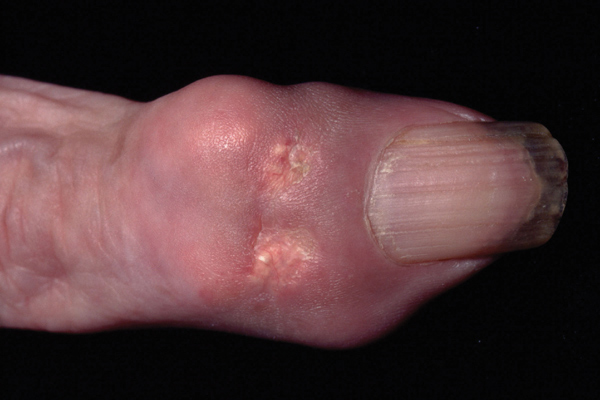
Early on during the disease, gouty flares are usually infrequent with long intercritical asymptomatic periods. At later stages, as the disease progresses without treatment, gouty attacks tend to become more frequent, may be oligo- or polyarticular, and associated with tophi formation (palpable urate nodules usually located near joints).
Diagnosis
Identification of MSU crystals in the synovial fluid of an inflamed joint or from tophi allows a definite diagnosis of gout to be made.10 MSU crystals appear as rod-like negative birefringent crystals under the polarised microscope. Hyperuricaemia does not confirm or exclude gout as the majority of people with hyperuricaemia are asymptomatic, while serum uric acid levels tend to decrease during acute attacks.
The EULAR guidelines state that for a typical presentation of gout, such as recurrent podagra associated with hyperuricaemia, gout can be reasonably diagnosed on clinical grounds, although not definitely without crystal confirmation.10
Radiologic findings including the characteristic punched-out erosive lesions are usually absent until late in the evolution of the disease. In addition, they lack specificity which limits their role in the diagnosis of gout.10
Atypical presentations of gout, such as absence of podagra, normal serum uric acid, oligo- or polyarticular gout, gout in women, and gout in unusual locations, pose a diagnostic challenge and may require a rheumatology opinion. Care should be taken in diagnosing a new onset inflammatory arthritis in someone who is known to have gout, when the likelihood is that of another gouty flare.
Treatment of acute gout
Non-pharmacological treatment
Joint rest and local application of ice packs can help to reduce pain and inflammation during acute gouty attacks.11
Pharmacological treatment
NSAIDs: The British Society for Rheumatology (BSR) recommends that short-acting NSAIDs should be used at maximal dose as the first drug of choice if not contraindicated.12 In patients at risk of gastrointestinal complications, co-prescription of a proton pump inhibitor or the use of COX-2 selective agents should be considered. Examples of NSAIDs used in acute gout include:
• diclofenac 150 mg daily in divided doses
• indometacin 150 mg daily in divided doses
• naproxen 1,000 mg daily in divided doses
• etoricoxib 120 mg in a single daily dose
Colchicine: Colchicine can be an effective alternative in patients with contraindications to NSAIDs. The BSR recommends using 0.5 mg two to four times daily, with cautious dose escalation to reduce side effects, especially nausea and diarrhoea.12 Colchicine can be particularly useful in patients with heart failure in whom NSAIDs are contraindicated but should be avoided in patients with severe renal impairment.
Corticosteroids: Joint aspiration and injection of intra-articular steroids is one of the most effective ways of treating acute monoarthritic gout. Examples of common doses of intra-articular steroids would be 80 mg methylprednisolone or triamcinolone for a large joint such as the knee, or 40 mg methylprednisolone or triamcinolone for a smaller joint, such as the elbow or wrist.13 Alternatively, a short course of oral steroids, such as 30-40 mg daily for two to four days with rapid tapering off, can be used. Intramuscular steroids, such as methylprednisolone 80-120 mg can also offer rapid pain relief, and are commonly co-prescribed with uric acid lowering drugs to prevent acute flares of gout on initiation of allopurinol.
Prophylaxis of recurrent attacks
The objective of uric acid lowering therapy is to reduce serum urate levels to a level where new crystal formation does not occur and existing crystals will dissolve, thereby eliminating gouty attacks.8
There is no general agreement on the optimal time to initiate uric acid lowering therapy. In general, it is initiated if a patient suffers two or more attacks in one year12 especially if lifestyle modifications and elimination of risk factors fail to control gout. Many rheumatologists will start uric acid lowering therapy in hyperuricaemic patients whose first attack is very severe or polyarticular gout. In both these cases, patients are at increased risk of subsequent attacks. Uric acid lowering therapy should also be initiated in patients with chronic gout arthropathy, tophi, radiological lesions, and uric acid renal calculi.
The BSR guidelines recommend that serum uric acid level should be reduced below 300 µmol/L.12 Lower levels of serum uric acid result in faster disappearance of the crystals.10
Non-pharmacological measures
Patients should be advised to lose weight when appropriate and do regular exercise. Patients should restrict their intake of alcohol (especially beer), and drink plenty of water. They should also limit their intake of purine-rich food such as red meat, offal and seafood. Loop and thiazide diuretics should be avoided if possible, especially when the indication is hypertension (where alternatives exist) rather than heart failure. Patient education is a very important part of the management of gout and written information such as the Arthritis Research UK Gout booklet (see Useful information box, p20) should be provided to patients.11
Pharmacological treatment
The initiation of urate lowering therapy should be delayed for one to two weeks after an acute gout attack has settled13 and co-administration of low-dose colchicine or NSAIDs must be initiated to reduce the risk of acute gouty flares that may be triggered by the reduction of serum uric acid. The optimal duration of prophylaxis with colchicine and NSAIDs is unknown but should be for a few weeks at least and up to six months for colchicine.8
Allopurinol, a competitive inhibitor of xanthine oxidase, is the main available drug to reduce serum uric acid.16 It is inexpensive and for most patients is a safe and effective option.8 It should be started at a low dose of 50-100 mg daily and then increased in 100 mg increments every few weeks until the therapeutic target of uric acid is reached (< 300 µmol/L). The most common dose in the UK is 300 mg daily, although the dose can be escalated up to 900 mg daily if tolerated.11 Allopurinol is usually well tolerated but can be associated with side effects, especially rash (in up to 5% of cases)14 and gastrointestinal symptoms mainly diarrhoea. Hypersensitivity syndrome is a rare but serious adverse effect of allopurinol. It presents as a Stevens-Johnson-like syndrome consisting of desquamating rash, fever, hepatitis, eosinophilia, and deteriorating renal failure. Allopurinol toxicity is more common in patients with impaired renal function or in older patients, so dose reduction is essential in these groups.
Febuxostat is a new non-purine selective xanthine oxidase inhibitor that has been approved by NICE for the treatment of hyperuricaemia in gout patients who are intolerant of allopurinol.15 Most of febuxostat metabolism occurs in the liver and therefore dose modification is not needed in patients with mild to moderate renal failure.16 However, its use in patients with severe renal impairment, ischaemic heart disease, and heart failure is not currently recommended.11
Febuxostat 80 mg and 120 mg daily were found to be superior to allopurinol 300 mg daily in reducing serum uric acid levels in a controlled study.17 Following initiation, allopurinol and febuxostat should not be stopped during gouty flares.
Finally, a number of uricosuric drugs such as probenecid and sulfinpyrazone can be considered when allopurinol or febuxostat are poorly tolerated. These drugs are usually initiated in secondary care. They work by increasing the renal clearance of uric acid and, therefore, should be given with caution in patients with uric acid renal calculi.
Conclusion
Gout is the the main cause of inflammatory monoarthritis. It is quite common and affects up to 2% of the adult population. Raised serum uric acid is the main metabolic drive that leads to gout. However, hyperuricaemia is much more common than gout and other risk factors do play a role in the pathogenesis of gout including excessive alcohol consumption and increased intake of purine-rich foods. Gout is also known to be associated with chronic comorbidities, in particular the metabolic syndrome.
A definite diagnosis of gout can be made by identifying urate crystals in the synovial fluid of an inflamed joint or bursa. However, it is not uncommon to make the diagnosis of gout on clinical grounds, especially with a typical presentation e.g. podagra.
Effective therapies are available to treat acute gouty flares including NSAIDs, colchicine, and local or systemic steroids. Prevention of gout requires lowering urate levels. Allopurinol is the main uric acid lowering drug and should be started with a cover of NSAIDs, colchicine, or steroids. The dose should be titrated up slowly by 100 mg increments every few weeks until serum
urate is reduced to below 300 µmol/L. Febuxostat, 80 mg or 120 mg daily, is an alternative that has been approved by NICE for gout patients with intolerance to allopurinol.
All patients should be advised to lose weight, limit their intake of alcohol and purine-rich foods, and take regular exercise to reduce the risk of gout and the metabolic syndrome.
REFERENCES
1 Wallace KL, Riedel AA, Joseph-Ridge N, Wortmann R. Increasing prevalence of gout and hyperuricemia over 10 years among older adults in a managed care population. J Rheumatol 2004; 31(8):1582-1587
2 Lawrence RC, Helmick CG, Arnett FC et al. Estimates of the prevalence of arthritis and selected musculoskeletal disorders in the United States. Arthritis Rheum 1998; 41(5):778-799
3 Mikuls TR, Farrar JT, Bilker WB et al. Gout epidemiology: results from the UK General Practice Research Database, 1990-1999. Ann Rheum Dis 2005; 64(2):267-272
4 Singh JA, Reddy SG, Kundukulam J. Risk factors for gout and prevention: a systematic review of the literature. Curr Opin Rheumatol 2011; 23(2):192-202
5 Bhole V, de Vera M, Rahman MM et al. Epidemiology of gout in women: Fifty-two-year followup of a prospective cohort. Arthritis Rheum 2010; 62(4):1069-1076
6 Choi HK, Atkinson K, Karlson EW et al. Purine-rich foods, dairy and protein intake, and the risk of gout in men. N Engl J Med 2004; 350(11):1093-1103
7 Choi HK, Willett W, Curhan G. Coffee consumption and risk of incident gout in men: a prospective study. Arthritis Rheum 2007; 56(6):2049-2055
8 Zhang W, Doherty M, Bardin T et al. EULAR evidence based recommendations for gout. Part II: Management. Report of a task force of the EULAR Standing Committee for International Clinical Studies Including Therapeutics (ESCISIT). Ann Rheum Dis 2006; 65(10):1312-1324
9 Hak AE, Curhan GC, Grodstein F, Choi HK. Menopause, postmenopausal hormone use and risk of incident gout. Ann Rheum Dis 2010; 69(7):1305-1309
10 Zhang W, Doherty M, Pascual E et al. EULAR evidence based recommendations for gout. Part I: Diagnosis. Report of a task force of the Standing Committee for International Clinical Studies Including Therapeutics (ESCISIT). Ann Rheum Dis 2006; 65(10):1301-1311
11 Roddy E, Mallen CD. Gout. Management in primary care. BMJ 2011; 342:d117
12 Jordan KM, Cameron JS, Snaith M et al. British Society for Rheumatology and British Health Professionals in Rheumatology guideline for the management of gout. Rheumatology (Oxford) 2007; 46(8):1372-1374
13 Wenham C, Conaghan P. Treating gout: a clinical update. Managing pain in practice 2010; 1(2):4-6
14 Schlesinger N. Management of acute and chronic gouty arthritis: present state-of-the-art. Drugs 2004; 64(21):2399-2416
15 National Institute for Health and Clinical Excellence TA164. Febuxostat for the management of hyperuricaemia in people with gout. NICE. London. 2008
16 Burns CM, Wortmann RL. Gout therapeutics: new drugs for an old disease. Lancet 2011; 377(9760):165-177
17 Becker MA, Schumacher HR, Jr, Wortmann RL et al. Febuxostat compared with allopurinol in patients with hyperuricemia and gout. N Engl J Med 2005; 353(23):2450-2461
ARTICLE IN PDF
 Download this article in PDF format
EXTERNAL WEBLINKS
NICE Evidence
PubMed