SEARCH

Stonier T, Simson N, Challacombe B. Diagnosing testicular lumps in primary care. Practitioner 2017;261(1803):13-17
Diagnosing testicular lumps in primary care
24 Apr 2017
AUTHORS
Dr Tom Stonier BSc (Hons) MBChB, Urology CT1, Department of Urology, Princess Alexandra Hospital, Harlow, UK
Dr Nick Simson BMBS (Hons) BMedSci (Hons), Urology CT1, Department of Urology, Princess Alexandra Hospital, Harlow, UK
Mr Ben Challacombe MS FRCS(Urol), Consultant Urological Surgeon, Department of Urology, Guy’s & St Thomas’, NHS Foundation Trust, London, UK
Article
Abstract
Although the incidence of testicular cancer has increased over the past few decades, testicular tumours are still rare and many GPs will only see one or two new diagnoses in their career. In one UK study, out of 845 patients who had been referred with testicular lumps or pain, only 33 (4%) were diagnosed with testicular cancer. Epididymal cysts, or spermatoceles when containing sperm, were the most common finding, and were found in 228 patients (27%). The second most common finding was hydrocele, a fluid collection between the parietal and visceral layers of the tunica vaginalis, which was found in 96 patients (11%). The vast majority (95-98%) of testicular cancers are germ cell tumours. In Western Europe, five-year survival for testicular cancer is approximately 95%. It is almost 100% for stage 1 seminomas, but falls to 48% in the poorest prognostic group – non-seminomatous tumours with metastases at diagnosis.When examining scrotal swellings, the key question is whether the lump is intra- or extra-testicular, as palpable intra-testicular lesions are highly likely (around 90%) to be malignant, whereas those lying outside the testis are usually benign. NICE recommends that men with non-painful enlargement or change in shape or texture of the testis should be referred via the two-week wait cancer pathway. Any painful or tender mass within the body of the testis which is not suggestive of infection should also be referred. GPs should refer patients for an urgent ultrasound if they are aged 20-40 with a hydrocele; if there is uncertainty as to whether the mass is intra-testicular or extra-testicular; or if the testis cannot be fully palpated.
Testicular lumps provide a diagnostic challenge. Although each potential diagnosis has distinctive features the underlying differential of a testicular malignancy can cause concern for patient and GP alike. Although the incidence of testicular cancer has increased over the past few decades,1,2 testicular tumours are still rare, diagnosed in only 4% of men undergoing scrotal ultrasound in one study.3 Many GPs will only see one or two new diagnoses in their career.
Furthermore, there has been an improvement in the time to diagnosis and subsequently treatment thanks to raised public awareness.4 The potential differential diagnoses are wide ranging, with epididymal cysts and hydroceles the most common findings.3,5
Causes
It is important that a testicular torsion is immediately excluded. The classical history is of acute pain coupled with an exquisitely tender testicle, abnormal lie, nausea/vomiting and an absent cremaster reflex, that will progress to a swollen testis as venous drainage is prevented.
In a UK study of 845 men undergoing ultrasound for scrotal swelling, epididymal cysts, or spermatoceles when containing sperm, were the most common finding, occurring in 27% of patients.3 Epididymal cysts arise from dilatation of the efferent ductules at the head of the epididymis. Spermatoceles are common after vasectomy as the vas deferens is obstructed but only cause symptoms in 3-5% of patients. Most episodes resolve with conservative treatment, but < 1% require surgical treatment.6 Benign cysts of the tunica albuginea can also occur and are rarely symptomatic or require treatment.
The other most common finding, hydrocele, is a fluid collection between parietal and visceral layers of the tunica vaginalis.5,7 Hydroceles may be congenital in which case the process vaginalis remains patent forming a communication with the peritoneal cavity, this usually closes spontaneously when the testis descends into the scrotum and most are closed by the age of two.8 In around 12-14% of patients, the process vaginalis persists into adulthood and may predispose them to both hydroceles and hernias.9,10 Acquired hydroceles may occur any time during adulthood and can be idiopathic/primary or secondary to infection, systemic disease, trauma, and rarely tumour or inguinal surgery.11
A third common diagnosis is a varicocele, an abnormal dilatation of the pampiniform venous plexus and internal spermatic vein, reportedly found in 8-22% of healthy males.12,13 They are most commonly found on the left side, probably because of the anatomy of the left gonadal vein and its angular drainage into the left renal vein.14 Akbay and colleagues found 89.7% of cases to be unilateral on the left side, 10.8% bilateral, and 0.38% on the right side.12
Testicular cancer is rare, accounting for only 1% of all male malignancies and 5% of urological tumours.15 However, it is still the most commonly diagnosed cancer in males ages 15-40.16 The typical histology of a testicular tumour is germ cell in 95-98% of cases.17,18 These tumours can be subdivided into seminomas, most common in the fourth decade, and non-seminomatous tumours more common in the third decade. Patients can be reassured that cure rates from this cancer are approximately 95% in Western Europe.18 Five-year survival is close to 100% for stage 1 seminomas, and falls to 48% in the poorest prognostic group – non-seminoma with metastases at diagnosis.18 The cure rates have improved dramatically in the past 30 years since the introduction of cisplatin-based combination chemotherapy.19
Inguinal hernias occur when part of the intra-abdominal tissue (bowel or omentum) protrudes through the abdominal wall in the region of the inguinal canal. The most common type is an indirect inguinal hernia in which the contents of the hernia follow the path of the spermatic cord, through the internal and external spermatic ring, and in some cases this may descend into the scrotum causing a lump. Classically the clinician cannot get above the lump on examination.
Other than torsion, a tender, testicular swelling might suggest epididymo-orchitis. In younger patients this is most commonly caused by an STI, while in older patients it is more likely to be caused by a urinary tract pathogen often secondary to instrumentation or an underlying abnormality (such as an enlarged prostate).20 The varying underlying aetiology explains the bimodal distribution seen in one retrospective study in Israel in which it was most frequently diagnosed in men aged 16-30 and 51-70.21
History and examination
A routine history should be taken. It is important to ascertain how long the testicular lump has been present. Associated symptoms such as a pulling or dragging sensation or pain should be noted, to understand how much functional bother the patient is suffering. Any signs of infection, tenderness, fever and discharge may point towards a diagnosis of epididymo-orchitis.
Although a painless testicular mass (within the body of the testis itself) is pathognomonic of testicular cancer, this was the presenting complaint in only half the patients in a Spanish registry study, and scrotal pain was the initial symptom in 20% of cases.22,23
Rarer signs and symptoms such as gynaecomastia and back pain have been reported as the presenting sign in 7% and 11% of testicular malignancies respectively and so should be asked about.23 A history of cryptorchidism/ undescended testis increases the risk by up to eight times, giving a lifetime risk of 4%.24,25 Occasionally minor trauma to a testis containing a tumour results in an acute presentation with a haematoma around the mass.
Other than a family history of testicular cancer in a first-degree relative or previous cancer in the contralateral testis, risk factors are rare and unlikely to guide the diagnostic process.17,26,27 Patients with ‘testicular dysgenesis syndrome’ (cryptorchidism, hypospadias, decreased spermatogenesis diagnosed by infertility) are also at risk of testicular cancer, but this is rare.28
Examination of the scrotum should be performed with the patient standing, which will exacerbate hernias, hydroceles and varicoceles, and lying. Diagnosis in testicular swellings can be made in a stepwise manner, as shown in box 1. The key question is whether the lump is intra- or extra-testicular, as palpable intra-testicular lesions are highly likely (around 90%) to be malignant, while those lying outside the testis are much more likely to be benign.29,30
The use of a pen torch will give good information on whether a lump is solid or fluid filled, such as a hydrocele which will transilluminate brilliantly.
Lymph node examination should be performed in all patients with suspected testicular tumour. Importantly these neoplasms spread most commonly to the para-aortic lymph nodes, which may be hard to palpate in the abdomen unless large.31,32 Inguinal lymphadenopathy meanwhile may provide evidence of an infective process of the scrotum.
The classic presentation for each diagnosis is shown in table 1. However variations of textbook definitions should be considered, and importantly it should be remembered that an underlying tumour may be obscured by another pathology.
For example an overlying hydrocele may obscure complete palpation of the testis and so should not provide reassurance.33
Investigation
The key message is that if a testicular tumour is suspected then no investigation should delay a two-week wait cancer referral.34
Urinalysis should be performed in patients with suspected epididymo-orchitis, in order to diagnose an underlying urinary tract infection. If positive, urine microscopy, culture and sensitivity testing should be carried out.20
Measuring testicular tumour markers, alpha-fetoprotein and human chorionic gonadotrophin, at the time of referral can be considered, but should not delay referral according to the Royal College of Surgeons commissioning guide.34 It should also be noted that these are only raised in 50% of patients with testicular cancer and so negative values should not be used as reassurance.22,35
An ultrasound of the testes is the gold standard investigation for any scrotal swelling, with sensitivity for a testicular tumour close to 100%.36 Patients who meet the criteria for a two-week wait referral should not be delayed by waiting for an ultrasound scan but should be referred directly.34 Urgent ultrasound, as arranged by the GP, is warranted in patients:
• Aged 20-40 years with a hydrocele
• Where there is uncertainty if the mass is intra-testicular or extra-testicular
• Where the testis cannot be fully palpated34
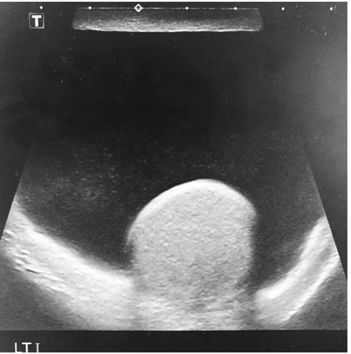
All extratesticular scrotal swellings of uncertain origin should have a routine ultrasound.34 Again an ultrasound is highly sensitive for the other differential diagnoses, such as hydrocele as shown in figure 1.
It is a traditionally held view that the finding of a varicocele should lead to a renal ultrasound in order to exclude a renal tumour.13 However, a systematic review found only 2.3% of cases of renal tumour presented with a varicocele, and concluded that this typically occurs at a late stage at which point a retroperitoneal tumour would likely have manifest itself in other ways.13 NICE still recommends further investigation with ultrasound of an acute varicocele in men over 40.37
At the two-week wait clinic patients will have serum tumour markers, testicular ultrasound followed by CT chest, abdomen and pelvis. Sperm banking should be offered if a positive diagnosis is found.18 Inguinal/radical orchidectomy should occur on the next available operating list.
Referral
Any suspicion of testicular torsion warrants immediate review by a senior urologist as surgery is required within four to six hours of symptom onset.38
NICE recommends that all patients with a non-painful enlargement or change in shape or texture of the testis be referred via the two-week wait cancer pathway to a urologist.39 Reassuringly for patients the conversion rates for these referrals are still low. Any painful or tender mass within the body of the testis which is not suggestive of infection should also be referred. Kumaraswamy and colleagues found that only 8% of 241 men referred for suspected testicular cancer under the two-week rule had sonographic findings suggestive of testicular cancer.40
Proven asymptomatic hydroceles, epididymal cysts, spermatoceles and varicoceles with no underlying tumour (on ultrasound) do not necessitate referral to a urologist.34 Patients in whom these are causing functional symptoms or discomfort should be referred routinely.
Clearly any evidence of a strangulated or obstructed inguinal hernia warrants an emergency referral to a general surgeon.41,42 Irreducible or partially reducible inguinal hernias warrant urgent referral to a general surgery outpatient clinic, otherwise reducible asymptomatic hernias can be referred routinely.41,42 The Royal College of Surgeons commissioning guide suggests diagnostic imaging should not be arranged at a primary care level.43
Treatment
Treatment for testicular cancer involves inguinal (radical) orchidectomy, sometimes followed by chemotherapy,44 and in more advanced cases subsequent lymph node dissection.18 Patients commonly ask about impact on fertility and should be counselled that these treatments can affect fertility to varying extents but semen analysis and cryopreservation should be offered prior to treatment.18
Once an underlying testicular tumour is excluded hydrocele, epididymal cysts and spermatoceles can often be managed conservatively. Symptomatic and unsightly cysts (usually when larger than the testis itself) are excised through a scrotal incision. Aspiration and sclerotherapy, despite fewer initial complications, are less successful with lower patient satisfaction and generally only considered in patients unfit for surgery.45 Hydroceles are managed by opening the layers of the tunica vaginalis to drain the fluid, followed by either excision of the tunica sac, plication of the sac (Lord’s procedure) or eversion of the sac (Jaboulay’s procedure).46
Varicoceles should again be managed with reassurance where possible.
In those with functional/pain problems embolisation or ligation can be considered for symptomatic treatment. Around 20% of patients will have fertility problems,47 and although a meta-analysis by Baazeem and colleagues found intervention improved sperm parameters such as count and motility, there was no improvement in pregnancy rates and so it is not currently recommended by NICE in order to improve fertility.48-50
Conclusion
Testicular swellings provide a diagnostic challenge and one that can often be resolved within primary care.
A testicular lump may be a source of great anxiety for the patient, and more often than not these fears can be allayed by a thorough examination and if necessary a timely ultrasound.
REFERENCES
1 Jemal A, Siegel R, Ward E et al. Cancer statistics, 2009. CA Cancer J Clin 2009;59(4):225-49
2 Huyghe E, Matsuda T, Thonneau P. Increasing incidence of testicular cancer worldwide: a review. J Urol 2003;170 (1):5-11
3 Moore JA, O'Neil C, Fawcett D. A one-stop clinic for men with testicular anxiety. Ann R Coll Surg Engl 2009; 91(1):23-4
4 Moore RA, Topping A. Young men's knowledge of testicular cancer and testicular self-examination: a lost opportunity? Eur J Cancer Care 1999;8(3):137-42
5 Micallef M, Torreggiani WC, Hurley M et al. The ultrasound investigation of scrotal swelling. Int J STD AIDS 2000;11(5):297-302
6 McDonald SW. Vasectomy review: sequelae in the human epididymis and ductus deferens. Clin Anat 1996;9(5):337-42
7 Kapur P, Caty MG, Glick PL. Pediatric hernias and hydroceles. Pediatr Clin North Am 1998;45(4):773-89
8 Skoog SJ. Benign and malignant pediatric scrotal masses. Pediatr Clin North Am 1997;44(5):1229-50
9 van Wessem KJ, Simons MP, Plaisier PW, Lange JF. The etiology of indirect inguinal hernias: congenital and/or acquired? Hernia 2003;7(2):76-9
10 van Veen RN, van Wessem KJ, Halm JA et al. Patent processus vaginalis in the adult as a risk factor for the occurrence of indirect inguinal hernia. Surg Endosc 2007;21(2):202-5
11 Parks K, Leung L. Recurrent hydrocoele. J Fam Med Prim Care 2013;2(1):109-10
12 Akbay E, Cayan S, Doruk E et al. The prevalence of varicocele and varicocele-related testicular atrophy in Turkish children and adolescents. BJU Int 2000;86(4):490-3
13 El-Saeity NS, Sidhu PS. "Scrotal varicocele, exclude a renal tumour". Is this evidence based? Clin Radiol 2006;61(7):593-9
14 Rubenstein RA, Dogra VS, Seftel AD, Resnick MI. Benign intrascrotal lesions. J Urol 2004;171(5):1765-72
15 Purdue MP, Devesa SS, Sigurdson AJ, McGlynn KA. International patterns and trends in testis cancer incidence. Int J Cancer 2005;115(5):822-27
16 Chia VM, Quraishi SM, Devesa SS et al. International trends in the incidence of testicular cancer, 1973-2002. Cancer Epidemiol Biomarkers Prev 2010;19(5):1151-19
17 Bosl GJ, Motzer RJ. Testicular germ-cell cancer. N Engl J Med 1997;337(4):242-53
18 Albers P, Albrecht W, Algaba F et al. Guidelines on testicular cancer: 2015 Update. Eur Urol 2015;68(6):1054-68
19 Feuer EJ, Frey CM, Brawley OW et al. After a treatment breakthrough: a comparison of trial and population-based data for advanced testicular cancer. J Clin Oncol 1994;12(2):368-77
20 Walker NA, Challacombe B. Managing epididymo-orchitis in general practice. Practitioner 2013;257(1760):21-5
21 Kaver I, Matzkin H, Braf ZF. Epididymo-orchitis: a retrospective study of 121 patients. J Fam Pract 1990;30(5):548-52
22 Germà-Lluch JR, Garcia del Muro X, Maroto P et al. Clinical pattern and therapeutic results achieved in 1490 patients with germ-cell tumours of the testis: the experience of the Spanish Germ-Cell Cancer Group (GG). Eur Urol 2002;42(6):553-63
23 Moul JW. Timely diagnosis of testicular cancer. Urol Clin North Am 2007;34(2):109-17; abstract vii.
24 Pettersson A, Richiardi L, Nordenskjold A et al. Age at surgery for undescended testis and risk of testicular cancer. N Engl J Med 2007;356(18):1835-41
25 Thorup J, McLachlan R, Cortes D et al. What is new in cryptorchidism and hypospadias-a critical review on the testicular dysgenesis hypothesis. J Pediatr Surg 2010;45(10):2074-86
26 Osterlind A, Berthelsen JG, Abildgaard N et al. Risk of bilateral testicular germ cell cancer in Denmark: 1960-1984. J Natl Cancer Inst 1991;83(19):1391-95
27 Dieckmann KP, Pichlmeier U. The prevalence of familial testicular cancer: an analysis of two patient populations and a review of the literature. Cancer 1997;80(10):1954-60
28 Jorgensen N, Rajpert-De Meyts E, Main KM, Skakkebaek NE. Testicular dysgenesis syndrome comprises some but not all cases of hypospadias and impaired spermatogenesis. Int J Androl 2010;33(2):298-303
29 Beccia DJ, Krane RJ, Olsson CA. Clinical management of non-testicular intrascrotal tumors. J Urol 1976;116(4):476-79
30 Woodward PJ, Schwab CM, Sesterhenn IA. From the archives of the AFIP: extratesticular scrotal masses: radiologic-pathologic correlation. Radiographics 2003;23(1):215-40
31 Donohue JP, Zachary JM, Maynard BR. Distribution of nodal metastases in nonseminomatous testis cancer. J Urol 1982;128(2):315-20
32 Ray B, Hajdu SI, Whitmore WF Jr. Proceedings: Distribution of retroperitoneal lymph node metastases in testicular germinal tumors. Cancer 1974;33(2):340-48
33 Roy CR, Peterson NE. Positive hydrocele cytology accompanying testis seminoma. Urology 1992;39(3):292-93
34 Royal College of Surgeons (RCS) and British Association of urological surgeons (BAUS). Commissioning guide: Asymptomatic scrotal swelling. October 2013 (Reviewed November 2016). www.baus.org.uk/patients/surgical_outcomes/radical_prostatectomy/default.aspx [Last accessed 3 April 2017]
35 Wanderas EH, Tretli S, Fossa SD. Trends in incidence of testicular cancer in Norway 1955-1992. Eur J Cancer 1995;31a(12):2044-48
36 Kim W, Rosen MA, Langer JE et al. US MR imaging correlation in pathologic conditions of the scrotum. Radiographics 2007;27(5):1239-53
37 National Institute for Health and Care Excellence. Clinical Knowledge Statement: Varicocele management. October 2012. https://cks.nice.org.uk/varicocele#!scenario [Last accessed 3rd April 2017]
38 Kallerhoff M, Gross AJ, Botefur IC et al. The influence of temperature on changes in pH, lactate and morphology during testicular ischaemia. Br J Urol 1996;78(3):440-45
39 National Institute for Health and Care Excellence. NG12. Suspected cancer: recognition and referral. NICE. London. 2015. www.nice.org.uk/guidance/ng12 [Last accessed 3 April 2017]
40 Kumaraswamy P, Cox R, O'Rourke JS, Willis RG. Audit of two-week rule referrals for suspected testicular cancer in Cornwall, 2003-2005. Ann R Coll Surg Engl 2009;91(3):239-44
41 Bay-Nielsen M, Kehlet H, Strand L et al. Quality assessment of 26,304 herniorrhaphies in Denmark: a prospective nationwide study. Lancet 2001;358(9288):1124-28
42 Nilsson H, Stylianidis G, Haapamaki M et al. Mortality after groin hernia surgery. Ann Surg 2007;245(4):656-60
43 Royal College of Surgeons (RCS) and British Hernia Society. Commissioning guide: Groin hernia. October 2013 (Update November 2016)
44 Masters JR, Koberle B. Curing metastatic cancer: lessons from testicular germ-cell tumours. Nat Rev Cancer 2003;3(7):517-25
45 Beiko DT, Kim D, Morales A. Aspiration and sclerotherapy versus hydrocelectomy for treatment of hydroceles. Urology 2003;61(4):708-12
46 Ku JH, Kim ME, Lee NK, Park YH. The excisional, plication and internal drainage techniques: a comparison of the results for idiopathic hydrocele. BJU Int 2001;87(1):82-84
47 The influence of varicocele on parameters of fertility in a large group of men presenting to infertility clinics. World Health Organization. Fertil Steril 1992;57(6):1289-93
48 Baazeem A, Belzile E, Ciampi A et al. Varicocele and male factor infertility treatment: a new meta-analysis and review of the role of varicocele repair. Eur Urol 2011;60(4):796-808
49 Nork JJ, Berger JH, Crain DS, Christman MS. Youth varicocele and varicocele treatment: a meta-analysis of semen outcomes. Fertil Steril 2014;102(2):381-7 e6
50 National Institute for Health and Care Excellence. CG156. Fertility problems: assessment and treatment. NICE. London. 2013 (Updated August 2016) www.nice.org.uk/guidance/CG156 [Last accessed 3 April 2017]
ARTICLE IN PDF
 Download this article in PDF format
PRESET SEARCHES
PubMed
NICE evidence search
US National Cancer Institute
EXTERNAL WEBLINKS
British Association of Urological Surgeons
The Urology Foundation