SEARCH

Kirby R. Optimising the management of early prostate cancer. The Practitioner 2014;258 (1770):15-18
Optimising the management of early prostate cancer (2014)
22 Apr 2014
AUTHORS
Professor Roger Kirby MA MS MD FRCS (Urol), Professor of Urology, Medical Director, The Prostate Centre, London, UK
Article
One in every eight men in the UK will be diagnosed with prostate cancer during their lifetime, making it the most common solid tumour to afflict middle- and older-aged men.
Since the risk of prostate cancer is so strongly age-related, and the population of the UK is growing more elderly, the prevalence of this disease, which is also linked to a Western lifestyle, seems certain to rise.
Other risk factors for prostate cancer include an Afro-Caribbean ethnic origin and a positive family history. The more first-degree relatives affected the greater the risk of developing the disease. More than 70 familial prostate cancer susceptibility genes, including the important breast cancer gene BRCA2, have now been identified.1
Most currently diagnosed prostate cancers are described as early, that is to say that they have not spread beyond the capsule of the gland itself. The dilemma then is whether or not a given patient requires treatment, and if so, which modality of therapy is optimum.
Assessment
A suspicion of a diagnosis of prostate cancer is usually based on either induration or nodularity of the prostate on digital rectal examination or, more commonly, a rise in serum prostate specific antigen (PSA) level. The usual cut-off point for PSA is taken as 4 ng/ml, but in men below 65, a value of more than 2.5 ng/ml should raise suspicion.
A progressive rise in PSA over time may also indicate the possibility of the presence of a cancer within the prostate.
Any man in whom there is a clinical suspicion of early prostate cancer should be referred for urgent urological assessment and investigation. Delayed or missed diagnosis of cancer constitutes one of the most common causes of complaints or litigation against GPs.
Investigations
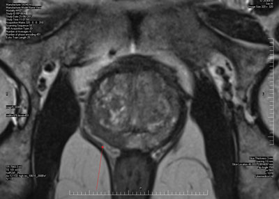
Recently the methodology for the investigation and diagnosis of early prostate cancer has changed significantly. Instead of proceeding immediately with transrectal ultrasound-guided (TRUS) prostate biopsies, increasingly urologists are requesting multiparametric magnetic resonance imaging (MpMRI) of the gland before deciding whether or not a biopsy is indicated. 2
The rationale for this is that if the MpMRI looks blameless a biopsy may be avoided. Conversely, if there is a convincing abnormality seen on the scan, this suspicious area can be targeted, see figure 1.
MpMRI essentially evaluates the motion of water molecules within tissues, when subjected to a powerful external magnetic field, as well as tumour neovascularity. Diffusion weighted images and apparent diffusion coefficient images are able to evaluate disordered tissue cellularity and its effect on water movement, and, along with dynamic contrast enhanced images are capable of identifying tumour within the prostate. Furthermore, the more aggressive the tumours are, the more marked are the changes. As a consequence small, clinically insignificant cancers do not show up on MpMRI.
There is also a current preference for using the transperineal (TP) route as opposed to a transrectal (TR) approach for prostate biopsies.
This reflects anxieties about the risk of infective complications, including E. coli septicaemia, after TR biopsy. This risk can be minimised by amikasin infusion immediately before the biopsy and dosage with oral ciprofloxacin 24 hours before and for five days afterwards.3
TP biopsy requires a general anaesthetic, usually as a day case procedure, which significantly increases the cost of the investigation. It also carries a greater chance of acute urinary retention after the procedure, and patients need to be informed about this risk.
Histological analysis of the biopsy cores will confirm or exclude the presence of adenocarcinoma.
The Gleason grading of any cancer identified is an important part of the decision-making process concerning the need for active treatment, as opposed to surveillance alone. It also dictates the need for further staging investigations. Because of the heterogeneity in the histological appearance of prostate cancer the two most dominant patterns are scored from 1 to 5 and the two most predominant patterns summated: e.g. 3 + 3 = 6, 4 + 3 = 7 etc. Gleason pattern 6 cancers are regarded as low risk, Gleason 7 intermediate risk and 8-10 high risk. The percentage of any core infiltrated by cancer and number of cores involved also provide useful staging information.
In patients diagnosed with intermediate- or high-risk prostate cancer further investigations are required to determine the local extent of the disease and to exclude the presence of metastases. If, as is increasingly the case, an MRI has been performed before the biopsy, information about the local extent of the disease and the presence or absence of either seminal vesicle involvement or spread to pelvic lymph nodes will already have been obtained.
If not, an MRI will be required, but only after several months have elapsed, to allow the bruising and reaction to the biopsy within the prostate to subside.
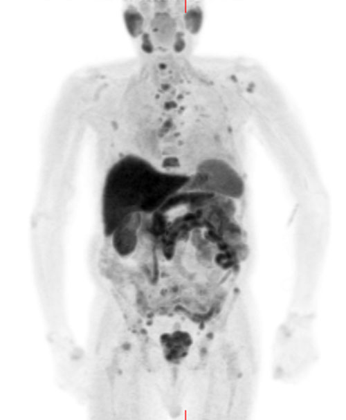
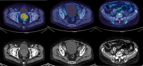
More distant metastases in, for example, the spine and pelvis which are the most common locations for secondary spread of adenocarcinoma of the prostate, can be excluded by a technetium bone scan. Increasingly though, because of its higher sensitivity and specificity and its ability to detect soft tissue and bony metastases, a choline positron emission tomography (PET) scan is requested, see figures 2 and 3. This is a more expensive alternative.
Management
Advice about the necessity, or not, for treatment of early prostate cancer, and consideration of the various treatment options available is increasingly risk-based and should take into account not only the stage and grade of the tumour but also patient preferences.
Low-risk patients
In low-risk cases (PSA < 10 ng/ml and Gleason score 6 or less) the majority of men are now advised to undergo a period of active surveillance to determine whether or not intervention by surgery or radiotherapy is required.
The recently revised NICE guidelines 4 have proposed four monthly PSA determinations and accompanying DRE with reassessment at 12 months by repeat biopsy, preceded by an MpMRI.
In the absence of evidence of progression, follow-up should continue with regular PSA determinations either in primary or secondary care. Conversely, if the cancer progression is evident, a discussion will need to be had about more definitive treatment.
Thus far patients treated by surgery or radiotherapy after a period of active surveillance do not seem to have worse outcomes than those treated at the time of initial diagnosis.
Intermediate-risk patients
In intermediate-risk cases (PSA 10-20 ng/ml or Gleason score 7), following an initial discussion about active surveillance, most urologists advise definitive treatment to prevent the development of local complications and/or metastatic spread.
The most reliable way of eliminating early prostate cancer is to remove the gland before the cancer has spread. Increasingly this is achieved by keyhole surgery, with or without robotic assistance.
With careful technique and an experienced surgeon, the risk of more than temporary stress urinary incontinence should be in the order of 2-5%. The chances of erectile dysfunction (ED) however are considerably greater and this needs to be discussed with patients. ED can be quite effectively managed with regular phosphodiesterase type 5 inhibitor therapy and prostaglandin E1 injections.
Alternatively, the patient can be offered treatment by radiotherapy, using either external beam radiation, usually preceded by at least three months androgen blockade, or brachytherapy.
Other newer focal therapies, which target the tumour rather than the entire prostate, such as high-intensity focused ultrasound, are regarded as experimental and should only be employed within the context of a clinical trial.
High-risk patients
In high-risk cases (PSA > 20 ng/ml or Gleason score 8-10) active surveillance is not advised. Instead surgery, or alternatively radiotherapy plus hormone therapy, is recommended.
The choice between these options will depend on patient preference and on the local extent of the tumour. If, for example, there is evidence of bulky local disease and/or seminal vesicle involvement the pendulum will swing in the direction of radiotherapy. If it looks as though the tumour can be removed in its entirety, with negative surgical margins, then radical prostatectomy may be possible. However, patients in this category should be informed that subsequent multimodality treatment with radiotherapy and hormones may often be required.
Conclusions
The diagnosis and management of early prostate cancer continues to generate much discussion and controversy. Very recently several studies have reported superior outcomes for surgery compared with either radiotherapy or watchful waiting, 5,6 but every treatment option carries the risk of side-effects, which need to be carefully explained to the patient and his partner. The recent vogue for active surveillance, which features very prominently in the recently updated NICE guidelines, will reduce the incidence of these and should significantly reduce the potential for overtreatment of low-risk cancers that are destined never to affect the patient within his lifespan.
Anxieties persist that our current methods of initial diagnosis and staging and subsequent identification of disease progression are still suboptimal. Use of genomic markers, such as the Prolaris test which identifies and quantifies markers of cell cycle progression thereby providing prognostic information, may prove useful refinements in the future.7 Extensive current research seems likely to identify other clinically helpful biomarkers in the near future.8
REFERENCES
1 Kirby RS, Eeles RA, Kote-Jarai Z et al. Screening for prostate cancer: the way ahead. BJU Int 2010;105:295-7
2 Dickinson L, Ahmed HU, Allen C et al. Magnetic resonance imaging for the detection, localisation, and characterisation of prostate cancer: recommendations from a European consensus meeting. Eur Urol 2011;59:477-94
3 Patel U, Dasgupta P, Amoroso P et al. Infection after transrectal ultrasonography-guided prostate biopsy: increased relative risks after recent international travel or antibiotic use. BJU Int 2012;109:1781-4
4 National Institute for Health and Care Excellence. CG175. Prostate cancer. NICE. London. 2014
5 Sooriakumaran P, Nyberg T, Akre O. Comparative effectiveness of radical prostatectomy and radiotherapy in prostate cancer: observational study of mortality outcomes. BMJ 2014;348:13
6 Bill-Axelson A, Holmberg L, Garmo H et al. Radical prostatectomy or watchful waiting in early prostate cancer. N Engl J Med 2014;370:932-942
7 Cuzick J, Berney DM, Fisher G et al. Prognostic value of a cell cycle progression signature for prostate cancer death in a conservatively managed needle biopsy cohort. Br J Cancer 2012;106:1095-99
8 Kirby RS, Giling P (Eds). Fast Facts: Prostate Cancer 8E. Health Press. Abingdon, UK. 2013
ARTICLE IN PDF
 Download this article in PDF format
EXTERNAL WEBLINKS
Prostate Cancer UK
www.prostatecanceruk.org
The Urology Foundation
www.theurologyfoundation.org
UPDATED EVIDENCE
US: National Cancer Institute
PDQ® (Physician Data Query) is NCI's cancer database of peer-reviewed, regularly updated evidence-based, referenced summaries
Prostate prevention
Prostate screening
Prostate treatment
UK: NICE- evidence search results
Prostate cancer
Metastatic
Cochrane Reviews
Prostate cancer
PubMed
PubMed is a database of the US National Library of Medicine/National Institutes of Health
Metastatic prostate cancer
Benign prostatic hyperplasia and prostate cancer
BPH relation to prostate cancer
Palliative care at home
Causes of prostate cancer - reviews
Early prostate cancer - reviews
British National Formulary
Malignant disease and immunosuppression
Cancer Research UK
Prostate cancer statistics
Early diagnosis in primary care
UK Risk Assessment Tools
QCancer
Qcancer men
UK Early diagnosis programmes
UK NCAT
Macmillan - early diagnosis
BMJ Learning CPD module
Managing patients at risk of prostate cancer 1 hour