SEARCH

Aning J, Gillatt D. When should patients with varicocoele be referred? Practitioner 2012; 256 (1750):17-20
When should patients with varicocoele be referred?
24 Apr 2012
AUTHORS
Mr Jonathan Aning BMBS DM FRCS (Urol)
Urology Specialist Registrar
Professor David Gillatt MB ChB ChM FRCS
Consultant Urological Surgeon, Bristol Urological Institute, Southmead Hospital, Bristol
Article
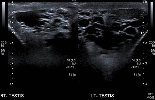
Varicocoele is a common urological condition present in 15% of men. The term varicocoele describes a collection of enlarged veins in the scrotum. Figure 1 illustrates the ultrasound appearance of a varicocoele.
Varicocoeles are rare in children; the majority of patients who seek a medical opinion are aged between 15 and 30 and asymptomatic.
Up to 40% of men who present with subfertility1 may have a varicocoele but although there is an association between varicocoele and impaired semen parameters the majority of men will be able to achieve a natural conception without intervention. Most men as a consequence do not require treatment for this condition.
Causes
Varicocoele is defined as an abnormal tortuous dilation of testicular veins in the pampiniform plexus. The testicular veins normally travel in the spermatic cord and are neither visible nor palpable.
The precise mechanism by which varicocoeles develop is poorly understood, however the clinical condition is acknowledged to result from venous reflux. The left testicular vein normally drains into the left renal vein, the right testicular vein drains directly into the inferior vena cava, the absence of valves in these vessels results in venous reflux and varicocoele formation. The absence of valves is more common on the left side thus approximately 90% of varicocoeles are left sided.
Increased renal vein or testicular vein pressure is another recognised cause of varicocoele, this may result from the angle of venous drainage or multiple pathologies including: retroperitoneal mass; venous thrombus; vena cava occlusion and rarely nutcracker phenomenon (where the renal vein is compressed between the aorta and the superior mesenteric artery).
Presentation and assessment
Most men will present because they are concerned about one of the following problems:
• An asymptomatic scrotal swelling that has been present for a while
• An asymptomatic scrotal swelling associated with a small testicle
• Infertility
• Symptoms of pain or discomfort related to a scrotal swelling
• A sudden finding of a varicocoele
A thorough history and examination
is essential to make a clinical diagnosis and determine the probable cause.
The history should include:
• the amount of time the swelling has been present for
• an assessment of degree of bothersome symptoms
• details of any previous surgery
• fertility
It is important to be alert to the patient with an acutely presenting varicocoele or a solitary right-sided varicocoele as this may be the first presentation of a retroperitoneal malignancy (usually a renal tumour) especially if the patient is over the age of 40 and the varicocoele does not disappear on lying down.
A full abdominal examination including the scrotum should be performed and the patient examined in supine and standing positions.
The Valsalva manoeuvre (ask the patient to strain down) in both positions may make the veins more pronounced. The classical clinical examination finding of a varicocoele is described as the sensation of a bag of worms present in the scrotum when the patient is standing or during the Valsalva manoeuvre which disappears on lying down.
Varicocoeles may be graded according to the Hudson classification,2 (illustrated in table 1) and it is important to document the testicular volume.
Measurement of testicular volume is important because 70-80% of the testicular mass consists of seminiferous tubules and the volume represents a measure of the likely quality of spermatogenesis. Normal adult testicular volume is approximately 20 cm3. 3
Several methods of estimating testicular volume exist, all have limitations. Ultrasound assessment is thought to be the most accurate, closely followed by the volume displacement method (measuring the volume of water displaced when a testicle is placed in a container of water). These may be difficult to perform in everyday general practice.
Simpler methods include the use of orchidometers which are practical aids to measure testicle size, perhaps the most well known being the Prader model.4 This consists of beads of increasing size with known volume which may be compared with the patient’s testicle. Alternatively measurement of the testis, with callipers or a ruler, in three dimensions with the values input into Lambert's equation of L x W x H x 0.71 will give an estimate of volume.5
In essence though it is only clinically relevant for a GP to identify whether the patient’s testicle is small/normal size and in adolescents whether the testicle is failing to develop to an adult size.
Management and referral
The majority of varicocoeles occur in young healthy men and clinical examination alone is enough to make the diagnosis. Referral to a urologist and further investigations in most of these cases are not required and the patient should be reassured. Supportive underwear may aid patients with small varicocoeles complaining of a mild dragging sensation in the scrotum.
Patients in whom an intra-abdominal malignancy is suspected or with significant pain related to their varicocoele should be referred to a urologist for review. In these patients a colour Doppler ultrasound scan of the scrotum in lying and standing positions is an appropriate first-line investigation. This may be arranged in primary care at the same time as the referral in order to facilitate the initial secondary care consultation. The radiology department protocol in most centres will automatically include imaging the abdomen to ensure there is no evidence of retroperitoneal pathology when the investigation is requested for varicocoele.
Patients with a confirmed renal mass on imaging or a palpable abdominal mass meet the criteria to be referred urgently under the two-week wait rule.
If an adult patient with a varicocoele presents with infertility, a semen analysis should be performed. If the result is abnormal, a repeat sample should be sent and if the abnormality is confirmed, the patient should be referred to a urologist with an andrology interest or the couple should be referred to their local specialist fertility centre.
NICE guidance does not recommend the surgical treatment of varicocoeles for infertility.6 The controversy regarding varicocoele treatment and infertility is detailed below.
There is no evidence that treatment of a varicocoele at a younger age will improve fertility compared with intervention later. Adolescents with normal testicular volume on the ipsilateral side of the varicocoele should be offered monitoring with measurement and semen analysis.
The European Association of Urology guidelines recommend varicocoelectomy in children and adolescents in the following cases: 7
• Varicocoele associated with a small testis with persistent failure of development documented by serial clinical examination
• Patients with a varicocoele and an additional testicular condition affecting fertility
• Bilateral palpable varicocoeles
• Older adolescents with pathological sperm quality
• Symptomatic varicocoeles
• Patients with psychological effects from the appearance of a large varicocoele
These patients should be referred for urological review.
Varicocoele and infertility
A quarter of men who have a varicocoele may have abnormal semen parameters.8 Decreased sperm motility and oligospermia are the most common abnormalities seen.
The mechanism by which varicocoeles cause impaired testicular function is poorly understood. Prominent theories include abnormally high scrotal temperature secondary to the increased vascular flow around the testicle and loss of the counter current heat exchange function performed by the normal pampiniform plexus. This temperature increase, which may be as little as 0.6°C, leads to an alteration of Leydig cell function which results in increased sperm DNA damage from oxidative stress.
Reflux of renal and adrenal metabolites from the renal vein and hypoxia from venous stasis may also contribute to impaired function. In reality the cause is likely to be multifactorial.
Varicocoelectomy has been shown to improve semen parameters and reverse sperm damage in several studies.9, 10 Randomised controlled trials have demonstrated that varicocoelectomy for subclinical varicocoeles is ineffective.11 Varicocoelectomy in men with normal semen parameters who present with infertility is not clinically indicated.12 Most adolescents with varicocoeles will have no problem fathering a child in later life and overtreatment is a real risk.13
Controversy remains in the urological community as to whether varicocoele repair is warranted in subfertile men.
A clear correlation between either intervention or observation alone and increased spontaneous pregnancy rate has not been proven.
Unfortunately, the data published to date demonstrate contradictory results and have significant limitations. Two key meta-analyses have been published recently.
In 2009, Evers and colleagues published a Cochrane review which concluded that there was no evidence that varicocoele repair improved a couple’s chance of conception.14 However, this meta-analysis included several trials where patients had subclinical varicocoeles with normal semen parameters which when corrected for suggested a different statistical outcome favouring intervention.
In a more recent meta-analysis of four randomised controlled trials on varicocoelectomy in men with a clinical varicocoele, oligospermia and unexplained fertility a trend in favour of surgical correction was observed; however this was not statistically significant.15
Emerging evidence suggests that the timing of varicocoele surgery is important and repair should be considered primarily in patients with a clinical varicocoele, oligospermia, duration of infertility of at least two years and otherwise unexplained infertility in the couple.16
The concern regarding varicocoelectomy is that there may be more effective measures to overcome infertility. It is therefore reasonable that all patients with a varicocoele presenting as part of
a couple with fertility problems are referred to a urologist with a specialist interest in fertility and an expert in assisted reproduction techniques and be counselled regarding the relative success rates of all options: varicocoele surgery versus assisted reproduction technology.
Treatment
The principles of therapy are to ligate or occlude the internal spermatic veins while preserving arterial flow to the testis from the testicular artery. Surgical techniques or interventional radiology using embolisation or sclerotherapy form the mainstay of treatment.
All techniques have a failure rate, the primary intervention offered by a centre is dependent on their experience and outcomes. The main complications of intervention are postoperative haematoma, testicular atrophy and hydrocoele formation.
Surgery
Open or laparoscopic techniques may be used to ligate the spermatic veins at different levels above and below the inguinal canal. Although multiple techniques for varicocoelectomy have been documented, currently favoured techniques are laparoscopy and microsurgical techniques.
Laparoscopy confers the benefits of minimally invasive surgery and allows the testicular vein to be taken proximally where it inserts into the renal vein or inferior vena cava, where there are fewer vessels to ligate (recurrence rates 3-7%).17 Microsurgical varicocoelectomy, using magnification during surgery for example an operating microscope, allows optimal visualisation of small vessels and reduced risk of arterial ligation (recurrence rate 0.8-4%).18 Lymphatic-sparing surgery minimises the risk of postoperative hydrocoele.
Postoperative advice will vary between centres but generally dressings may be left on for up to 48 hours, patients may return to nonstrenuous work after two days with analgesia and it is wise to advise patients to refrain from intercourse for a week.
Interventional radiology
Embolisation is the least invasive method of varicocoele treatment. It may be performed under sedation and is favoured by many centres. A catheter is inserted via the right femoral vein or internal jugular vein and passed into the spermatic vein under radiological screening. Venography is used to identify collateral vessels, embolisation is then performed using coils (see figure 2) or less commonly a sclerosant (recurrence rate up to 10%).19 Misplacement of coil or coil migration are recognised additional complications.
Conclusion
The management of men with varicocoeles is largely conservative. Patients suspected of having underlying malignant pathology or with significant discomfort from a large varicocoele should be referred for specialist review.
The data regarding the relationship between varicocoele and infertility is inconclusive. NICE does not recommend the surgical treatment of varicocoele for subfertile men. Patients or couples with infertility should be referred to a urologist with a specialist interest in fertility and an expert in assisted reproduction techniques, ideally to be seen in a joint clinic. Patients can then be counselled regarding the relative success rates of all options for varicocoele surgery versus assisted reproduction.
REFERENCES
1 Cockett ATK, Takihara H, Consentino MJ. The varicocoele. Fertil Steril 1984;41:5-11
2 Hudson RW, Perez Marrero RA, Crawford VA et al. Hormonal parameters in incidental varicocoeles and those causing infertility. Fertil Steril 1986;45(5):692-700
3 Kim ED, Lipshultz LI, Howard SS. Male Infertility. In Gillenwater JY, ed. Adult and Paediatric Urology. Lippincott Williams & Wilkins. Philadelphia, PA. 2002
4 Prader A. Testicular size: assessment and clinical importance. Triangle 1966;7:240-243
5 Lambert B. The frequency of mumps and of mumps orchitis and the consequences of sexuality and fertility. Acta Genet Stat Med 1951;2(suppl 1):1-166
6 National Institute for Health and Clinical Excellence. CG11. Fertility: assessment and treatment for people with fertility problems. NICE. London. 2004
7 Tekgul S, Riedmiller H, Dogan HS et al. European Association of Urology Guidelines on Paediatric Urology. www.uroweb.org/gls/pdf/21_Paediatric_Urology_LR%20[correctie%20Hoebeke].pdf
8 The influence of varicocoele on parameters of fertility in a large group of men presenting to infertility clinics. World Health Organization. Fertil Steril 1992;57(6):1289-93
9 Zini A, Dohle G. Are varicocoeles associated with increased deoxyribonucleic acid fragmentation? Fertil Steril 2011;96(6):1283-7
10 Agrawal A, Deepinder F, Cocuzza M et al. Efficacy of varicocoelectomy in improving semen parameters: a new meta-analytical approach. Urology 2007;70(3):532-8
11 Grasso M, Lania C, Castelli M et al. Low grade left varicocoele in patients over 30 years old: the effect of spermatic vein ligation on fertility. BJU Int 2000;85(3):305-7
12 Breznik R, Vlaisavljevic V, Borko E. Treatment of varicocoele and male fertility. Arch Androl 1993;30(3):157-160
13 Zargooshi J. Sperm count and sperm motility in incidental high-grade varicocoele. Fertil Steril 2007; 88(5):1470-3
14 Evers JH, Collins J, Clarke J. Surgery or embolisation for varicocoeles in subfertile men. Cochrane Database Syst Rev 2009;(1):CD000479
15 Baazeem A, Belzile E, Ciampi A et al. Varicocoele and male factor infertility treatment: a new meta-analysis and review of the role of varicocoele repair. Eur Urol 2011;60(4):796-808
16 Giagulli VA, Carbone MD. Varicocoele correction for infertility: which patients to treat? Int J Androl 2011;34(3): 236-241
17 Miersch WD, Shoeneich G, Winter P et al. Laparoscopic varicocoelectomy: indication, technique and surgical results. Br J Urol 1995;76(5): 636-8
18 Jungwirth A, Gogus C, Hauser G et al. Clinical outcome of microsurgical subinguinal varicocoelectomy in infertile men. Andrologia 2001;33(2):71-74
19 Lenk S, Fahlenkamp D, Gliech V et al. Comparison of different methods of treating varicocoele. J Androl 1994; 15(Suppl):34S-37S
 Download this article in PDF format