SEARCH

Lee JYJ, Keane MG, Pereira S. Diagnosis and treatment of gallstone disease. Practitioner 2015;259(1783):15-19
Diagnosis and treatment of gallstone disease
22 Jun 2015
AUTHORS
Dr Janice YJ Lee BSc MBBS, Year 6 Medical Student, UCL Medical School, London, UK
Dr Margaret G Keane MSc MRCP, Clinical Research Fellow in Gastroenterology and Hepatology, UCL Institute for Liver and Digestive Health, London, UK
Dr Stephen Pereira BSc(Hons) PhD FRCP, Reader in Gastroenterology and Hepatology, UCL Institute for Liver and Digestive Health, London, UK
Article
Gallstone disease is common, affecting 10 to 15% of adult Western populations.1 An ultrasound survey of British adults found gallstones in 12% of men and 22% of women over 60 years.2 There has also been an increase in hospital admissions and operations for gallstone disease over recent years. In England, admissions increased by 30% in men and 64% in women from 1989-90 to 1999-2000.3 Cholecystectomy now represents 3% of all gastrointestinal surgical procedures performed in the UK.4
Causes
Bile contains cholesterol, bile pigments and phospholipids at varying concentrations. Gallstones form when there is an imbalance in the composition of bile resulting in precipitation of one or more of its components. Between 37 and 86% of gallstones are cholesterol-rich stones, 2 to 27% are pigment stones and 4 to 16% are mixed stones.5,6
The most common type of gallstones found in Western populations are cholesterol-rich stones. These form when the liver produces bile with excess cholesterol which precipitates as microcrystals in the gallbladder.7 Gallbladder mucin hypersecretion and impaired gallbladder motility contribute to retention and aggregation of crystals into macroscopic stones.
Risk factors
A number of constitutional and environmental factors have been associated with gallstone formation, see table 1. Gallstone disease increases with age, being exceptionally rare in neonates and children but becoming noticeably more common after the age of 40 years.8 Women have a higher prevalence of gallstones than men,9 which is attributed to exposure to the female sex hormones oestrogen and progesterone.
Oestrogens increase cholesterol uptake and biosynthesis by stimulating hepatic lipoprotein receptors and hepatic HMG-CoA reductase activity, while progestins have been shown to impair gallbladder emptying leading to stasis. Exogenous hormones in the form of the oral contraceptive pill and oestrogen replacement therapy also increase the risk of gallstone formation.10
The incidence of gallstones varies globally. Native Americans and Hispanics have the highest incidence of gallstone disease, with familial studies and the identification of several human Lith genes indicating a genetic predisposition.11,12
Different types of gallstones are also more prevalent in certain populations and parts of the world. Cholesterol-rich gallstones are more common in Europe and North America. This has been attributed to obesity and diets containing a high proportion of refined carbohydrates and fat. Pigment stones are more common in those with a predisposition to haemolysis e.g. those with sickle cell disease and in Asians. However, as Asian diets have become more westernised cholesterol-rich stones are becoming more frequent in this population.8,13
Low-calorie diets and rapid weight loss are also associated with the development of cholesterol-rich gallstones. Weight loss exceeding 1.5 kg/week (e.g. after bariatric surgery) is particularly associated with cholesterol hypersecretion and an increased risk of gallstone formation.14
A history of dieting and significant weight fluctuations also increases the risk of stone formation.15
Some studies have suggested that gallstone formation and disease can be affected by statin use, but outcomes have not been consistent. A recent meta-analysis, which included all studies from 1980, found that moderate to high doses of statin did decrease the risk of gallstone disease but the authors acknowledged that further multicentre studies were needed on the topic to confirm their findings.16
The risk of gallstone formation is higher in several chronic gastrointestinal diseases. For example, the prevalence of gallstones is increased by approximately 25% in patients with liver cirrhosis compared with the general population and the risk increases with severity of liver disease.17 Most of the stones formed in patients with cirrhosis are pigment stones, which is thought to relate to altered pigment secretion in liver failure and reduced gallbladder motility caused by increased oestrogen levels.
Total parenteral nutrition (TPN) is associated with the development of microlithiasis and gallstones.18 A possible mechanism for this is the loss of enteric stimulation of the gallbladder in the absence of eating, leading to gallbladder stasis and stone formation.
Gastrointestinal diseases that affect the ileum such as Crohn’s disease or ileal resection are also associated with gallstones. Reduced active bile acid absorption in the terminal ileum reduces the enterohepatic circulation of bile acids. This causes a rise in hepatic low-density lipoprotein uptake, upregulation of enzymes of bile acid and cholesterol synthesis, and an increased ratio of hepatic cholesterol to bile acid secretion.
These patients thus have supersaturated bile and a predisposition to forming cholesterol microcrystals and hence cholesterol-rich gallstones.19
Rarely, gallstones can also form in the common bile duct (choledocholithiasis) or the intrahepatic bile ducts (hepatolithiasis).
Risk factors associated with the development of these primary bile duct stones are bacterial infection (usually by Escherichia coli and Klebsiella spp), biliary parasites (Clonorchis sinensis, Opisthorchis species) and biliary obstruction from any cause.7
Presentation
Of those with gallstones, around 1 to 4% will develop symptoms annually.
The majority of patients (> 80%) will remain asymptomatic throughout their lifetime and the likelihood of developing symptoms diminishes with time.1 Gallstone disease can be associated with a number of presentations, see table 2.20 The most common are discussed in detail.
Biliary colic and cholecystitis
Biliary-type pain occurs in the epigastrium or right upper quadrant of the abdomen and is often triggered by eating fatty foods. Contrary to its name of biliary ‘colic’, it often does not fluctuate but persists from a few minutes up to 24 hours. It is frequently associated with nausea or vomiting but not with a change in bowel habit.
Acute cholecystitis is defined as inflammation of the gallbladder, which is usually caused by infection secondary to an obstructed cystic duct. In addition to biliary-type pain, patients may have fever, anorexia, nausea and vomiting. The attack can resolve spontaneously but recurrence is common. Repeated attacks can lead to chronic cholecystitis.
Around 10% of cases of acute cholecystitis occur in an acalculous gallbladder. Often this occurs in severely ill patients but has also been reported in those with HIV or on long-term TPN.21
Choledocholithiasis and ascending cholangitis
If a gallstone migrates into the common bile duct (CBD) it may be asymptomatic but often causes pain, resulting from impaction at the ampulla, or abnormal liver biochemical tests. If a stone impacts at the gallbladder neck or the cystic duct it can also cause CBD obstruction by compression (Mirizzi’s syndrome). Patients with these complications of gallstone disease typically develop pain, obstructive jaundice (dark urine, pale stools) and fever signifying cholangitis.
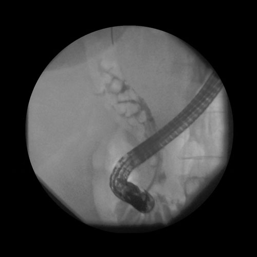
The most common pathogens in the UK are Escherichia coli, Klebsiella spp, Enterobacter spp, enterococci and streptococci. Up to a fifth of patients with ascending cholangitis will develop sepsis syndrome requiring urgent assessment, antibiotics and biliary decompression (see figure 1).22
Acute pancreatitis
There are a number of causes of acute pancreatitis but in Western populations the two most common causes are alcohol and migrating gallstones.
Acute gallstone pancreatitis is thought to be the result of a gallstone or sludge impacting at the region of the ampulla leading to back pressure on the pancreas, triggering the premature release of pancreatic enzymes causing acute pancreatitis.
Patients typically experience persistent severe epigastric pain radiating to the back and associated with vomiting. A number of clinical scores e.g. Ranson or Glasgow scores have been developed to differentiate mild from severe disease.23
Gallstones are common and the range of related symptoms is broad. Different attributable symptoms are associated with different symptomatic outcomes following cholecystectomy.
In a study, which pooled symptomatic outcomes based on presenting symptom, 92% of patients with biliary colic had relief of their symptoms after cholecystectomy, whereas only 65% had an improvement in food intolerance at 12 months.24
Investigation
Blood tests and ultrasound
Liver function tests (LFTs) and an abdominal ultrasound should be offered to patients with symptoms suggestive of gallstone disease (e.g. abdominal pain, jaundice, fever). They should also be considered in patients with less typical but chronic abdominal or gastrointestinal symptoms that have been unresponsive to other management.25
Additional imaging
Abdominal X-ray and computerised tomography (CT) scan are of limited diagnostic utility in gallstone disease. Cholesterol-rich stones, which make up the majority of gallstones in Europe and North America, are typically radiolucent and may be impossible to detect by these imaging modalities. However, if the diagnosis is uncertain or complications of gallstone disease such as gallbladder perforation or pancreatitis are suspected, a CT scan may be necessary.
Magnetic resonance cholangio-pancreatography (MRCP) should be performed in a patient with gallbladder stones and dilated bile ducts and/or abnormal LFTs, particularly if at ultrasound the distal bile duct was not visualised because of body habitus or overlying bowel gas.26
If there is a high index of suspicion for gallbladder disease but gallstones are not identified, patients could be considered for a hepatobiliary iminodiacetic acid (HIDA) or fatty meal-stimulated gallbladder ultrasound to look for biliary dyskinesia (ejection fraction < 40%).
Although a single small randomised controlled trial has shown some patients with biliary dyskinesia have symptomatic improvement after cholecystectomy,27a recent Cochrane review advised further studies were needed on the topic to support this indication for laparoscopic cholecystectomy.28
Endoscopic retrograde cholangio-pancreatography (ERCP) is not advised for the investigation of suspected CBD stones, but it can be used to treat confirmed choledocholithiasis before, or at the time of, laparoscopic cholecystectomy.25
Endoscopic ultrasound (EUS) is a non-invasive test that has high sensitivity for detecting gallstones.
In patients with gallbladder stones and dilated bile ducts or abnormal LFTs, an MRCP is usually advised. If it is contraindicated or unavailable locally,
an EUS is an excellent minimally invasive alternative to look for bile duct stones.29 A systematic review comparing EUS ± ERCP with ERCP alone for suspected choledocholithiasis demonstrated that ERCP could be avoided in up two thirds of patients; with a significant reduction in post-ERCP related pancreatitis.30
Management
Asymptomatic gallbladder stones
Gallstones within a normal gallbladder and without symptoms do not need any specific treatment with the exception of a minority of groups who are at high risk of complications or malignancy; porcelain gallbladder, immunosuppressed patients (e.g. after transplantation), elderly patients with insulin dependent diabetes, possibly those with rapid weight loss or weight cyclers and those that live in very remote regions of the world.31
Acute cholecystitis
Patients with acute cholecystitis should be referred for laparoscopic cholecystectomy, unless the patient’s comorbidities prevent this. The timing of laparoscopic cholecystectomy is debated but it is generally advisable for it to be performed either at presentation or after the acute cholecystitis episode has settled.
Delaying surgery potentially exposes patients to gallstone-related complications, so the latest NICE guidelines advise surgery within one week where clinically feasible.25 Evidence for timing is based on a small single randomised controlled trial32 and outcomes from a larger trial by the Dutch pancreatitis group (PONCHO trial) are awaited.
While awaiting surgery patients should avoid foods that may trigger their symptoms.25 Approximately a quarter of patients will develop increasingly severe symptoms necessitating surgical intervention while an inpatient.
Medical therapy for cholecystitis
Medical therapies such as extracorporeal shock wave lithotripsy (ESWL) or bile acid dissolution therapy with ursodeoxycholic acid (UCDA) are associated with much lower rates of clinical success when compared with surgery. Only 27% of patients receiving UCDA will achieve dissolution of their gallstones, while approximately half of carefully selected patients (one to three radiolucent stones, patent cystic duct) will be stone-free after ESWL.
Recurrence following these therapies is also high; more than 40% of patients will have recurrence of gallstones within four years of dissolution with UCDA or ESWL.33,34 Sustained weight loss and increased physical activity can help prevent the development of gallstones, but are unlikely to be of benefit once symptoms have developed.35
In patients with acute cholecystitis and multiple comorbidities preventing surgery, a percutaneous cholecystostomy (temporary external drainage of the gallbladder contents through a tube inserted under radiological guidance) may be considered, although a systematic review of the benefits of this intervention remains equivocal.36
If some of the comorbidities were related to the acute illness, cholecystectomy can be reconsidered, but typically patients are managed conservatively unless further symptoms develop.37,38
Gallbladder empyema
During an episode of acute cholecystitis the cystic duct may become obstructed and infection can build up in the gallbladder causing an empyema. Urgent drainage by cholecystostomy is indicated to prevent gallbladder perforation and subsequent peritonitis.25
Choledocholithiasis
In patients with CBD stones, bile duct clearance by ERCP (see figure 1) should be undertaken prior to, or at, laparoscopic cholecystectomy. This often involves endoscopic sphincterotomy, with or without balloon sphincteroplasty followed by removal of the stones with either a basket or a balloon catheter.
There is no evidence of any difference in efficacy, morbidity or mortality between laparoscopic CBD exploration (LCBDE) at the time of surgery or peri-operative ERCP, although the former is associated with a shorter hospital stay.39 If the bile duct cannot be cleared with ERCP, a biliary stent can be used to ensure biliary drainage until definitive endoscopic or surgical clearance is possible.25
Occasionally, especially in large stone disease, standard stone extraction techniques need to be supplemented with mechanical lithotripsy or extracorporeal shockwave lithotripsy.
If these forms of lithotripsy fail, electrohydraulic and laser lithotripsy can effect duct clearance.39
Acute pancreatitis
In patients with acute pancreatitis and evidence of ongoing bile duct obstruction and/or cholangitis, ERCP and biliary sphincterotomy is recommended within 24-72 hours of the onset of symptoms. In the absence of cholangitis or jaundice, an MRCP or EUS can be performed to exclude persisting choledocholithiasis.
In patients with acute pancreatitis attributed to gallstones, both the British Society of Gastroenterology (BSG) and the American College of Gastroenterology recommend laparoscopic cholecystectomy before or within two weeks of discharge.40,41
In patients with necrotising pancreatitis, cholecystectomy is often delayed until the local inflammation and collections have resolved.
Special situations
Pregnancy
Complications of gallstone disease are the most common reasons for non-gynaecological surgery during pregnancy. Gallstones are present in around 12% of pregnant women and complications such as ascending cholangitis and acute pancreatitis are associated with high rate of recurrence, exposing both fetus and mother to a risk of increased morbidity and mortality without intervention.
However, several international case series have demonstrated that ERCP and cholecystectomy where needed are safe during pregnancy.42,43,44 The BSG supports ERCP and LCBDE in pregnant patients with symptomatic CBD stones.39
Cirrhosis
There is no difference in the frequency of symptoms or complications in cirrhotic patients compared with the general population, but if patients do develop complications they tend to be more severe.45 Laparoscopic cholecystectomy is recommended over open surgery because it results in less blood loss, lower transfusion requirements and shorter hospital stay.46 The overall morbidity and mortality after cholecystectomy, however is higher in later stages of cirrhosis (Child-Pugh Class B/C) than in non-cirrhotic patients.47
Conclusions
Gallstone disease is common and prevalence is increasing. Although harmless in the majority of individuals, its complications can be a significant source of morbidity and mortality.
Patients with right upper quadrant pain; abnormal LFTs with or without fever should be investigated for suspected gallstone disease with routine blood tests and an abdominal ultrasound.
Laparoscopic surgical resection is the safest and most cost-effective intervention for the management of symptomatic gallstones. Medical management strategies are much less effective and are reserved for those whose comorbidities prevent surgical management.
REFERENCES
1 Halldestam I, Enell EL, Kullman E, Borch K. Development of symptoms and complications in individuals with asymptomatic gallstones. Br J Surg 2004;91(6):734-8
2 Heaton KW, Braddon FE, Mountford RA et al. Symptomatic and silent gall stones in the community. Gut 1991;32(3):316-20
3 Kang JY, Ellis C, Majeed A et al. Gallstones — an increasing problem: a study of hospital admissions in England between 1989/1990 and 1999/2000. Aliment Pharmacol Ther 2003;17(4):561-9
4 Williams JG, Roberts SE, Ali MF et al. Gastroenterology services in the UK. The burden of disease, and the organisation and delivery of services for gastrointestinal and liver disorders: a review of the evidence. Gut 2007;56 Suppl 1:1-113
5 Qiao T, Ma RH, Luo XB et al. The systematic classification of gallbladder stones. PLoS One 2013;8(10):e74887
6 Schafmayer C, Hartleb J, Tepel J et al. Predictors of gallstone composition in 1025 symptomatic gallstones from Northern Germany. BMC Gastroenterol 2006;6:36
7 Stinton LM, Myers RP, Shaffer EA. Epidemiology of gallstones. Gastroenterol Clin North Am 2010;39(2):157-69
8 Shaffer EA. Gallstone disease: Epidemiology of gallbladder stone disease. Best Pract Res Clin Gastroenterol 2006;20(6):981-96
9 de Bari O, Wang TY, Liu M et al. Cholesterol cholelithiasis in pregnant women: pathogenesis, prevention and treatment. Ann Hepatol 2014;13(6):728-45
10 Cirillo DJ, Wallace RB, Rodabough RJ et al. Effect of estrogen therapy on gallbladder disease. JAMA 2005;293(3):330-9
11 Sarin SK, Negi VS, Dewan R et al. High familial prevalence of gallstones in the first-degree relatives of gallstone patients. Hepatology 1995;22(1):138-41
12 Katsika D, Grjibovski A, Einarsson C et al. Genetic and environmental influences on symptomatic gallstone disease: a Swedish study of 43,141 twin pairs. Hepatology 2005;41(5):1138-43
13 Maclure KM, Hayes KC, Colditz GA et al. Weight, diet, and the risk of symptomatic gallstones in middle-aged women. N Engl J Med 1989;321(9):563-9
14 Weinsier RL, Wilson LJ, Lee J. Medically safe rate of weight loss for the treatment of obesity: a guideline based on risk of gallstone formation. Am J Med 1995;98(2):115-7
15 Syngal S, Coakley EH, Willett WC et al. Long-term weight patterns and risk for cholecystectomy in women. Ann Intern Med 1999;130(6):471-7
16 Kan HP, Guo WB, Tan YF et al. Statin use and risk of gallstone disease: A meta-analysis. Hepatol Res 2014 Oct 9. doi: 10.1111/hepr.12433
17 Conte D, Fraquelli M, Fornari F et al. Close relation between cirrhosis and gallstones: cross-sectional and longitudinal survey. Arch Intern Med 1999;159(1):49-52
18 Angelico M, Della Guardia P. Review article: hepatobiliary complications associated with total parenteral nutrition. Aliment Pharmacol Ther 2000;14 Suppl 2:54-7
19 Pereira SP, Bain IM, Kumar D, Dowling RH. Bile composition in inflammatory bowel disease: ileal disease and colectomy, but not colitis, induce lithogenic bile. Aliment Pharmacol Ther 2003;17(7):923-33
20 Glasgow RE, Cho M, Hutter MM, Mulvihill SJ. The spectrum and cost of complicated gallstone disease in California. Arch Surg 2000;135(9):1021-5; discussion 5-7
21 Elwood DR. Cholecystitis. Surg Clin North Am 2008;88(6):1241-52
22 Takada T, Strasberg SM, Solomkin JS et al. TG13: Updated Tokyo Guidelines for the management of acute cholangitis and cholecystitis. J Hepatobiliary Pancreat Sci 2013;20(1):1-7
23 Bell D, Keane MG, Pereira SP. Acute pancreatitis. Medicine 2015;43(3):174-81
24 Berger MY, van der Velden JJ, Lijmer JG et al. Abdominal symptoms: do they predict gallstones? A systematic review. Scand J Gastroenterol 2000;35(1):70-6
25 National Institute for Health and Care Excellence. CG188. Gallstone disease. NICE. London. 2014 www.nice.org.uk/guidance/cg188/resources/guidance-gallstone-disease-pdf
26 Warttig S, Ward S, Rogers G. Diagnosis and management of gallstone disease: summary of NICE guidance. BMJ 2014;349:g6241
27 Yap L, Wycherley AG, Morphett AD, Toouli J. Acalculous biliary pain: cholecystectomy alleviates symptoms in patients with abnormal cholescintigraphy. Gastroenterology 1991;101(3):786-93
28 Gurusamy KS, Junnarkar S, Farouk M, Davidson BR. Cholecystectomy for suspected gallbladder dyskinesia. Cochrane Database Syst Rev 2009(1):CD007086
29 Tse F, Liu L, Barkun AN et al. EUS: a meta-analysis of test performance in suspected choledocholithiasis. Gastrointest Endosc 2008;67:235-44
30 Petrov MS, Savides TJ. Systematic review of endoscopic ultrasonography versus endoscopic retrograde cholangiopancreatography for suspected choledocholithiasis. Br J Surg 2009;96(9):967-74
31 WGO Practice Guideline: Asymptomatic Gallstone Disease. www.worldgastroenterology.org/asymptomatic-gallstone-disease.html
32 Salman B, Yüksel O, Irkörücü O et al. Urgent laparoscopic cholecystectomy is the best management for biliary colic. A prospective randomized study of 75 cases. Dig Surg 2005;22(1-2):95-9
33 O'Donnell LD, Heaton KW. Recurrence and re-recurrence of gall stones after medical dissolution: a longterm follow up. Gut 1988;29(5):655-8
34 Carrilho-Ribeiro L, Pinto-Correia A et al. A ten-year prospective study on gallbladder stone recurrence after successful extracorporeal shock-wave lithotripsy. Scand J Gastroenterol 2006;41(3):338-42
35 Leitzmann MF, Rimm EB, Willett WC et al. Recreational physical activity and the risk of cholecystectomy in women. N Engl J Med 1999;341(11):777-84
36 Gurusamy KS, Rossi M, Davidson BR. Percutaneous cholecystostomy for high-risk surgical patients with acute calculous cholecystitis. Cochrane Database Syst Rev 2013;8:CD007088
37 Schmidt M, Sondenaa K, Vetrhus M et al. A randomized controlled study of uncomplicated gallstone disease with a 14-year follow-up showed that operation was the preferred treatment. Dig Surg 2011;28(4):270-6
38 Schmidt M, Sondenaa K, Vetrhus M et al. Long-term follow-up of a randomized controlled trial of observation versus surgery for acute cholecystitis: non-operative management is an option in some patients. Scand J Gastroenterol 2011;46(10):1257-62
39 Williams EJ, Green J, Beckingham I et al. Guidelines on the management of common bile duct stones (CBDS). Gut 2008; 57:1004-21
40 UK guidelines for the management of acute pancreatitis. Gut 2005;54 Suppl 3:iii1-9
41 Tenner S, Baillie J, DeWitt J, Vege SS. American College of Gastroenterology guideline: management of acute pancreatitis. Am J Gastroenterol 2013;108(9):1400-15;16
42 Sungler P, Heinerman PM, Steiner H et al. Laparoscopic cholecystectomy and interventional endoscopy for gallstone complications during pregnancy. Surg Endosc 2000;14(3):267-71
43Glasgow RE, Visser BC, Harris HW et al. Changing management of gallstone disease during pregnancy. Surg Endosc 1998;12(3):241-6
44 Paramanathan A, Walsh SZ, Zhou J, Chan S. Laparoscopic cholecystectomy in pregnancy: An Australian retrospective cohort study. Int J Surg 2015;18:220-3
45 Orozco H, Takahashi T, Mercado MA et al. Long-term evolution of asymptomatic cholelithiasis diagnosed during abdominal operations for variceal bleeding in patients with cirrhosis. Am J Surg 1994;168(3):232-4
46 Gurusamy KS, Davidson BR. Surgical treatment of gallstones. Gastroenterol Clin North Am 2010;39(2):229-44
47 Keane MG, Lai C, Pereira SP. Detecting patients with cirrhosis in primary care. Practitioner 2014;258(1773):15-20
ARTICLE IN PDF
 Download this article in PDF format
PRESET SEARCHES
Immediate CPD prompts
These handy templates* prompt immediate drafting of personal learning, reflection and planning. Save your record straight into your personal development folder on your device or Cloud storage system.
*Adobe reader needed for iPad
EXTERNAL WEBLINKS
Useful information
British Society of Gastroenterology
Williams EJ, Green J, Beckingham I et al. Guidelines on the management of common bile duct stones (CBDS). Gut 2008;57:1004-21
Pancreatic Society of Great Britain and Ireland UK guidelines for the management of acute pancreatitis. Gut 2005;54 Suppl 3:iii1-9
National Institute for Health and Care Excellence NICE CG188. Gallstone disease www.nice.org.uk/guidance/cg188
RCS and the Association of Upper Gastrointestinal Surgeons of Great Britain and Ireland Commissioning Guide for Gallstone Disease, 2013. www.rcseng.ac.uk/healthcare-bodies/docs/published-guides/gallstones