SEARCH

El Menabawey T, Martin H, Johnson GJ. Tailor treatment to the patient with gallstone disease. Practitioner July/August 2020;264(1839):15-19
Tailor treatment to the patient with gallstone disease
24 Jul 2020
AUTHORS
Dr Tareq El Menabawey MBBS MSc MRCP, Senior Clinical Fellow in Hepatobiliary Medicine
Dr Harry Martin BA (Hons) BM (Hons) MRCP, Senior Clinical Fellow in Hepatobiliary Medicine
Dr Gavin J Johnson MSc MD FRCP, Consultant Gastroenterologist
Department of Gastroenterology, University College Hospitals NHS Foundation Trust, London, UK
Competing interests: Dr Gavin Johnson has done consultancy educational work for Boston Scientific, Mylan, Pentax, Olympus and Cook Medical. The other authors have no competing interests.
Article
Gallstones affect around 15% of adults in the UK. Between 50 and 70% of patients with gallstones are asymptomatic at diagnosis and only 10-25% of these individuals will go on to develop symptomatic gallstone disease. The vast majority (90-95%) of gallstones are cholesterol stones. Obesity is associated with an increased risk of symptomatic gallstones. There is also a strong independent association between cholesterol gallstones and type 2 diabetes. Other risk factors for cholesterol stones include female gender, pregnancy, oral hormone replacement therapy, advancing age, rapid weight loss, bariatric surgery, Crohn’s disease and cirrhosis. The most common symptom of gallstones is biliary colic. This is a steady pain in either the epigastrium or right upper quadrant (that may radiate to the interscapular region or right shoulder). It typically builds to a peak within 30 min, and lasts for between 30 min and 8 hours. It is frequently triggered by fatty foods and may be associated with nausea and vomiting but not fever or abdominal tenderness. Patients with symptoms suggestive of gallstones should be offered liver function tests (LFTs) and an abdominal ultrasound. Patients with proven gallstones and uncomplicated biliary pain should be referred for routine laparoscopic cholecystectomy. It is imperative that a laparoscopic cholecystectomy should only be carried out when the bile duct is clear of stones. As ultrasound may fail to detect common bile duct stones, NICE recommends MRCP for patients who have a dilated bile duct and/or abnormal LFTs. Any patients found to have choledocholithiasis should have bile duct clearance with an ERCP prior to surgery. Bile duct clearance is also recommended for patients with incidental asymptomatic choledocholithiasis.
Gallstone disease affects up to 15% of patients in the UK. 1 It is one of the most common reasons for gastroenterology admissions in Europe.2 In the Western world, most gallstones are cholesterol stones (90-95%).3 The remainder are either pigment, calcium or mixed stones.4 Around 50-70% of patients with gallstones are asymptomatic at the time of diagnosis, the gallstones are detected incidentally, and most have a benign course. Overall, 10-25% of patients will go on to develop symptomatic gallstone disease.5,6
The most common symptom of gallstones is abdominal pain, historically thought of as ‘biliary colic’, although a recent study found the pain was equally likely to be epigastric (43%) or in the right upper quadrant (41%).7 This is a steady pain that is non-paroxysmal in nature lasting for more than 30 minutes but less than 8 hours, and classically is maximal within 30 minutes.
It is frequently triggered by fatty foods and is experienced as a severe pain that may be associated with nausea and vomiting but not fever or abdominal tenderness. The pain can radiate to the interscapular region or right shoulder.
A proportion of patients will present with atypical symptoms such as dyspepsia. As asymptomatic gallstones and dyspepsia are common in the general population they frequently coexist but patients who undergo cholecystectomy for dyspepsia attributed to gallstones do not have a good clinical outcome and symptoms may persist in up to 20% of cases post cholecystectomy.8
A recent systematic review and meta-analysis found that the majority (65.7%) of patients with symptomatic uncomplicated gallstones had symptoms similar to functional dyspepsia, either in association with biliary colic or as the predominant symptom. Between 37 and 40% of patients had persistent abdominal symptoms following cholecystectomy. The authors concluded that ‘cholecystectomy for dyspeptic symptoms alone is unjustified.9
If patients present with atypical symptoms an alternative underlying cause may be present and should be investigated accordingly. Differential diagnoses for gallstone disease are listed in table 1. Patients may also present with complications of gallstone disease, see table 2. These include cholecystitis, obstructive jaundice, cholangitis and gallstone pancreatitis.
Causes and risk factors
Cholesterol and pigment gallstones originate from an interaction of genetic, environmental and metabolic abnormalities. In Western populations brown pigment stones are found in the bile duct post cholecystectomy and in primary sclerosing cholangitis but in South East Asian populations they occur with chronic infection.3
Most stones are cholesterol stones and several factors increase the risk of cholesterol stone formation. Obesity is associated with an increased risk of symptomatic gallstones necessitating cholecystectomy.
Other factors associated with metabolic syndrome including insulin resistance, dyslipidaemia (low HDL and hypertriglyceridaemia), sedentary lifestyle and fast food consumption are also associated with the formation of cholesterol gallstones.6 Insulin resistance and type 2 diabetes are strongly associated independent of obesity. The European Association for the Study of the Liver (EASL) guideline recommends that ‘healthy lifestyle and food, regular physical activity and maintenance of an ideal body weight might prevent cholesterol gallbladder stones and symptomatic gallstones.’3
Patients should be advised to reduce their consumption of refined carbohydrates and saturated fats, and increase their fibre intake.
Rapid weight loss (> 1.5 kg/week) is also a risk factor for cholesterol stone formation10 whereas moderate weight loss (< 1.5 kg/week) is protective against new gallstone formation.7 Bariatric surgery is also a risk factor, which may have implications should those patients go on to develop common bile duct stones and conventional access for endoscopic therapy with an endoscopic retrograde cholangiopancreatography (ERCP) is not possible, for example following Roux-en-Y gastric bypass.
Other risk factors for cholesterol stones include female gender, pregnancy, hormone replacement therapy (HRT) and advancing age.
The risk is substantially lower with transdermal compared with oral HRT. 11 There is a lower incidence in black Americans, and those of African, Chinese, Indian, Japanese and Thai ethnicity. Crohn’s disease and cirrhosis are also risk factors for cholesterol stone formation.4 Low alcohol intake and high coffee consumption are associated with gallstone formation in males.12
Haemolysis and chronic bacterial or parasitic infections are the main risk factors for pigment stones and are preventable causes of gallstones.13
Diagnosis
Investigations are needed primarily to confirm the diagnosis of gallstones and also to identify patients with gallstones within the bile duct. It is imperative that a laparoscopic cholecystectomy should only be carried out when the bile duct is clear of stones (otherwise a post-cholecystectomy bile leak via the cystic duct stump is more likely).
Patients with symptoms suggestive of gallstones should be offered liver function tests and an abdominal ultrasound in line with NICE guidance on gallstone disease and British Society of Gastroenterology (BSG) guidance on common bile duct stones.14,15 Patients with confirmed gallstones on ultrasound presenting with typical symptoms should be referred to secondary care for surgical assessment and consideration of cholecystectomy.
A Cochrane systematic review on the performance of ultrasound and liver function tests (LFTs) for the diagnosis of common bile duct stones noted that the sensitivity for ultrasound varies widely but the summary sensitivity was 0.73 and the specificity 0.91.16 Therefore, patients in whom gallstones are strongly suspected despite normal ultrasound findings warrant further investigation.
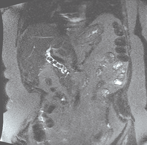
To determine if patients have common bile duct stones, NICE recommends magnetic resonance cholangiopancreatography (MRCP), see figure 1, for those who have abnormal LFTs and/or an ultrasound scan demonstrating dilated ducts as either of these features might indicate a common bile duct stone.15
A Cochrane systematic review of MRCP reported a sensitivity of 0.93 and specificity of 0.96 for identifying common bile duct stones.17 Endoscopic ultrasound is equivalent to MRCP at identifying common bile duct stones.18 MRCP has the advantages of being more widely available, minimally invasive and enables imaging of the intrahepatic ducts.
Endoscopic ultrasound is less widely available but may be preferential or necessary in patients who are claustrophobic, have cardiac or metal implants or who are morbidly obese.14 Endoscopic ultrasound may also be advantageous if there is a high index of suspicion of a common bile duct stone as the patient can proceed immediately to an ERCP to clear the bile duct at the same time.
Complications
Complications of gallstones can range from pain alone to life-threatening sepsis and end organ damage. Patients with asymptomatic gallbladder stones need no intervention as most remain asymptomatic throughout life.
Biliary pain
Biliary pain can arise from gallbladder, cystic duct and bile duct stones (choledocholithiasis). This is the most common presentation of gallstone disease accounting for up to 75% of symptomatic gallstone presentations.1
Cholecystitis
Cholecystitis is the most common complication of gallstones. It occurs in about 10% of patients with symptomatic gallstones19 and is the underlying cause in 3-9% of patients presenting to accident and emergency with acute abdominal pain.3 The most common cause of cholecystitis is a gallstone blocking the cystic duct.
The presentation of cholecystitis is similar to that of biliary pain. Typically, it lasts longer (more than 6 hours) and has accompanying signs of fever and raised inflammatory markers. Examination may reveal a positive Murphy's sign; halted inspiration during deep inspiration on palpation of the right upper quadrant. This occurs as a result of the inflamed gallbladder coming into contact with the examiner’s hand causing significant discomfort.
Choledocholithiasis
The presentation of choledocholithiasis is indistinguishable from gallbladder stones. However, it is vital that patients with choledocholithiasis are identified before referral for cholecystectomy as bile duct clearance should occur prior to surgery. For patients with incidental, asymptomatic choledocholithiasis bile duct clearance is also recommended, as without intervention up to 25% will develop complications.3
Cholangitis
Patients with choledocholithiasis can also present with infection of the biliary tree (cholangitis). Raised intraductal pressure secondary to obstruction causes the spaces between the cells lining the duct to widen resulting in contaminated bile coming into contact with the bloodstream.20 This can result in life-threatening bacteraemia and biliary sepsis. Patients typically present with pain often in the presence of fever and jaundice (Charcot’s triad). The sensitivity of Charcot’s triad is very low (around 26%). Pain can be absent, especially in elderly patients, and only two-thirds of patients are jaundiced. The Tokyo diagnostic criteria specify fever or lab evidence of an inflammatory response plus jaundice or abnormal LFTs plus imaging evidence of biliary obstruction.21 Delirium and septic shock may also be present (Reynold’s pentad) and indicate a more severe clinical presentation.
Pancreatitis
Around 50% of acute pancreatitis episodes in the UK are due to gallstone disease.22 History and examination may be indicative of acute pancreatitis and two out of the following three criteria need to be met for diagnosis:
• Typical history
• Elevated serum amylase or lipase
(> 3 times the upper limit of normal)
• Imaging (CT, MRI or ultrasound) consistent with acute pancreatitis.23
An ALT > 150 IU/L has a 95% positive predictive value for identifying gallstones as the causative insult.24
Management
Management of gallstone complications depends on the presentation although the principles of treatment are the same in all cases; to ensure that the bile ducts are clear followed by cholecystectomy.
Patients with proven gallstones and uncomplicated biliary pain should be referred for routine laparoscopic cholecystectomy. Several meta-analyses have demonstrated that early cholecystectomy for patients with acute cholecystitis has comparable outcomes but shortens length of hospital stay.25,26 The EASL recommends cholecystectomy within 72 hours in cases of cholecystitis.3
While the natural history of bile duct stones is not fully understood the evidence suggests patients with choledocholithiasis should have stone clearance prior to cholecystectomy if tolerated. A large retrospective cohort analysis from Sweden found that patients who had duct clearance had half the risk of complications compared with those who did not (12.7% vs 25.3%).27
The most common approach to bile duct clearance is ERCP performed using a side viewing endoscope. Commonly a biliary sphincterotomy ± sphincteroplasty (balloon dilatation of the sphincter) is performed to increase the size of the bile duct exit into the duodenum and stones are then removed using an extraction balloon or basket. The main risks of ERCP are bleeding (1.3%), perforation (0.6%), cholecystitis/cholangitis (1.4%) and post procedure pancreatitis (3.5-9.7%);28 this should be a consideration if patients present to primary care with pain after their procedure.
Bile duct stones may also be cleared at the time of cholecystectomy with intraoperative ERCP or laparoscopic bile duct exploration and clearance. These methods are equally efficacious but there are a decreasing number of surgeons experienced at performing laparoscopic bile duct exploration and so preoperative ERCP is the more common approach in the UK.3
There are a cohort of predominantly elderly patients who become unwell from cholecystitis or cholangitis and may not be candidates for laparoscopic cholecystectomy. Traditionally, percutaneous gallbladder drainage performed in interventional radiology has been the next modality of choice for an acutely inflamed gallbladder that is not settling with antibiotics, but it is associated with a high rate of morbidity and complications.29
Recently, internal drainage has been made possible by endosopic ultrasound-guided drainage using lumen apposing metal stents sited to allow drainage of the gallbladder into the stomach or duodenum. One study suggests a much lower complication rate compared with percutaneous gallbladder drainage and favourable outcomes.30
The treatment of gallstone pancreatitis is supportive. It is beyond the scope of this article and has been covered by an earlier article in this journal.31 Gallstone pancreatitis is a strong indication for cholecystectomy and a multicentre, randomised controlled trial showed that same admission cholecystectomy reduced the rate of recurrent gallstone related complications in patients with mild gallstone pancreatitis with a very low risk of postoperative complications. The EASL suggests cholecystectomy should be performed during the same admission as the initial pancreatitis episode.3
Conclusions
Gallstones are a very common cause of patients presenting to primary care. Most patients will remain asymptomatic and can be reassured if their gallstones are detected incidentally. However, a proportion will go on to develop complications some of which can be very severe. Symptomatic patients should be referred for consideration of cholecystectomy and any patients found to have choledocholithiasis should have bile duct clearance with an ERCP.
Patients at risk of gallstone development may have risk factors that also increase their operative risk for cholecystectomy such as obesity and should be encouraged to reduce their risk through lifestyle modification.
REFERENCES
1 National Institute for Health and Care Excellence. QS104. Gallstone disease. NICE. London. 2015 [Last accessed 13 July 2020] www.nice.org.uk/guidance/qs104
2 Farthing M, Roberts SE, Samuel DG et al. Survey of digestive health across Europe: Final report. Part 1: The burden of gastrointestinal diseases and the organisation and delivery of gastroenterology services across Europe. United European Gastroenterol J 2014;2(6):539-43
3 European Association for the Study of the Liver (EASL). EASL Clinical Practice Guidelines on the prevention, diagnosis and treatment of gallstones. J Hepatol 2016;65(1):146-81
4 Gurusamy KS, Davidson BR. Gallstones. BMJ 2014;348:g2669
5 Halldestam I, Enell E-L, Kullman E, Borch K. Development of symptoms and complications in individuals with asymptomatic gallstones. Br J Surg 2004;91(6):734-38
6 Sakorafas GH, Milingos D, Peros G. Asymptomatic cholelithiasis: is cholecystectomy really needed? A critical reappraisal 15 years after the introduction of laparoscopic cholecystectomy. Dig Dis Sci 2007;52(5):1313-25
7 Resnick E, Shteingart S, Melamud B et al. Enzyme pattern of biliary colic: A counterintuitive picture. World J Hepatol 2016;8(36):1629-36
8 Beckingham IJ. ABC of diseases of liver, pancreas, and biliary system. Gallstone disease. BMJ 2001;322(7278):91-94
9 Latenstein CSS, de Jong JJ, Eppink JJ et al. Prevalence of dyspepsia in patients with cholecystolithiasis: a systematic review and meta-analysis. Eur J Gastroenterol Hepatol 2019;31(8):928-34
10 Weinsier RL, Wilson LJ, Lee J. Medically safe rate of weight loss for the treatment of obesity: a guideline based on risk of gallstone formation. Am J Med 1995;98(2):115-17
11 Liu B, Beral V, Balkwill A et al. Gallbladder disease and use of transdermal versus oral hormone replacement therapy in postmenopausal women: prospective cohort study. BMJ 2008;337:a386
12 Völzke H, Baumeister SE, Alte D et al. Independent risk factors for gallstone formation in a region with high cholelithiasis prevalence. Digestion 2005;71(2):97-105
13 Van Erpecum KJ. Pathogenesis of cholesterol and pigment gallstones: an update. Clin Res Hepatol Gastroenterol 2011;35(4):281-87
14 Williams E, Beckingham I, El Sayed G et al. Updated guideline on the management of common bile duct stones (CBDS). Gut 2017;66(5):765-82
15 National Institute for Health and Care Excellence. CG188. Gallstone disease: diagnosis and management. NICE. London. 2014 www.nice.org.uk/guidance/cg188
16 Gurusamy KS, Giljaca V, Takwoingi Y et al. Ultrasound versus liver function tests for diagnosis of common bile duct stones. Cochrane Database Syst Rev 2015 Feb 26;(2):CD011548
17 Giljaca V, Gurusamy KS, Takwoingi Y et al. Endoscopic ultrasound versus magnetic resonance cholangiopancreatography for common bile duct stones. Cochrane Database Syst Rev 2015 Feb 26;(2):CD011549
18 de Lédinghen V, Lecesne R, Raymond JM et al. Diagnosis of choledocholithiasis: EUS or magnetic resonance cholangiography? A prospective controlled study. Gastrointest Endosc 1999;49(1):26-31
19 Friedman GD. Natural history of asymptomatic and symptomatic gallstones. Am J Surg 1993;165(4):399-404
20 Sung JY, Costerton JW, Shaffer EA. Defense system in the biliary tract against bacterial infection. Dig Dis Sci 1992;37(5):689-96
21 Kiriyama S, Kozaka K, Takada T et al. Tokyo Guidelines 2018: diagnostic criteria and severity grading of acute cholangitis (with videos). J Hepatobiliary Pancreat Sci 2018;25(1):17-30
22 National Institute for Health and Care Excellence. NG104. Pancreatitis. NICE. London. 2018 www.nice.org.uk/guidance/ng104
23 Working Group IAP/APA Acute Pancreatitis Guidelines. IAP/APA evidence-based guidelines for the management of acute pancreatitis. Pancreatology 2013;13(4 Suppl 2):e1–15
24Tenner S, Dubner H, Steinberg W. Predicting gallstone pancreatitis with laboratory parameters: a meta-analysis. Am J Gastroenterol 1994;89(10):1863-36
25 Menahem B, Mulliri A, Fohlen A et al. Delayed laparoscopic cholecystectomy increases the total hospital stay compared to an early laparoscopic cholecystectomy after acute cholecystitis: an updated meta-analysis of randomized controlled trials. HPB 2015;17(10):857-62
26 Lyu Y, Cheng Y, Wang B et al. Early versus delayed laparoscopic cholecystectomy for acute cholecystitis: an up-to-date meta-analysis of randomized controlled trials. Surg Endosc 2018;32(12):4728-41
27 Möller M, Gustafsson U, Rasmussen F et al. Natural course vs interventions to clear common bile duct stones: data from the Swedish Registry for Gallstone Surgery and Endoscopic Retrograde Cholangiopancreatography (GallRiks). JAMA Surg 2014;149(10):1008-13
28 Dumonceau J-M, Kapral C, Aabakken L et al. ERCP-related adverse events: European Society of Gastrointestinal Endoscopy (ESGE) Guideline. Endoscopy 2020;52(2):127-49
29 Small AJ, Irani S. EUS-guided gallbladder drainage vs. percutaneous gallbladder drainage. Endosc Ultrasound 2018;7(2):89-92
30 Teoh AYB, Serna C, Penas I et al. Endoscopic ultrasound-guided gallbladder drainage reduces adverse events compared with percutaneous cholecystostomy in patients who are unfit for cholecystectomy. Endoscopy 2017;49(2):130-38
31 Goodchild G, Johnson GJ. Early recognition and prompt referral key in acute pancreatitis. Practitioner 2018;262(1819):23-26
32 National Institute for Health and Care Excellence. Clinical knowledge summaries: gallstones. Revised June 2019 cks.nice.org.uk/gallstones [Last accessed 22 June 2020]
ARTICLE IN PDF
 Download this article in PDF format