SEARCH

Johnson L, Tun GSZ, Lobo AJ. Optimising the treatment of inflammatory bowel disease. Practitioner July/August 2019;263(1828):11-16
Optimising the treatment of inflammatory bowel disease
07 Aug 2019
AUTHORS
Ms Lara Johnson, Medical Student, University of Sheffield, Sheffield, UK
Dr Gloria SZ Tun MBBS MRCP, Clinical Fellow in Gastroenterology, Academic Department of Gastroenterology, Sheffield Teaching Hospitals NHS Foundation Trust, Sheffield, UK
Professor Alan J Lobo MD FRCP, Consultant Gastroenterologist, Academic Department of Gastroenterology, Sheffield Teaching Hospitals NHS Foundation Trust, Sheffield, UK
Competing interests
Professor Alan J Lobo has received speaker fees from, or been an advisory board member or carried out consultancy work for, Takeda, Janssen, MSD, Vifor, Shield Therapeutics, Abbvie, Pfizer, Janssen, and Medtronic.
Article
Abstract
Inflammatory bowel disease (IBD) is a chronic inflammatory condition which runs a relapsing and remitting course. The aetiology remains unclear but is thought to involve genetic susceptibility, altered gut microbiota, changes in immunological response and environmental triggers. Ulcerative colitis (UC) is more common than Crohn’s disease (CD). UC almost always affects the rectum and extends proximally and continuously to the colon to a variable extent. CD most commonly affects the terminal ileum or colon but can affect any part of the gastrointestinal tract from the mouth to the anus. The vast majority (90%) of people with UC report bloody stools compared with less than 50% of those with CD. Diarrhoea in UC is commonly associated with urgency and there may be cramping lower abdominal pain. CD is characterised by a triad of abdominal pain, diarrhoea and weight loss. Perianal disease is more common in CD and can consist of skin tags, fissures, abscesses and fistulae. IBD can also affect other parts of the body in around 25% of cases. If IBD is suspected, the severity of symptoms should be assessed and risk factors such as smoking history and family history reviewed. A full blood count, U&E, inflammatory markers (CRP and ESR) and haematinics should be requested. Patients should be asked about foreign travel and a stool sample sent for microscopy, culture and a Clostridium difficile toxin assay. IBD patients with colonic disease should be offered ileocolonoscopy eight years after symptom onset to assess cancer risk and determine the frequency of further colorectal screening.
Inflammatory bowel disease (IBD) mainly comprises ulcerative colitis (UC) and Crohn’s disease (CD) with approximately 5% of patients having an unspecified subtype known as IBD-unclassified (IBD-U).1
IBD is a chronic inflammatory condition that runs a relapsing and remitting course, of varying severity.1,2,3 The aetiology remains unknown but includes genetic susceptibility, altered gut microbiota, changes in immunological response and environmental triggers.4,5 UC is more common with a prevalence of 240 per 100,000 compared with CD which has a prevalence of 157 per 100,000.6,7
UC almost always affects the rectum, and extends proximally and continuously to the colon to a variable extent.3 Approximately 50% of people with UC have disease limited to the rectum (proctitis) or rectum and sigmoid colon (proctosigmoiditis), with only 3.3% having rectal sparing.1,3 CD most commonly affects the terminal ileum or colon but can affect any part of the gastrointestinal (GI) tract from the mouth to the anus.8
Treatment options have expanded rapidly with the introduction of novel agents producing benefits in terms of symptom improvement but also reduced tissue damage. The biologics have resulted in a transformation in the treatment of IBD but also new challenges in terms of understanding drug toxicity and the causes of loss of response to these agents, for example, due to the development of antidrug antibodies.
Organisation of care
Prompt diagnosis and management requires early recognition, assessment and referral in primary care and is likely to affect clinical outcomes. In addition, GPs play an important role in monitoring treatment and assessment of disease flares.
A multidisciplinary approach in secondary care with gastroenterologists, specialist IBD nurses, colorectal surgeons with an interest in IBD and additional specialties including dietetic, psychology and pharmacy input is accepted as the standard of care,1 but not all components are available to every patient. Additional specialty input may also be needed from obstetrics and stoma care at different points in an individual’s management.1
Presentation
IBD may affect people at any age.9 The most common age of onset for CD is the early to mid-20s.10 UC has an initial peak at 15-25 years and a second smaller peak between 55 and 65.3
Current UK guidance suggests that people presenting to primary care with a six-week history of diarrhoea, with or without blood or mucus, and/or abdominal pain, discomfort or bloating should be suspected of having IBD. However, symptoms such as abdominal pain, bloating or altered bowel habit also occur in people with a wide range of other conditions, including irritable bowel syndrome.11 Individual symptoms may not be strongly predictive of the presence of IBD but it has been suggested that combinations of clinical features
(rectal bleeding with diarrhoea, thrombocytosis, low MCV, low Hb or raised inflammatory markers; change in bowel habit with low MCV, thrombocytosis or low Hb; and diarrhoea with thrombocytosis) may raise the positive predictive value (PPV) for either IBD or colorectal cancer above 3%.12 In practice, a threshold of 3-4 weeks will help differentiate self-limiting causes such as infection and in the presence of a negative stool culture or severe symptoms, earlier referral should be considered.13
Smoking is an important risk factor in CD, whereas in UC the disease predominantly affects former smokers and nonsmokers.1
Chronic diarrhoea is a common symptom in UC and CD.2,3 Weight loss, rectal bleeding and nocturnal diarrhoea are clinical features that indicate a need for further investigation.1
The vast majority (90%) of people with UC report bloody stools14 compared with < 50% of those with CD.15 Diarrhoea in UC is commonly associated with urgency both during the day and night. Urgency reflects rectal involvement and the stool frequency and presence of blood are markers of disease severity. This may be associated with lower cramping abdominal pain.9
The severity of an acute episode or flare of UC is usually described using the criteria of Truelove and Witts,3 shown in table 1.
CD is characterised by a triad of abdominal pain, diarrhoea and weight loss. Perianal disease is more common in CD and can consist of skin tags, fissures, abscesses and fistulae.15 Associated symptoms of pain and discharge are intrusive and debilitating but may be under-reported by people with CD and under-appreciated by clinicians. Upper GI CD is rare in adults: symptoms may include upper abdominal pain, nausea and vomiting.1
Fatigue is a common and debilitating symptom in IBD.1 It may be related to a range of causes including anaemia or iron deficiency, sleep disturbance from pain or diarrhoea and depression but there may be no underlying explanation.1
IBD can also affect other parts of the body in approximately 25% of cases.16,17 Patients may suffer from arthropathies, skin manifestations such as erythema nodosum and pyoderma gangrenosum, ocular conditions such as episcleritis, scleritis and uveitis, aphthous oral ulcers, osteopenia, primary sclerosing cholangitis and autoimmune hepatitis.16,17
Assessment
The differential diagnoses for chronic abdominal pain and diarrhoea are shown in table 2.
A history of foreign travel, diarrhoeal illnesses and other contacts should be sought. Antibiotics may be associated with self-limiting diarrhoea or more severe Clostridium difficile colitis.
A range of drugs including proton pump inhibitors, NSAIDs, acarbose and sertraline can be associated with microscopic colitis.18,19
If IBD is suspected, the severity of symptoms should be assessed and risk factors such as smoking history and family history reviewed. In primary care the following investigations should be considered:
• Stool microscopy and culture, and Clostridium difficile toxin assay to exclude infective causes1,3
• Microscopy and culture for amoebic or Shigella dysentery should be carried out if travel history is suggestive1
• Blood tests: full blood count (FBC), urea and electrolytes (U&E), inflammatory markers: CRP and ESR and haematinics
• Anaemia may be due to iron deficiency (frequently unrecognised in established IBD), B12 or folate deficiency or anaemia of chronic disease
• CRP, ESR and platelet count may be raised due to the acute inflammatory phase response
• Faecal calprotectin: Faecal calprotectin is a protein released by intestinal neutrophils. It is a non-invasive test, both sensitive and specific for intestinal inflammation that can be used to investigate people who have had chronic symptoms for more than six weeks,11 who are low risk for malignancy and where others causes have been excluded.1 The British Society of Gastroenterology (BSG) suggests a minimum of 100-250 µg/g to trigger colonoscopy investigation1 though the yield in primary care can be low.20 Faecal calprotectin is not sufficiently sensitive as an assessment for colorectal adenoma or carcinoma.1
A normal faecal calprotectin is defined by the assay as < 50 µg/g stool. However, at this threshold, although the negative predictive value (NPV) is 98% the PPV is low at 20%.20 The BSG suggests using a higher threshold to trigger colonoscopy investigation.1 The York Faecal Calprotectin Pathway for primary care using a higher threshold of 100 µg/g for ‘normal’ had an NPV of 97% and a PPV of 40%. The pathway advises repeat faecal calprotectin within 2 weeks if >100 µg/g, routine referral to gastroenterology for a faecal calprotectin of 100-250 µg/g and urgent referral to gastroenterology if >250 µg/g.
Referral and diagnosis
All people with suspected IBD should be seen within four weeks from referral for a specialist assessment,11 or more urgently if symptoms are indicative of a more severe disease course.11 Patients should be referred directly to a consultant gastroenterologist specialising in IBD to ensure that there is no delay in diagnosis and treatment can be commenced quickly.11 Treatment is usually initiated in secondary care. Patients fulfilling the Truelove and Witts’ criteria for a severe flare require hospital admission.
The diagnosis of IBD is based on clinical features, imaging findings, macroscopic features on endoscopy and microscopic features on histology. The gold standard for diagnosis is histology following endoscopy or surgical resection.1
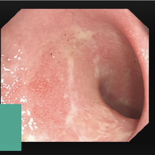
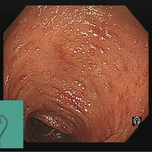
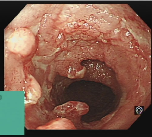
UC is most commonly initially diagnosed with flexible sigmoidoscopy (see figures 1-3) and histological assessment of mucosal biopsies.
This may be confirmed with ileocolonoscopy often carried out within the first year of diagnosis to define the full extent of disease.1
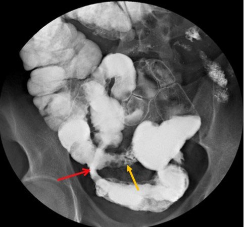
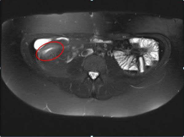
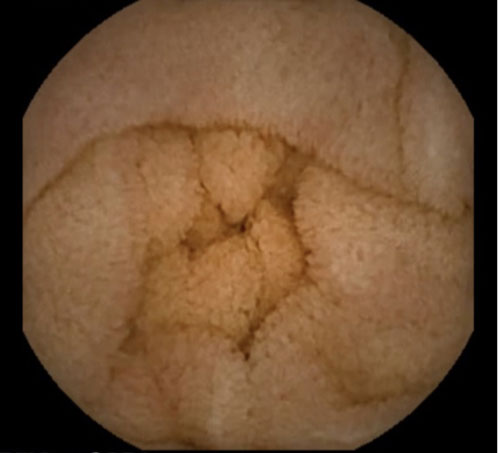
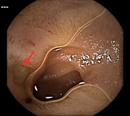
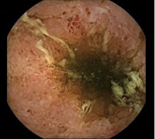
In CD the first-line investigation is an ileocolonoscopy with mucosal biopsies from the colon and ileum.1 Small bowel investigations such as barium meal (see figure 4), MR enterography (see figure 5) and capsule endoscopy (see figure 6), based on local availability and expertise, further aid clinical diagnosis.15,22
Pharmacological therapy
Treatment approaches for IBD cover two overlapping phases: induction and maintenance of remission.2
The choice of treatment is guided by the extent and site of involvement, severity of disease and associated complications such as the presence of perianal disease.
Currently, a step up approach is often used. The debate remains as to whether a more aggressive top down approach with earlier use of anti-TNF agents and immunosuppression may result in fewer long-term complications.
Ulcerative colitis
A summary of induction therapy for UC is shown in table 3.
Maintenance of remission1,3
• 5-ASA: Proctitis or proctosigmoiditis — topical or oral 5-ASA alone or both topical 5-ASA and oral 5-ASA. Left-sided and extensive UC — oral 5-ASA
• Thiopurines: azathioprine or mercaptopurine: Used as first line in steroid-dependent disease — two or more inflammatory exacerbations
that have required systemic corticosteroids in the past 12 months or patients who have had a single severe flare episode1
• Biologic therapies (infliximab, adalimumab, golimumab, vedolizumab) and tofacitinib. These can be given with an immunosuppressant or as monotherapy.
Crohn’s disease
Induction of remission1,2
• Time limited course of corticosteroids. Systemic (oral) glucocorticosteroid. Oral budesonide can be considered if there is distal ileal, ileocaecal or right-sided colonic disease and if this is a first presentation or a single inflammatory exacerbation in a 12-month period, and conventional glucocorticosteroids are declined, contraindicated or not tolerated. Budesonide is associated with fewer side effects than systemic corticosteroids but may be less effective.
• Enteral nutrition. Exclusive enteral nutrition for 8 weeks may be used for mild to moderate disease but adherence is difficult and this approach is predominantly used in children with IBD.
• Addition of a thiopurine (azathioprine or mercaptopurine) or methotrexate
• Biologic therapies: Infliximab, adalimumab, vedolizumab, ustekinumab. These can be administered in combination with an immunosuppressant or as monotherapy.
Maintenance of remission1,2
• Thiopurines
• Methotrexate if thiopurines are not tolerated, contraindicated or ineffective. Consider continued use if methotrexate was needed to induce remission.
• Biologic therapies: infliximab, adalimumab, golimumab, vedolizumab, ustekinumab.
Surgery
Surgery may be required in UC as an emergency following admission for acute severe UC or electively for refractory or relapsing disease that is impairing quality of life or following the development of dysplasia or colorectal cancer.
Surgical options comprise panproctocolectomy either with a permanent ileostomy or an ileoanal pouch to preserve intestinal continuation and avoid a stoma.
In 10% of those who undergo pouch surgery however, the surgery will be unsuccessful and a permanent stoma will be needed. Quality of life is good following pouch surgery. Median stool frequency is 5 times in 24 hours but without the urgency associated with active proctitis.1,3
In CD, surgery may be required for medically refractory active disease or complications such as chronic stricturing disease or an intra-abdominal abscess. Surgical control of sepsis including the insertion of setons into fistula tracks is central to the management of perianal CD before treatment with biologic agents.1,2
Monitoring and follow-up
Treatment targets have moved from solely patient reported outcomes, clinical improvement and remission towards endoscopic and mucosal healing.1 There is increasing evidence to suggest that the ongoing presence of endoscopic and histological inflammation or ulceration leads to further flares, a lack of sustained remission and a need for repeat corticosteroids and colectomy. In addition, endoscopic and histological remission reduces the risk of colorectal carcinoma in those with extensive colitis from UC or CD.1
Live vaccines are contraindicated if the patient is on an immunosuppressant and should be avoided for at least three months after stopping the therapy. Immunomodulators should be withheld for four weeks after live vaccine administration.1
The following vaccinations are recommended for patients taking glucocorticoids, thiopurines, methotrexate, anti-TNF agents or tofacitinib:1
• Annual influenza vaccination
• Pneumococcal vaccination. It is affected by immunosuppression and should be given at least two weeks before starting treatment. A single booster should be given at five years
• IBD patients seronegative for hepatitis B should be offered the vaccine and anti-HBs response should be measured.1
Drug monitoring
Clear shared care policies between primary care and the IBD centre on drug monitoring should be established and communicated to the patient. Requirements for specific medications are shown in table 4. (A full list of drug side effects can be found in the BNF.)
Adherence to medications is vital and if non adherence is suspected, this should be explored and GPs may need to liaise with secondary care.1
Smoking cessation
Smoking cessation should be encouraged, and patients should be referred to smoking cessation services. In CD, smoking increases the risk of further flares and surgery. In UC, patients should be warned that there
is a risk of flare after cessation. However, the overall benefits of smoking cessation should be discussed.1
Mental health
Anxiety and depression are common in IBD and often not identified or addressed. Access to psychological support is recommended in national guidance.1
It should be treated in the same way as depression in other situations, but addressing disease activity is likely to help, as well as specific issues such as body image especially in relation to a stoma or planned surgery.
Colorectal cancer screening
Colonic involvement in IBD is a risk factor for colorectal cancer.1
The risk of developing colorectal cancer is cumulative and starts to increase from ten years after symptom onset.23 Those with colonic disease from IBD should be offered ileocolonoscopy eight years after symptom onset to assess cancer risk to determine the frequency of further colorectal screening and to evaluate extent of disease.1
Flare management
Approximately 50% of IBD patients have at least one flare annually and their GP is often the first to assess them.24Adherence to medication and potential triggers should be considered, and NSAIDs stopped.24
A stool culture should be collected with a Clostridium difficile assay, FBC, U&E and CRP/ESR blood tests1,24 and a faecal calprotectin test performed.1,24
Prompt recognition of a severe IBD flare (see table 1) is fundamental, as this will require emergency admission6 and the on-call gastroenterologist and medical team should be contacted.22,24 The definition of acute severe UC has been outlined above. Severe CD is defined as poor general health and one or more of: weight loss, fever, severe abdominal pain or frequent diarrhoea stools (> 3-4 per day).2
If hospital admission is not necessary, patients can be treated with appropriate medications to induce remission. The IBD team should be alerted whenever oral steroids have been administered24 and informed if a patient has had two or more courses within 12 months or a relapse occurred within six weeks of previous treatment.9
If a patient is under 16, has a stoma or fistula, has had surgery, is on biologic therapy or immunomodulators, medication changes should be made in collaboration with the specialist team.24
Conclusion
Management of patients with IBD can be complex because of the variation in the course and severity of the disease, between individuals and over time. Optimal patient care requires coordination between primary and secondary care regarding the medications used, drug monitoring, possible side effects and management of relapses. Clear referral pathways and good communication between healthcare professionals and the patient are vital to ensure timely treatment.
REFERENCES
1 Lamb CA, Kennedy NA, Raine T et al. British Society of Gastroenterology consensus guidelines on the management of inflammatory bowel disease in adults. British Society of Gastroenterology. 2019 Gut (in press)
2 National Institute for Health and Care Excellence. NG129. Crohn’s disease: management. NICE. London. 2019 www.nice.org.uk/guidance/ng129 [Last accessed 9 July 2019]
3 National Institute for Health and Care Excellence. NG130. Ulcerative colitis: management. NICE. London. 2019 www.nice.org.uk/guidance/ng130 [Last accessed 9 July 2019]
4 Ye BD, McGovern DP. Genetic variation in IBD: progress, clues to pathogenesis and possible clinical utility. Expert Rev Clin Immunol 2016;12(10):1091-1107
5 Sartor RB and Mazmanian SK. Intestinal microbes in inflammatory bowel diseases. Am J Gastroenterol Suppl 2012;1:15-21
6 Tun GSZ, Harris A, Lobo AJ. Ulcerative colitis: management in adults, children and young people – concise guidance. Clin Med 2017;17(5):429-33
7 Tun GSZ, Cripps S, Lobo AJ. Crohn’s disease: management in adults, children and young people – concise guidance. Clin Med 2018;18(3):1-6
8 Farmer RG, Hawk WA and Turnbull RB. Clinical patterns in Crohn's disease: A statistical study of 615 cases. Gastroenterology 1975;68:627-35
9 Royal College of General Practitioners. Inflammatory Bowel Disease Toolkit. www.rcgp.org.uk/ibd [Last accessed 9 July 2019]
10 Cosnes J, Gower-Rousseau C, Seksik P et al. Epidemiology and natural history of inflammatory bowel diseases. Gastroenterology 2011;140:1785-94
11 IBD Standards: Section 2: Pre-diagnosis: Timeframe for referral. IBD UK. https://ibduk.org/ibd-standards/pre-diagnosis/timeframe-for-referral [Last accessed 9 July 2019]
12 Stapley SA, Rubin GP, Alsina D et al. Clinical features of bowel disease in patients aged e336-e344
13 Fine KD, Schiller LR. AGA technical review on the evaluation and management of chronic diarrhea. Gastroenterology 1999;116:1464-86
14 Dignass A, Eliakim R, Magro F et al. Second European evidence-based consensus on the diagnosis and management of ulcerative colitis Part 1: Definitions and diagnosis. J Crohns Colitis 2012;6:965-90
15 Van Assche G, Dignass A, Panes J et al. The second European evidence-based consensus on the diagnosis and management of Crohn's disease: Definitions and diagnosis. J Crohns Colitis 2010;4:7-27
16 Assche GV, Dignass A, Reinisch W et al. The second European evidence-based consensus on the diagnosis and management of Crohn's disease: Special situations. J Crohns Colitis 2010;4:63-101
17 Assche GV, Dignass A, Bokemeyer B et al. Second European evidence-based consensus on the diagnosis and management of ulcerative colitis Part 3: Special situations. J Crohns Colitis 2013;7:1-33
18 Masclee G, Coloma P, Kuipers E, Sturkenboom M. Increased risk of microscopic colitis with use of proton pump inhibitors and non-steroidal anti-inflammatory drugs. Am J Gastroenterol 2015;110(5):749-59
19 Lucendo AJ. Drug exposure and the risk of microscopic colitis: A critical update. Drugs R D 2017;17(1):79-89
20 Turvill J, O’Connell S, Brooks A et al. Evaluation of a faecal calprotectin care pathway for use in primary care. Prim Health Care Res Dev 2016;17:428-36
21 IBD Standards: Section 2: Pre-diagnosis: Referral pathways: https://ibduk.org/ibd-standards/pre-diagnosis/referral-pathways [Last accessed 2 August 2019]
22 IBD Standards: Section 1: The IBD Service: Investigations. IBD UK. https://ibduk.org/ibd-standards/the-ibd-service/investigations [Last accessed 9 July 2019]
23 Eaden JA, Abrams KR, Mayberry JF. The risk of colorectal cancer in ulcerative colitis: a meta-analysis. Gut 2001;48:526-35
24 IBD Standards: Section 4: Flare management: Flare pathways. IBD UK. www.ibduk.org/ibd-standards/flare-management/flare-pathways [Last accessed 29 July 2019]
25 Truelove SC, Witts LJ. Cortisone in ulcerative colitis: final report on a therapeutic trial. Br Med J 1955;ii:1041-48
26 Joint Formulary Committee. British National Formulary (online) London: BMJ Group and Pharmaceutical Press. www.medicinescomplete.com [Last accessed 12 July 2019]
27 Beaugerie L, Brousse N, Bouvier AM et al. Lymphoproliferative disorders in patients receiving thiopurines for inflammatory bowel disease: a prospective observational cohort study. Lancet 2009;374(9701):1617-25
28 Peyrin-Biroulet L, Khosrotehrani K, Carrat F et al. Increased risk for nonmelanoma skin cancers in patients who receive thiopurines for inflammatory bowel disease. Aliment Pharmacol Ther 2016;43(2):252-61
29 Bourrier A, Carrat F, Colombel JF et al. Excess risk of urinary tract cancers in patients receiving thiopurines for inflammatory bowel disease: a prospective observational cohort study. Gastroenterology 2011;141(5):1621-28
30 Dignass A, Assche GV, Lindsay JO et al. The second European evidence-based consensus on the diagnosis and management of Crohn's disease: Current management. J Crohns Colitis 2010;4:28-62
31 National Institute for Health and Care Excellence. TA329. Infliximab, adalimumab and golimumab for treating moderately to severely active ulcerative colitis after the failure of conventional therapy. NICE. London. 2015 www.nice.org.uk/guidance/ta329/ [Last accessed 9 July 2019]
32 National Institute for Health and Care Excellence. TA342. Vedolizumab for treating moderately to severely active ulcerative colitis. NICE. London. 2015 www.nice.org.uk/guidance/ta342 [Last accessed 29 July 2019]
33 National Institute for Health and Care Excellence. TA456. Ustekinumab for moderately to severely active Crohn’s disease after previous treatment. NICE. London. 2017 www.nice.org.uk/guidance/ta456
34 National Institute for Health and Care Excellence. TA547. Tofacitinib for moderately to severely active ulcerative colitis. NICE. London. 2018 www.nice.org.uk/guidance/ta547
35 Winthrop KL, Melmed GY, Vermeire S et al. Herpes zoster infection in patients with ulcerative colitis receiving tofacitinib. Inflam Bowel Dis 2018;24(10):258-65
ARTICLE IN PDF
 Download this article in PDF format
CLICK ON IMAGES
PRESET SEARCHES
PubMed
Inflammatory bowel disease review
Inflammatory bowel disease children
NICE evidence search
EXTERNAL WEBLINKS
Royal College of General Practitioners Inflammatory Bowel Disease Toolkit
Crohn’s & Colitis UK
NICE clinical guidelines
NG129. Crohn’s disease: management.
www.nice.org.uk/guidance/ng129
NG130. Ulcerative colitis: management