SEARCH

Davies MJ, Wijesurendra RS, Betts TR. Detecting and managing atrial fibrillation in primary care. Practitioner October 2021;265(1852):17-20
Detecting and managing atrial fibrillation in primary care
27 Oct 2021
AUTHORS
Dr Mark J Davies PhD MRCP, Clinical Fellow in Electrophysiology and Devices
Dr Rohan S Wijesurendra DPhil MRCP, Clinical Lecturer in Cardiovascular Medicine
Professor Tim R Betts MD FRCP, Consultant Cardiologist and Professor of Cardiovascular Medicine
Oxford Heart Centre, Oxford University Hospital NHS Trust, Oxford, UK
Competing interests: None
Article
Atrial fibrillation (AF) is often diagnosed as an incidental finding during a routine medical check. The prevalence increases with age and the presence of CVD further increases the risk of AF. Around 15-30% of patients with AF are asymptomatic. When patients present to their GP the most common symptoms of AF include: palpitations, chest pain, breathlessness and a reduction in exercise/functional capacity. To establish a diagnosis of AF, rhythm documentation with an ECG showing at least 30 seconds of AF is required. The risk of stroke in patients with AF is increased five-fold. Stroke risk is independent of the degree of symptoms and the burden of AF. As soon as AF is diagnosed, the CHA2DS2-VASc score should be calculated, and anticoagulation initiated in appropriate patients. A vitamin K antagonist e.g. warfarin, should only be offered to patients with a new diagnosis of AF if a direct oral anticoagulant is contraindicated, not tolerated, or not suitable. Anticoagulation should not be withheld solely because of advanced age or risk of falls. Rhythm control should be considered in new onset AF, when there is a reversible cause for AF, when heart failure is present and thought to be due to AF, and in young patients or others in whom a rhythm control strategy appears suitable. In other patients (typically the elderly and truly asymptomatic), rate control is an appropriate first-line strategy. In patients with persistent AF that has lasted longer than 48 hours, electrical cardioversion is usually the option of choice for rhythm control. Left atrial catheter ablation is indicated for patients with symptomatic paroxysmal or persistent AF in whom drug treatment is unsuccessful, unsuitable, or not tolerated, and may also be considered as an alternative to antiarrhythmic drugs in patients with paroxysmal AF.
Atrial fibrillation (AF) is the most common sustained cardiac arrhythmia seen in primary care. It is often diagnosed as an incidental finding during a routine medical check.
The prevalence of AF increases with age, particularly from the age of 50, with an overall prevalence of approximately 2%.1 The prevalence is around 10% in people aged ≥ 80 years. The presence of cardiovascular disease (CVD) further increases the risk of AF.2
AF is characterised by the loss of effective atrial contractility and an irregular ventricular response. The main consequences of AF are a potential reduction in cardiac output and thrombus formation within the atria.3,4 Improving the quality of life for those with intrusive debilitating symptoms is central to management.
Assessment and management of each patient’s individual thromboembolic risk is key.4 The risk of stroke in patients with AF is increased five-fold compared with those without AF and this risk can be significantly reduced with appropriate antithrombotic treatment.
Both NICE and the European Society of Cardiology (ESC) have recently updated their guidelines on the diagnosis and management of AF.5,6
Presentation
GPs may have 20-30 cases of AF, on average, on their active workload and can expect to be the first point of contact in up to 5 new cases per year.1 As AF is associated with a significant risk of thromboembolic events capturing cases through opportunistic screening is of great importance.
Around 15-30% of patients with AF are asymptomatic, leading to diagnostic uncertainty and missed opportunities to intervene.6
Furthermore, the magnitude of symptoms attributed to AF may be misleading, particularly as the risk factors are common to several other cardiovascular and non-cardiac conditions.
The most striking presentations of AF, such as stroke or haemodynamic compromise, usually result in urgent emergency department attendance. However, the majority of AF cases may have a wide variety of less obvious manifestations, ranging from depression or acute confusional states to fatigue or listlessness, breathlessness or fluid retention.
When patients present to their GP the most common symptoms of AF include palpitations, chest pain, breathlessness and a reduction in exercise/functional capacity. There is a great deal of heterogeneity between patients.6
Although palpitations or an awareness of the heart beating out of rhythm is commonly reported, it has yet to be explained how some patients are unaware of the rapid irregular heart rhythm. Previous studies have suggested that the afferent neural network for palpitations may not originate in the heart, as demonstrated by the finding that despite the absence of cardiac innervation, one third of heart transplant recipients were accurately aware of their resting heart rate.7
Chest pain may occur during AF but is not indicative of ischaemic heart disease. During AF with rapid ventricular conduction there can be a reduction in coronary artery perfusion sometimes associated with a small rise in troponin levels, but this is not diagnostic of underlying coronary atheroma or myocardial infarction.
Around half of patients with AF experience a reduction in their exercise capacity. This can be characterised by increased breathlessness at rest or on exertion and fatigue. Loss of atrial contractile function and impaired diastolic filling secondary to a rapid or irregular ventricular rate reduces cardiac output by up to one third. The resulting increase in left-sided intracardiac pressures predisposes to pulmonary hypertension and/or congestion and raised NT-BNP levels. Thus, there is a strong association between heart failure with preserved ejection fraction and AF.8,9
Breathlessness and reduced functional capacity may also be a direct consequence of AF with patients presenting with an AF related cardiomyopathy (tachycardia cardiomyopathy). If the ventricular rate is persistently more than 100 bpm, day and night, for weeks on end, the left ventricle dilates and eventually systolic function becomes severely impaired and heart failure develops. Often these individuals have minimal symptoms from the AF, so it may go undetected. Fortunately, this is a reversible condition with the restoration of good rate control or ideally restoration of sinus rhythm (usually with cardioversion) combined with standard heart failure medications.
However, it is important to note that AF can be the first presentation of an alternative aetiology for cardiomyopathy. If on restoration of sinus rhythm an element of ventricular dysfunction prevails then this raises the suspicion of an underlying, non-arrhythmia related cardiomyopathy.
Risk factors
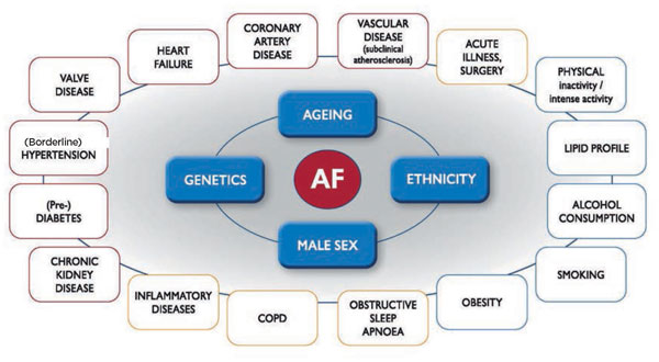
Risk factors which are associated with an increased propensity to developing AF are shown in figure 1.
Along with increased incidence with advancing age, patients with AF often have one or more risk factors that are classically associated with CVD.4,5
These include, but are not exclusively: hypertension even if well controlled; underlying heart disease i.e. valvular heart disease, ischaemic heart disease or cardiomyopathy; environmental factors i.e. smoking or alcohol; obesity and chronic comorbidities i.e. diabetes, obstructive sleep apnoea and thyroid dysfunction.
However, approximately 50% of patients will have no comorbidity.
Diagnosis
There has been much debate about how a diagnosis of AF should be established and what should be considered clinically relevant. Consensus agreement forms part of the ESC 2020 AF management guidelines.6 To establish a diagnosis of AF, rhythm documentation with an ECG showing at least 30 seconds of AF is required.
This can be in either a standard 12-lead ECG, single lead ECG or rhythm recording device showing a heart rhythm with an absence of repeating P waves, irregular R-R interval (when AV conduction is not impaired) and irregular fibrillatory atrial activation.
The guidelines make a distinction between clinical AF being confirmed on an ECG and subclinical AF. This also applies to patients with devices capable of recording heart rate/rhythm including pacemakers and implantable loop recorders and wearable devices.
Atrial high rate episodes or subclinical AF are usually short-lived but can be frequent.6 The guidelines reflect that a high burden of these episodes and/or duration (e.g. single episodes lasting > 6 min) may reflect a high predisposition to transition to clinical AF and warrant risk stratification for protection against thromboembolism.
Each patient should be assessed for their individual risk profile with shared decision making for initiation of stroke protection and AF treatment. A period of more intensified observation may be needed to detect clinical AF more promptly in these patients.
There are several options for rhythm analysis recording for AF detection. Many of these are patient-directed and symptom-responsive devices. These include patient-initiated intermittent ECG rhythm strips using a smartphone or dedicated connectable device. These wearable devices that can produce a diagnostic quality ECG rhythm strip often come packaged in watches, belts, skin patches or rings.10 They have the advantage of detecting subclinical and asymptomatic AF.
Assessment
The ESC guidelines describe a standard approach for diagnostic evaluation of AF patients that should include:
• A complete medical history
• Assessment of concomitant conditions
• AF pattern
• Stroke risk
• AF-related symptoms
• Thromboembolism
• LV dysfunction
A 12-lead ECG is recommended in all patients with suspected AF to confirm the diagnosis of AF, assess ventricular rate during AF, and check for the presence of conduction defects, ischaemia, or signs of structural heart disease. Blood tests (thyroid and kidney function, serum electrolytes, full blood count) and transthoracic echocardiography (LV size and function, LA size, valvular disease, and right heart size and systolic function) are needed to guide treatment.
The new guidelines recommend that all newly diagnosed AF patients should undergo echocardiographic studies. From a pragmatic standpoint, priority should be given to patients with suspected structural heart disease, such as AF related or de novo cardiomyopathies and/or suspected valvular abnormalities and younger patients (< 60 years of age).
Most AF patients benefit from annual review, mainly focused on updating thromboembolic risk stratification and monitoring of oral anticoagulation, which can usually be delivered within primary care.
Reasons for referral to cardiology services include:
• A diagnosis of underlying structural heart disease
• Difficulties achieving adequate rate control
• Advice on choice of rhythm control (medication, cardioversion, etc.)
• Challenges with anticoagulation
• Consideration of non-pharmacological intervention.
The expertise of a cardiac electrophysiologist may be required for patients with symptomatic AF despite trials of antiarrhythmic medications, associated atrial flutter and AF with Wolf-Parkinson-White syndrome as these patients may be candidates for catheter ablation procedures.
Management
Stroke prevention
Oral anticoagulation is the cornerstone of AF management for most patients in whom there is clear prognostic benefit in terms of stroke prevention.11 As soon as AF is diagnosed, the CHA2DS2-VASc score should be calculated, and anticoagulation initiated in appropriate patients (see tables 1 and 2). Stroke risk is independent of the degree of symptoms and the burden of AF.
Along with calculating a patient’s thromboembolic risk the updated NICE guidance recommends that all patients should have a bleeding risk profile.5 Many clinicians will be familiar with the HAS-BLED score but the latest guidelines favour the ORBIT bleeding score, which has a higher accuracy in predicting absolute bleeding risk (see table 3). NICE emphasises that an accurate knowledge of bleeding risk supports shared decision-making and has long-term clinical benefits, e.g. increasing patient confidence and willingness to accept treatment when the ORBIT risk is low and prompting discussion regarding modifiable risk reduction when patients have a high ORBIT score. Although ORBIT is deemed the best tool, other bleeding risk tools may still be used.
Apixaban,12 dabigatran,13 edoxaban,14 and rivaroxaban15 have all been tested for stroke prevention in AF in randomised trials. Relevant clinical factors to consider when choosing an agent include bleeding risk, presence and degree of renal impairment, availability of reversal agents and monitoring, and preference for a once or twice daily dosing regimen.11
It should be noted that in the clinical trials, where no direct oral anticoagulant (DOAC) reversal agents were available, mortality from bleeding was higher in the warfarin arms (where vitamin K was readily available) than the DOAC arms.12,14,15
A vitamin K antagonist (VKA) e.g. warfarin, should only be offered to patients with a new diagnosis of AF if a DOAC is contraindicated, not tolerated, or unsuitable. In patients with AF who are already taking a VKA and are stable, the option to switch to a DOAC should be discussed at their next routine appointment, taking into account their time in therapeutic INR range.
For most patients with AF, the benefit of anticoagulation outweighs the bleeding risk. Anticoagulation should not be withheld solely because of advanced age or risk of falls, and aspirin and other antiplatelet drugs are not effective for stroke prevention in patients with AF.6
If anticoagulation is contraindicated or not tolerated, left atrial appendage occlusion is an option that is now approved and funded by NHS England at a number of specialist centres. This involves deployment of a self-expanding occlusion device using a minimally invasive percutaneous technique.
Only a brief period (6-12 weeks) of dual antiplatelet therapy is needed post procedure, after which aspirin is continued for another 4 months, then stopped. It offers lifelong protection equivalent to oral anticoagulants, but without the increased bleeding risk.
Rate and rhythm control
The decisions regarding rate and rhythm control depend on symptoms and clinical context. Rhythm control should be considered in new onset AF, when there is a reversible cause for AF, when heart failure is present and thought to be due to AF, and in young patients or others in whom a rhythm control strategy appears more suitable.
In other patients (typically the elderly and truly asymptomatic), rate control is an appropriate first-line strategy, with no survival disadvantage compared with rhythm control and some potential advantages, such as lower risk of adverse drug effects.5,6
Options for rate control include a standard beta-blocker (e.g. bisoprolol), a rate-limiting calcium channel blocker (diltiazem or verapamil), and digoxin.
A lenient target heart rate of less than or equal to 110 bpm at rest can be accepted if this is sufficient to control symptoms.5,6 Atrioventricular node ablation and pacing can be considered in patients with permanent AF with difficult to control high ventricular rates and symptoms or left ventricular dysfunction.
Rhythm control should be considered for patients with AF in whom a rate control strategy has not adequately controlled symptoms, and it is reasonable to refer such patients for a specialist opinion and transthoracic echocardiography.
Medical options for rhythm control include flecainide, sotalol, amiodarone, and dronedarone. A ‘pill-in-the-pocket’ strategy, i.e. where the antiarrhythmic medication is taken at the beginning of an AF episode, can be used in patients with paroxysmal AF and is ideally suited to those who have infrequent attacks which would normally last for at least a few hours. It requires a single, 300 mg oral dose of flecainide.
In patients with persistent AF that has lasted longer than 48 hours, electrical cardioversion is usually the option of choice for rhythm control. Amiodarone therapy may be commenced prior to cardioversion to increase the probability of successful restoration of sinus rhythm and may also be continued after cardioversion to reduce the risk of AF recurrence, although usually for a limited period of a few months to avoid long-term side effects.
Left atrial catheter ablation (pulmonary vein isolation) is indicated for patients with symptomatic paroxysmal or persistent AF in whom drug treatment is unsuccessful, unsuitable, or not tolerated, and may also be considered as an alternative to antiarrhythmic drugs in patients with paroxysmal AF.6,16 It has also been shown to improve life expectancy and reduce hospital admissions in people with heart failure and symptoms or whose heart function deteriorates in AF. It can be performed as a day case procedure, sometimes under sedation but often with a general anaesthetic.
Risk factors and lifestyle interventions
Management of risk factors and concomitant CVD complements stroke prevention and reduces AF burden and symptom severity.6,17 Weight loss in obese patients with AF18 and alcohol abstinence in patients with AF who drink 10 units or more per week19 have been shown to improve AF-related symptoms and reduce AF recurrence in clinical trials. By contrast, it is unlikely that caffeine consumption causes or contributes to AF.20 All patients should be assessed for obstructive sleep apnoea. Hypertension and diabetes should be aggressively managed.
Monitoring and follow-up
After a diagnosis of AF has been confirmed and stroke risk stratification performed, control of symptoms will generally drive further management; routine monitoring of rhythm or AF burden is usually not required. Importantly, anticoagulation should not be stopped simply because sinus rhythm appears to be maintained. Any decision to discontinue anticoagulation should be based on reassessment of both stroke and bleeding risk, AF burden and a discussion of the patient’s preferences.
Conclusion
AF is a common condition that is increasing in frequency. Although the dysrhythmia is associated with significant morbidity and mortality, evidence-based AF management improves patient outcomes. In particular, anticoagulation in high-risk patients, as determined by the CHA2DS2-VASc score, reduces the risk of AF-related stroke. It is important to select appropriate rate control and rhythm control therapies carefully to minimise symptoms and adverse reactions.
REFERENCES
1 Chugh SS, Havmoeller R, Narayanan K et al. Worldwide epidemiology of atrial fibrillation: A global burden of disease 2010 study. Circulation 2014;129(8):837-47
2 Lip GYH, Nieuwlaat R, Pisters R et al. Refining clinical risk stratification for predicting stroke and thromboembolism in atrial fibrillation using a novel risk factor-based approach: The Euro Heart Survey on atrial fibrillation. Chest 2010;137(2):263-72
3 Gladstone DJ, Spring M, Dorian P et al. Atrial fibrillation in patients with cryptogenic stroke. N Engl J Med 2014;370(26):2467-77
4 Wolf PA, Abbott RD, Kannel WB. Atrial fibrillation as an independent risk factor for stroke: the Framingham study. Stroke 1991;22(8):983-88
5 National Institute for Health and Care Excellence. NG196. Atrial fibrillation: diagnosis and management. NICE. London. 2021
6 Hindricks G, Potpara T, Dagres N et al. 2020 ESC Guidelines for the diagnosis and management of atrial fibrillation developed in collaboration with the European Association for Cardio-Thoracic Surgery (EACTS). Eur Heart J 2021;42:373-498
7 Barsky AJ, Ahern DK, Brener J et al. Palpitations and cardiac awareness after heart transplantation. Psychosomatic Med 1998;60(5):557-62
8 Santos ABS, Kraigher-Krainer E, Gupta DK et al. Impaired left atrial function in heart failure with preserved ejection fraction. Eur J Heart Fail 2014;16(10):1096-1103
9 Zakeri R, Chamberlain AM, Roger VL, Redfield MM. Temporal relationship and prognostic significance of atrial fibrillation in heart failure patients with preserved ejection fraction: a community-based study. Circulation 2013;128(10):1085-93
10 Steinhubl SR, Waalen J, Edwards AM et al. Effect of a home-based wearable continuous ECG monitoring patch on detection of undiagnosed atrial fibrillation the mSToPS randomized clinical trial. JAMA 2018;320(2):146-55
11 Ruff CT, Giugliano RP, Braunwald E et al. Comparison of the efficacy and safety of new oral anticoagulants with warfarin in patients with atrial fibrillation: a meta-analysis of randomised trials. Lancet 2014;383(9921):955-62
12 Granger CB, Alexander JH, McMurray JJV et al. Apixaban versus warfarin in patients with atrial fibrillation. N Engl J Med 2011;365(11):981-92
13 Connolly SJ, Ezekowitz MD, Yusuf S et al. Dabigatran versus warfarin in patients with atrial fibrillation. N Engl J Med 2009;361(12):1139-51
14 Giugliano RP, Ruff CT, Braunwald E et al. Edoxaban versus warfarin in patients with atrial fibrillation. N Engl J Med 2013;369(22):2093-2104
15 Patel MR, Mahaffey KW, Garg J et al. Rivaroxaban versus warfarin in nonvalvular atrial fibrillation. N Engl J Med 2011;365(10):883-91
16 Imberti JF, Ding WY, Kotalczyk A et al. Catheter ablation as first-line treatment for paroxysmal atrial fibrillation: a systematic review and meta-analysis. Heart 2021;107(20):1630-36
17 Rienstra M, Hobbelt AH, Alings M et al. Targeted therapy of underlying conditions improves sinus rhythm maintenance in patients with persistent atrial fibrillation: Results of the RACE 3 trial. Eur Heart J 2018;39(32):2987-96
18 Abed HS, Wittert GA, Leong DP et al. Effect of weight reduction and cardiometabolic risk factor management on symptom burden and severity in patients with atrial fibrillation: a randomized clinical trial. JAMA 2013;310(19):2050-60
19 Voskoboinik A, Kalman JM, de Silva A et al. Alcohol abstinence in drinkers with atrial fibrillation. N Engl J Med 2020;382(1):20-28
20 Cheng M, Hu Z, Lu X et al. Caffeine intake and atrial fibrillation incidence: dose response meta-analysis of prospective cohort studies. Can J Cardiol 2014;30:448-54
ARTICLE IN PDF
 Download this article in PDF format
PRESET SEARCHES
PubMed
Atrial fibrillation treatment review
Stroke and atrial fibrillation
NICE evidence search