SEARCH

Salmon N, Tidman MJ. Managing actinic keratosis in primary care. Practitioner 2016; 260(1797)25-29
Managing actinic keratosis in primary care
24 Oct 2016
AUTHORS
Dr Nicola Salmon MBChB MRCP, Dermatology LAT
Dr Michael J Tidman MD FRCP(Edin), FRCP(Lond), Consultant Dermatologist
Department of Dermatology, Royal Infirmary of Edinburgh, Edinburgh, UK
Article
Abstract
Actinic, or solar, keratosis is caused by chronic ultraviolet-induced damage to the epidermis. In the UK, 15-23% of individuals have actinic keratosis lesions. Risk factors include: advanced age; male gender; cumulative sun exposure or phototherapy; Fitzpatrick skin phototypes l-ll; long-term immuno-suppression and genetic syndromes e.g. xeroderma pigmentosum and albinism. Actinic keratoses are regarded by some authorities as premalignant lesions that may transform into invasive squamous cell carcinoma (SCC) and by others as in situ SCC that may progress to an invasive stage. The risk of malignant change appears low; up to 0.5% per lesion per year. Up to 20-30% of lesions may spontaneously regress but in the absence of any reliable prognostic clinical indicators regarding malignant potential active treatment is considered appropriate. Actinic keratosis lesions may present as discrete hyperkeratotic papules, cutaneous horns, or more subtle flat lesions on sun-exposed areas of skin. The single most helpful diagnostic sign is an irregularly roughened surface texture: a sandpaper-like feel almost always indicates actinic damage. Dermatoscopy can be helpful in excluding signs of basal cell carcinoma when actinic keratosis is non-keratotic. It is always important to consider the possibility of SCC. The principal indication for referral to secondary care is the possibility of cutaneous malignancy. However, widespread and severe actinic damage in patients who are immunosuppressed is also a reason for referral.
Article
Actinic, or solar, keratosis is caused by chronic ultraviolet-induced damage to the epidermis. Histologically, lesions are characterised by epidermal keratinocytes demonstrating cytological abnormalities of varying degree, ranging from mild to severely atypical dysplasia.
Actinic keratosis tends to develop in fair-skinned individuals and the prevalence rates vary widely depending on latitude. In the UK, 15-23% of individuals have actinic keratoses,1,2 compared with 52-55% in Australia.3,4 Chronically immunosuppressed individuals are at particular risk of developing actinic keratosis.5
The risk factors for actinic keratosis are summarised in table 1.
Actinic keratoses are regarded by some authorities as premalignant lesions that may transform into invasive squamous cell carcinoma (SCC), and by others as in situ SCC that may progress to an invasive stage.5
The risk of malignant transformation is not precisely quantifiable from available data, but appears low — up to 0.5% per lesion per year.6 It is estimated that there is a 10% risk of SCC developing within ten years in an individual with eight actinic keratoses.7
Up to 20-30% of lesions may spontaneously regress,6 but in the absence of any reliable prognostic clinical indicators regarding malignant potential active treatment is considered appropriate.5
Presentation
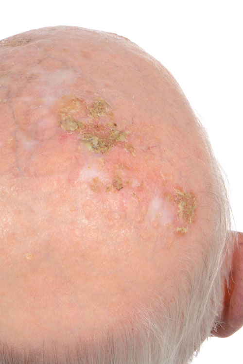
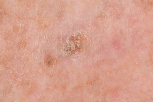
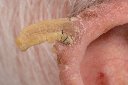
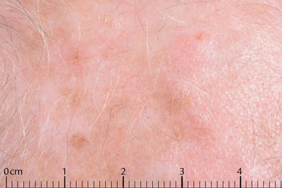
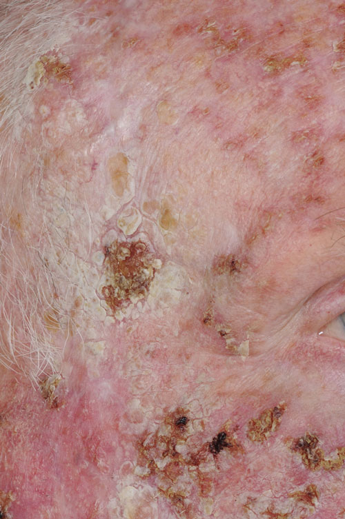
Actinic keratosis may present as discrete hyperkeratotic papules, see figure 1, cutaneous horns, see figure 2, or more subtle flat lesions, see figure 3, on chronically sun-exposed regions of adult skin. The surrounding skin often has features of sun damage (dappled pigmentation, solar lentigines, erythema and telangiectasia, and elastotic changes). It is not unusual to see multiple or even confluent lesions covering large areas of skin, for instance the entire pate in an elderly man, see figure 4, or the backs of the hands, commonly referred to as field change or field cancerisation.
Actinic keratoses are usually asymptomatic but can be irritating and occasionally tender especially on the scalp if there is an accumulation of pus.
The single most helpful diagnostic sign is an irregularly roughened surface texture: a sandpaper-like feel almost always indicates actinic damage.
Diagnosis
For the majority of actinic keratoses, the clinical features are sufficient to enable a confident diagnosis. Dermatoscopy can be helpful in excluding signs of basal cell carcinoma when actinic keratosis is non-keratotic. It is always important to consider the possibility of SCC,8 and, of the features highlighted in table 2, pain on gentle pressure is a particularly concerning sign. If there is clinical doubt as to the diagnosis, a biopsy for histological evaluation is indicated.
Management
The rationale for treatment is to minimise the risk of invasive SCC, although from the patient’s perspective there may also be a cosmetic benefit.
Any therapeutic approach should be tailored not only to the morphological type of actinic keratosis (single lesions versus field change) but also to the individual’s age, general health and personal preferences.9 Given the low risk of malignant change, some affected individuals may prefer the option of watchful vigilance.
Sun protection
Patients should be informed about realistic and sensible sun protection measures, see table 3, below, particularly when abroad and between 10 am and 2 pm when the sun is at its most vertical. Ideally patients should be given a written information leaflet such as that produced by the British Association of Dermatologists
(see Useful information box). Adherence to sun protection measures will minimise further actinic damage. However, it will not completely reverse the damage already done and thus will not entirely prevent more actinic keratoses developing. Additionally, patients should be taught to recognise features that might indicate neoplastic transformation, see table 2.
Sun protection measures should be weighed against the undoubted benefits of sun exposure.10 Assiduous avoidance of sunlight may significantly compromise vitamin D biosynthesis, and, if thought necessary, dietary advice or vitamin D supplements should be considered.
Cryotherapy
The destruction of actinic keratoses with liquid nitrogen is particularly appropriate for relatively small numbers of discrete lesions, whether keratotic or not. It is simple, quick and effective. However, it can be uncomfortable and cause an inflammatory reaction that may include blistering, but nonetheless is generally well tolerated. The resultant scabs may take two to three weeks to disappear, usually leaving no trace of sun damage, although hypopigmentation and scarring are potential adverse effects.
When cryotherapy is not available, is unacceptable to the patient, or is not appropriate because of field change, there are a number of topical options. These include 5-fluorouracil (5-FU), diclofenac, imiquimod and ingenol mebutate. All these medications cause an irritant reaction to a greater or lesser degree, see figure 5, below, and patients should be carefully counselled about this. Discomfort can be alleviated with a topical corticosteroid. Large areas of actinic keratosis may require piecemeal treatment.
5-fluorouracil
5-FU is a thymidylate synthase inhibitor, which causes DNA damage when applied to actinic keratoses.
There are two licensed fluorouracil products in the UK: 5% 5-FU cream, which is applied once or twice daily for three to four weeks, and can be safely applied to up to 500 cm2 of skin at any one time; and a 0.5% 5-FU/10% salicylic acid cutaneous solution, applied once daily for up to 12 weeks, limited to 25 cm2 at any one time. The latter has the advantage of the salicylic acid component reducing the hyperkeratotic element of actinic keratosis and thus improving the penetration of the 5-FU.
Diclofenac
Diclofenac gel (3%) has only moderate efficacy and requires an extended treatment period (twice daily for 60-90 days), but is less irritant than the other topical options. It can be applied to an area of up to 200 cm2.
Imiquimod
Imiquimod is an immune response modifier with an efficacy and side effects similar to 5-FU. There are two licensed products in the UK:
5% imiquimod cream, applied three nights a week for four weeks, covering up to 25 cm2 (if after a four-week treatment-free period lesions persist the course can be repeated); and 3.75% imiquimod cream, applied once daily at night (to up to 200 cm2) for two treatment cycles of a fortnight each, interrupted by a two-week treatment-free interval.
Ingenol mebutate
Ingenol mebutate gel is a relatively novel topical therapy, the mechanism of action of which is not fully understood. It is a macrocyclic diterpene ester licensed for the treatment of non-hyperkeratotic, non-hypertrophic actinic keratoses.11 There are two strengths: 0.015%, for use on the face and scalp, applied once daily for three consecutive days to up to 25 cm2 of skin; and 0.05%, for use on the trunk and limbs, applied once daily for two consecutive days (25 cm2 of skin).
Surgery
Curettage is particularly useful for treating very keratotic or hypertrophic actinic keratoses, see figure 2, and formal excision should be considered when neoplastic change is a possibility.
Individualising treatment
A detailed multinational guideline has recently been published.5 It strongly recommends that small numbers of discrete actinic keratoses are best treated by cryotherapy, with a lesser strength of recommendation for the use of 5-FU, imiquimod (3.75% and 5%), ingenol mebutate, and the 0.5% 5-FU/10% salicylic acid combination. Individual hyperkeratotic actinic keratoses can be removed by curettage.
The strongest recommendation for the treatment of patients with multiple actinic keratoses (> 6 per body region) or with field change is for ingenol mebutate and 3.75% imiquimod. There is less evidence and consensus support for the use of 5-FU, the 0.5% FU/10% salicylic acid combination, 5% imiquimod and 3% diclofenac in this patient group. Cryotherapy can be used for individual actinic keratoses but is not recommended for field change.
In immunosuppressed patients with actinic keratosis there is no recommendation at the strongest level, although cryotherapy, curettage, 5% 5-FU and 5% imiquimod receive weak recommendations.
Referral
The majority of patients with actinic keratosis can be diagnosed and adequately managed in the community. The principal indication for referral to secondary care is the possibility of cutaneous malignancy. However, widespread and severe actinic damage in immunosuppressed patients is also a reason for referral.
Photodynamic therapy, which involves applying a photosensitising cream (such as 5-aminolevulinic acid) to affected skin prior to exposure to visible light, and laser surgery can be used to treat multiple actinic keratoses.
Acitretin, a systemic retinoid, may be justified as chemoprophylaxis when there is a high cutaneous malignancy risk in immunocompetent and immunosuppressed individuals with severe actinic damage.
Conclusion
Patients with actinic keratosis are at risk of developing invasive SCC. Management should be tailored to the individual, but education about sun protection and warning features of malignancy is essential in all cases. Long-term continued vigilance and intermittent treatment of actinic keratosis are necessary in its effective management.
REFERENCES
1 Harvey I, Frankel S, Marks R et al. Non-melanoma skin cancer and solar keratoses. 1. Methods and descriptive results of the South Wales skin cancer study. Br J Cancer 1996;74:1302-7
2 Memon AA, Tomenson JA, Bothwell JS, Friedmann PS. Prevalence of solar damage and actinic keratosis in a Merseyside population. Br J Dermatol 2000;142:1154-59
3 Frost C, Green A and Williams G. The prevalence and determinants of solar keratoses at a subtropical latitude (Queensland, Australia). Br J Dermatol 1998;139:1033-39
4 Frost C, Williams G, Green A. High incidence and regression rates of solar keratoses in a Queensland community. J Invest Dermatol 2000;115:273-77
5 Werner R, Stockfleth E, Connolly S et al. Evidence and consensus-based (S3) guidelines for the treatment of actinic keratosis - International League of Dermatological Societies in cooperation with the European Dermatology Forum Short version. J Eur Acad Dermatol Venereol 2015;29:2069-79
6 Werner RN, Sammain A, Erdmann R et al. The natural history of actinic keratosis: a systematic review. Br J Dermatol 2013;169:502-18
7 Dodson JM, DeSpain J, Hewett JE, Clark DP. Malignant potential of actinic keratoses and the controversy over treatment. Arch Dermatol 1991;127:1029-31
8 Quaedvlieg P, Tirsi E, Thissen M, Krekels G. Actinic keratosis. How to differentiate the good from the bad ones? Eur J Dermatol 2006;16:335-39
9 de Berker D, McGregor J, Hughes B. Guidelines for the management of actinic keratoses. Br J Dermatol 2007;156:222-30
10 Wright F, Weller RB. Risks and benefits of UV radiation in older people: more of a friend than a foe? Maturitas 2015;81(4):425-31
11 Actinic keratosis: ingenol mebutate gel. Evidence summary: new medicine. [ESNM14] March 2013. nice.org.uk/guidance/esnm14
USEFUL INFORMATION
British Association of Dermatologists
www.bad.org.uk/for-the-public/skin-cancer/sunscreen-fact-sheet
DermNet New Zealand
www.dermnetnz.org/topics/actinic-keratosis/
British Skin Foundation
www.britishskinfoundation.org.uk/Home/AK.aspx
Primary Care Dermatology Society
www.pcds.org.uk/clinical-guidance/actinic-keratosis-syn.-solar-keratosis
 Download this article in PDF format