SEARCH

Panthagani AP, Tidman MJ. Diagnosis directs treatment in fungal infections of the skin. Practitioner 2015 - 259 (1786):25-29
Diagnosis directs treatment in fungal infections of the skin
21 Oct 2015
AUTHORS
Dr Anusha P Panthagani MBChB MRCP, Specialist Registrar
Dr Michael J Tidman MD FRCP(Ed) FRCP (Lond), Consultant Dermatologist
Department of Dermatology, Royal Infirmary of Edinburgh, Edinburgh, UK
Article
Cutaneous fungal infections are becoming more common worldwide,1 with increasing numbers being seen in general practice.
Fungi that cause superficial skin infection may be dermatophyte or non-dermatophyte organisms (including candida and other yeasts).
Dermatophyte fungi are confined to the keratin layer of the epidermis and include three genera: Microsporum, Epidermophyton and Trichophyton. These infections can be transmitted by human contact (anthropophilic), from the soil (geophilic) and by animal (zoophilic) spread.
This article will focus on the common dermatophyte infections seen in the surgery, together with pityriasis versicolor.
Dermatophyte infections (tinea or ringworm) are named according to the affected body site: tinea capitis, tinea faciei, tinea corporis, tinea cruris, tinea manuum, tinea pedis and tinea unguium.
Cutaneous fungal infections usually affect individuals who are otherwise entirely healthy, but immunocompromised individuals are particularly susceptible.
Local factors such as a warm, humid environment, excessive sweating, trauma and occlusive clothing can predispose to fungal growth. With global travel, superficial fungal infections that were once limited geographically are now being seen in the UK, for example Trichophyton soudanense and Trichophyton violaceum.2
Tinea pedis
Tinea pedis (athlete’s foot) involves the soles and toe webs and is the most prevalent dermatophyte infection, involving up to 10% of the world’s population at any given time.2 Trichophyton rubrum (T. rubrum) and
T. interdigitale (mentagrophytes) are the most common causative organisms.2
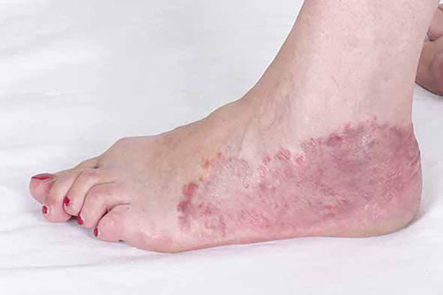
The clinical signs are fissuring, erythema, scaling and maceration of the lateral toe webs and dry scaling of the soles of the feet, see figure 1, above. Occasionally, small blisters may be present on the plantar surface.
When dry scaling involves the toe webs, erythrasma (caused by the bacterium Corynebacterium minutissimum, which glows a coral pink under ultraviolet light) should enter the differential diagnosis.
Tinea unguium
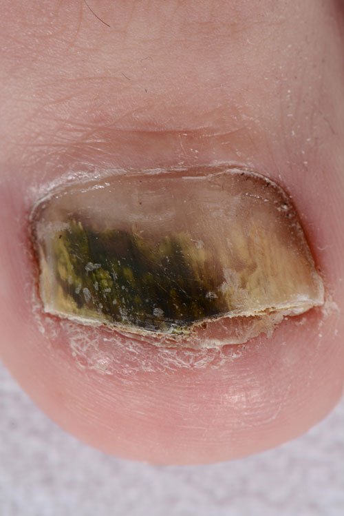
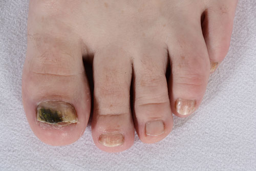
Tinea unguium (onychomycosis) refers to fungal nail infections, most commonly caused by T. rubrum and T. interdigitale. Usually, the nail plate is thickened and discoloured, and may be onycholytic, see figure 2. Occasionally, tinea unguium may present with white flaky patches on the nail plate (superficial white onychomycosis). The differential diagnosis should include psoriasis and peripheral vascular disease.
Tinea capitis
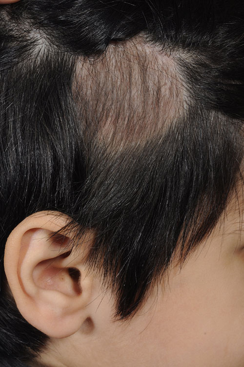
Tinea capitis, see figure 3, is an infection involving the hair follicles of the scalp and the surrounding skin caused by species of Microsporum and Trichophyton, with Trichophyton tonsurans accounting for 50-90% of scalp isolates in the UK.3 Children are particularly prone to tinea capitis.
The clinical features include patchy hair loss with varying degrees of scale, erythema and pustules. Infected hairs tend to break at the base, leaving stubble.
Occasionally, there is invasion of the visible epidermis, which leads to a very pronounced inflammatory response, resulting in a boggy, painful swelling with associated alopecia and regional lymphadenopathy. This is known as a kerion.
Tinea manuum
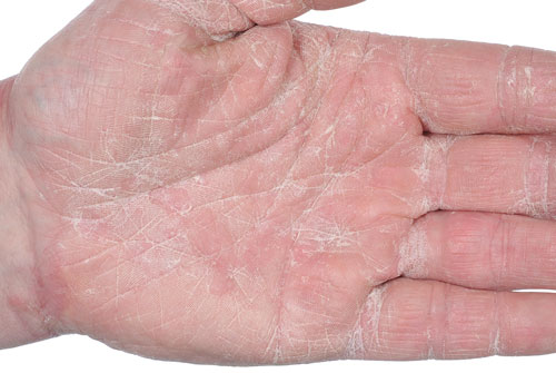
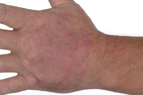
Any dermatophyte species affecting the body can affect the hands. The most common organism is T. rubrum but Epidermophyton floccosum and T. interdigitale are involved in a minority of cases. Tinea manuum usually presents as unilateral scaling of the palms and fingers with accentuation within the palmar creases, see figure 4.
Tinea corporis
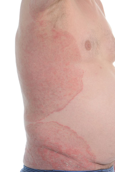
Tinea corporis infection affects the trunk, see figure 5, mainly in children and adolescents,4 and all genera of dermatophyte can cause it.
In the UK, the most common culprit is T. rubrum. Typical clinical features include a characteristic annular lesion with an erythematous or vesicular border, and a clearing centre.
Tinea cruris
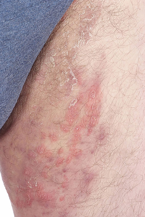
Tinea cruris infection involves the groin region and is more common in men than women. T. rubrum is the most common causative dermatophyte.4
The infection may begin with a well demarcated pruritic erythematous patch on the inner aspect of one or
both thighs and can extend to the pubic, suprapubic, perianal and buttock regions, see figure 6, opposite.
Again, erythrasma should be considered in the differential diagnosis.
Pityriasis versicolor
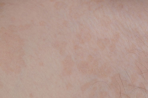
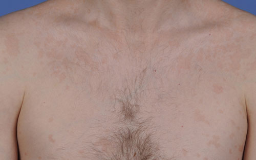
Pityriasis versicolor is caused by fungi of the genus Malassezia. The rash presents as well demarcated, hypo- or hyper-pigmented scaly macules, coalescing into large patches, see figure 7.
Diagnosis
Dermatophyte infections usually present as an erythematous, scaly eruption, which may or may not be itchy. Asymmetry is an important clinical clue to fungal infection, as is annular morphology.
Examination under ultraviolet (Wood’s light) can be helpful. Microsporum species tend to fluoresce a green colour, whereas Trichophyton species do not (except for T. schoenleinii). In pityriasis versicolor, the scales may fluoresce a yellowish colour.
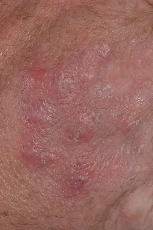
Inappropriate treatment of tinea infections with topical corticosteroids may alter the morphology of the eruption, to produce the characteristic appearance of tinea incognito, see figure 8. The lesions typically have a raised margin and a ‘lumpy, bumpy’ centre consisting of inflammatory nodules and areas of normal looking skin.
However, the gold standard for diagnosing cutaneous fungal infections is microscopy and culture of scale, hair or nail, and a definite diagnosis is desirable before commencing treatment, especially with oral therapy.
Microscopy of the skin samples is positive in up to 91% of fungal infections.5 Mycological culture can take two to three weeks, and even in positive areas can be negative.
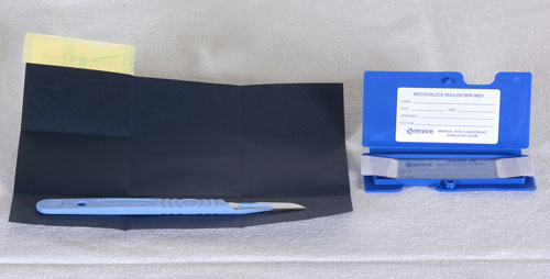
It is important to provide the laboratory with an adequate sample in order to minimise the risk of false-negative results. In the case of skin, the area is cleared with an alcohol wipe and scrapings are obtained with a scalpel and collected in folded dark paper squares. Commercially produced kits, see figure 9, are available from pathology laboratories. It is recommended that 5 mm2 of scrapings are collected.5 Some laboratories also encourage a sample of scale taken with an adhesive tape, which is then mounted onto a slide.
For suspected tinea capitis infections, it is preferable to take both scalp scrapings and plucked hairs.
It is important to take nail clippings, ideally using chiropody scissors, from the diseased part of the nail, and to include subungual debris. If there is superficial nail infection, remove samples of the diseased nail plate with a scalpel.
Management
Topical antifungals
Most localised tinea infections can be treated topically with antifungals. In order to prevent relapse, topical therapy should be continued one to two weeks after resolution of symptoms.
The imidazole antifungals (clotrimazole, ketoconazole and miconazole) are all effective against dermatophytes.
In asymptomatic patients with tinea unguium treatment may not be required, but if treatment is necessary oral therapy is more effective than topical therapy.
Topical antifungals in the form of a laquer (amorolfine and ciclopirox) or a solution (tioconazole) can be used for superficial and distal onychomycosis.6 These treatments need to be applied for at least six to twelve months and compliance may be an issue.
Systemic treatment
Systemic treatment may be initiated if topical treatment is unsuccessful. Oral imidazole or triazole antifungals such as terbinafine, itraconazole and fluconazole are frequently used as they have a broad spectrum of activity and require a shorter duration of treatment.
Treatment with griseofulvin, which is a fungistatic, needs to be continued until the fungal infection has resolved.
Onychomycosis usually necessitates systemic treatment. It is important to do baseline liver function tests before commencing treatment. Terbinafine is considered first line in a dose of 250 mg daily for six weeks for fingernail infections and twelve to sixteen weeks for toenail infections.6 Itraconazole can be considered at a dose of 200 mg once daily for twelve weeks or alternatively as pulse therapy in a dose of 400 mg per day for one week per month. Two pulses are recommended for the fingernails and three pulses for the toenails. Fluconazole is an alternative if the above two drugs cannot be tolerated.
Griseofulvin 500-1,000 mg per day over six to nine months has a lower efficacy and higher relapse rates compared with terbinafine and itraconazole.
Other interventions include debridement, photodynamic therapy, surgical avulsion followed by topical antifungal therapy, and laser treatment.
For tinea pedis, topical treatment for four weeks is recommended, although oral treatment may be indicated in severe infection. In a systematic review, terbinafine was more effective than griseofulvin, while the efficacy of terbinafine and itraconazole were similar.7 If there is any evidence of a concurrent bacterial infection, then antibiotics should be prescribed. Potassium permanganate soaks can be very useful in macerated weeping variants of tinea pedis.
Tinea capitis is treated systemically. Topical therapy alone is not recommended, but can be initiated to reduce transmission of spores.3 Povidine-iodine, ketoconazole 2%, selenium sulfide 1% shampoos have shown efficacy.3
Griseofulvin is the only licensed treatment for children,8 although terbinafine is widely used in children where the shorter duration of treatment can increase compliance. Itraconazole can be used as a second-line treatment as it is safe and effective.
If children are receiving appropriate systemic and adjunctive topical therapy, they should be allowed to attend school or nursery.3 Family screening should be considered and if positive mycology samples are present then treatment should be initiated.3
For pityriasis versicolor, treatment with a topical imidazole, ketoconazole shampoo or selenium sulfide shampoo can be used.8
The shampoo can be applied as a lotion and left on the affected area for ten minutes before it is washed off. This treatment should be administered daily for one week and repeated if necessary.
Pityriasis versicolor may be treated with oral fluconazole or itraconazole if topical treatment is ineffective. Topical terbinafine is effective for pityriasis versicolor but not oral terbinafine.8
The colour change of pityriasis versicolor will take time to resolve and may require gentle sun exposure. Recurrence is common.
Table 1, summarises the oral treatment options available for most of the superficial fungal infections discussed previously.
Conclusion
If there is recalcitrant disease which is not responding to standard therapy or the diagnosis is in doubt the patient should be referred to a dermatologist.
Cutaneous fungal infections are common and for some individuals they can be recurrent. This can cause considerable distress. It is important to take mycology samples and sometimes multiple samples may be necessary before initiation of treatment. Identification of the organism will direct treatment. Rarely, oral antifungals can cause significant adverse effects. including deranged liver function tests, drug-induced lupus and drug reactions including Stevens-Johnson syndrome and toxic epidermal necrolysis.
REFERENCES
1 Havlickova B, Czaika VA, Friedrich M et al. Epidemiological trends in skin mycoses worldwide. Mycoses 2008;51(Suppl 4):2-15
2 Hay RJ, Ashbee HR. (2010) Mycology, in Rook's Textbook of Dermatology, Eighth Edition (eds T. Burns,
S. Breathnach, N. Cox and C. Griffiths), Wiley-Blackwell. Oxford. UK
3 Fuller LC, Barton RC, Mohd Mustapa MF et al. British Association of Dermatologists’ guidelines for the management of tinea capitis 2014. Br J Dermatol 2014;171(3):454-63
4 Van Zuuren EJ, Fedorowicz Z, El-Gohary M. Evidence-based topical treatments for tinea cruris and tinea corporis: a summary of a Cochrane systematic review. Br J Dermatol 2015; 172:616-41
5 Health Protection Agency. Fungal Skin and Nail Infections: Diagnosis & Laboratory Investigation. Quick Reference Guide for Primary Care. London. 2011. www.gov.uk/government/uploads/system
6 Ameen M, Lear JT, Madan V et al. British Association of Dermatologists' guidelines for the management of onychomycosis 2014. Br J Dermatol 2014;171:937-58 doi: 10.1111/bjd.13358
7 Bell-Syer SE, Khan SM, Torgerson DJ. Oral treatments for fungal infections of the skin of the foot. Cochrane Database Syst Rev 2012; 10:CD003584
8 Joint Formulary Committee. British National Formulary 69. BMJ Group and Pharmaceutical Press. London. 2015
ARTICLE IN PDF
 Download this article in PDF format
PRESET SEARCHES
Immediate CPD prompts and frameworks
These handy templates* prompt immediate drafting of personal learning, reflection and planning. Save your record straight into your personal development folder on your device or Cloud storage system.
*Adobe reader needed for iPad