SEARCH

Ooi ET, Tidman MJ. Improving the management of seborrhoeic dermatitis. Practitioner 2014–258 (1768):23-26
Improving the management of seborrhoeic dermatitis
24 Feb 2014
AUTHORS
Dr Ee Ting Ooi MBChB MRCP(UK), Dermatology Specialty Trainee
Dr Michael J Tidman MD FRCP(Ed) FRCP (Lond), Consultant Dermatologist, Department of Dermatology, Royal Infirmary of Edinburgh, Edinburgh, UK
Article
Seborrhoeic dermatitis is a common inflammatory, erythematous, papulo- squamous skin disease.
It usually starts at puberty with a peak incidence at 40 years of age and is more common in males. Approximately 5% of the population have seborrhoeic dermatitis which is severe enough to require medical attention.1
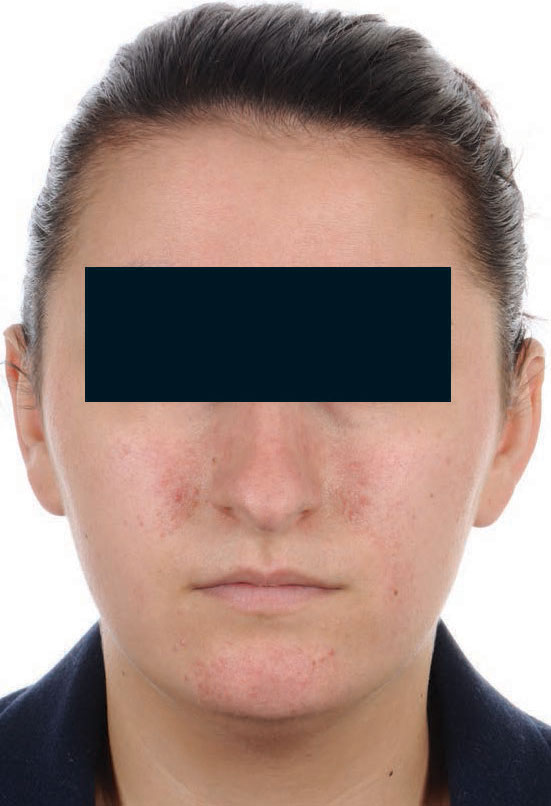
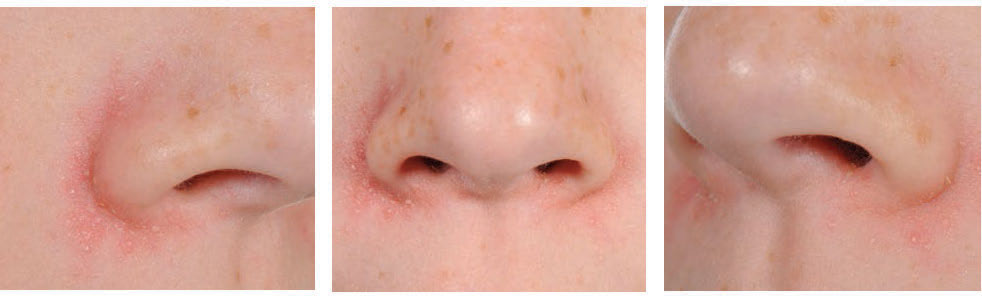
Patients with seborrhoeic dermatitis develop symmetrical, well demarcated, dull or yellowish red patches and plaques with overlying adherent, yellowish greasy scales.
Seborrhoeic dermatitis has a distinctive distribution in areas rich in sebaceous glands – the scalp, eyebrows, glabella, nasolabial and nasofacial folds, cheeks, peri-auricular skin, pre-sternal and interscapular areas
(see figure 1, and figure 2).
Some patients may develop isolated dandruff (visible desquamation from the scalp without scalp inflammation) which is best regarded as mild seborrhoeic dermatitis and not as a separate condition. Dandruff may also be a precursor to more severe seborrhoeic dermatitis.
Others may develop blepharitis with erythematous eyelids covered by small, white scales and/or yellow crusts and destruction of eyelash follicles. Seborrhoeic dermatitis may occur in flexures, especially the axillae, groin, anogenital skin, infra-mammary skin and the umbilicus, which may be complicated by maceration and intertrigo. Patients may experience mild itching.
Seborrhoeic dermatitis may be worsened by tiredness and stress. Exposure to sunlight may temporarily exacerbate the condition but this is generally followed by an overall improvement. Rarely, seborrhoeic dermatitis becomes generalised resulting in erythroderma.
Infantile seborrhoeic dermatitis
Infantile seborrhoeic dermatitis is a similar condition usually starting in the first month of life and resolving by four months of age.
Some consider cradle cap to be congenital infantile seborrhoeic dermatitis. Dermatologists are divided in their opinions as to whether or not infantile seborrhoeic dermatitis is a clinical entity distinct from seborrhoeic dermatitis because there is as yet no proven relationship between the two conditions and children with infantile seborrhoeic dermatitis are no more likely than their peers to develop seborrhoeic dermatitis. Children with infantile seborrhoeic dermatitis are almost invariably well and neither feeding nor sleep are disturbed.
Causes
Seborrhoeic dermatitis appears to be dependent on the presence of the yeast Malassezia furfur. M. furfur is a commensal, lipophilic yeast, naturally found on the skin surface in many animals. In humans, the yeast can be found in the acroinfundibulum of the hair follicle.
M. furfur has also been associated with pityriasis versicolor and pityrosporum folliculitis. At puberty, sebaceous glands become more active and colonisation with M. furfur increases. It is hypothesised that M. furfur then induces seborrhoeic dermatitis by activating complement through an alternative lipase-producing pathway.
The role of seborrhoea remains debatable. The sebum excretion rate from forehead skin was found to be normal in male patients and significantly reduced in female patients.2 However, adolescents with seborrhoeic dermatitis had a different composition of skin surface lipids compared with their peers. They had higher cholesterol, triglyceride and paraffin fractions and lower free fatty acids (FFA), wax esters and squalene fractions compared with control subjects.3 As FFAs are produced by bacterial lipolysis, this suggests that there may be an alteration of the bacterial microflora in seborrhoeic dermatitis.3
The pathogenesis of infantile seborrhoeic dermatitis is less clear cut. It may be caused by sebaceous glands being active at birth through stimulation by maternal androgens or nutritional factors e.g. biotin deficiency. However, these hypotheses have not been proven; and neither has the role of M. furfur.
The sebum excretion rate in infantile seborrhoeic dermatitis appears normal, although there may be an altered FFA composition. There is no evidence of a genetic component in infantile seborrhoeic dermatitis.
Risk factors
Patients with HIV infection have a higher incidence of seborrhoeic dermatitis and tend to be more severely affected.
This suggests that impaired cell-mediated immunity may facilitate yeast survival in the skin and contribute to a low T-cell dependent antibody response, resulting in the inflammatory response not being downregulated.3
Patients with neurological diseases, including parkinsonism and cranial nerve palsies, also have a higher incidence of seborrhoeic dermatitis. Interestingly, a patient with a unilateral facial nerve palsy has been reported to have developed unilateral seborrhoea and seborrhoeic dermatitis on the affected side.4
There may be a direct link between alterations in neurological pathways and the development of seborrhoeic dermatitis; or it may be that these diseases tend to cause seborrhoea, which in turn increases the colonisation of M. furfur.
Diagnosis
The diagnosis of seborrhoeic dermatitis can usually be made, without the need for any investigations, based on a complete clinical history and examination. However, if the presentation is not classic certain investigations may be required. Patients presenting with sudden onset severe seborrhoeic dermatitis should be screened for risk factors for HIV infection.
The differential diagnoses for seborrhoeic dermatitis are listed in box 1. In infants, there is significant overlap of clinical features of infantile seborrhoeic dermatitis with atopic eczema and psoriasis. Some authorities believe that infantile seborrhoeic dermatitis is actually no more than atopic eczema temporarily attenuated by the natural lubrication of sebum, and as soon as sebaceous secretion decreases and the scratch reflex matures, the typical eruption of atopic eczema appears.5
Primary irritant contact dermatitis can mimic infantile seborrhoeic dermatitis localised to the napkin area. In infants, psoriasis usually affects flexural sites, and differentiating between infantile seborrhoeic dermatitis and infantile inverse psoriasis may only be possible later in life.
Management
Recommended treatments for non-scalp disease are listed in table 1, above, and treatments for scalp disease in table 2.
Other imidazole creams may also be tried, e.g. 1% clotrimazole cream, 1% econazole cream or 2% miconazole cream.7 When symptoms are under control, maintenance with weekly use of topical imidazole creams may prevent recurrence, however repeated reapplication may induce tolerance. Shampoo containing 2% ketoconazole may also be used as a body wash and cotton buds moistened with baby shampoo may be used to cleanse eyelids.7
Patients should be advised to remove thick crusts or scales on hair-bearing sites by applying warm mineral/olive oil for several hours before washing with a medicated shampoo.7 Patients can also try coal tar, salicylic acid or tea tree oil-containing shampoos.3,7
When symptoms are under control, weekly use of medicated shampoos may prevent recurrence. Potent topical corticosteroid scalp application e.g. betamethasone valerate 0.1%, hydrocortisone butyrate 0.1% or mometasone furoate 0.1% can be prescribed concomitantly for four weeks.7 Alcohol-based preparations and hair tonics should be avoided.2
Mild cases of infantile seborrhoeic dermatitis do not require treatment. The mainstay of treatment for more severe scalp disease is softening and removing scales by massaging vegetable/mineral oils into the scalp before bathing in bath water containing a dispersing bath oil. Shampoo containing 2% ketoconazole may also be used; however shampoos containing salicylic acid or selenium sulphide should be avoided because of increased systemic absorption in infants.
Infants with non-scalp disease should be bathed every day with a water-dispersible emollient; over-the-counter soaps/detergents and vigorous cleansing should be avoided. Topical imidazole creams, e.g. 2% ketoconazole cream with or without a mild topical corticosteroid, e.g. 1% hydrocortisone cream, may be required.
Frequent napkin changes and attention to skin protection with simple barrier emollients or zinc oxide pastes is also important.
Referral
Patients should be referred in the following situations:7
• Diagnostic uncertainty – consider other differential diagnoses
• Failure to respond to first-line treatment after four weeks – consider secondary changes e.g. bacterial infection, flexural intertrigo, lichenification, otitis externa
• Severe/widespread disease
Conclusion
Patients with seborrhoeic dermatitis have a good prognosis, particularly infantile seborrhoeic dermatitis, which usually remits within a few weeks or months and does not recur.
Seborrhoeic dermatitis may require treatment for a number of years but the long-term prognosis remains good.
REFERENCES
1 Bieber T. Other types of dermatitis. In: Burgdorf WHC, Plewig G, Wolff HH, Landthaler M (Eds.) Braun-Falco's Dermatology. 3rd edition. Springer; 2009. p425-433
2 Berth-Jones J. Eczema, lichenification, prurigo and erythroderma. In: Burns T, Breathnach S, Cox N, Griffiths C (Eds.) Rook’s Textbook of Dermatology. 8th edition. Wiley-Blackwell; 2010. p23.29-23.34
3 Langtry JAA. Seborrhoeic dermatitis of adolescence. In: Irvine AD, Hoeger PH, Yan AC (Eds.) Harper’s Textbook of Pediatric Dermatology Volume 1. 3rd edition. Wiley-Blackwell; 2011. p41.1-41.5
4 Bettley FR, Marten RH. Unilateral seborrhoeic dermatitis following a nerve lesion. Arch Dermatol 1956;73:110-115
5 Gelmetti CM, Grimalt R. Infantile seborrhoeic dermatitis. In: Irvine AD, Hoeger PH, Yan AC (Eds.) Harper’s Textbook of Pediatric Dermatology Volume 1. 3rd edition. Wiley-Blackwell; 2011. p35.1-35.8
6 Williams J, Coulson I. Seborrheic eczema. In: Lebwohl MG, Heymann WR, Berth-Jones J, Coulson I (Eds.) Treatment of skin disease Comprehensive therapeutic strategies. 3rd edition. Saunders; 2010. p694-696
7 National Institute for Health and Care Excellence. Clinical Knowledge Summaries. Seborrhoeic dermatitis. http://cks.nice.org.uk/seborrhoeic-dermatitis
ARTICLE IN PDF
 Download this article in PDF format
EXTERNAL WEBLINKS
NICE Clinical Knowledge Summaries Summary of current evidence-based advice and practical guidance on diagnosing and managing seborrhoeic dermatitis