SEARCH

DeSouza K, Chowdhury S, Hughes S. Prompt diagnosis key in bladder cancer. Practitioner 2014;258 (1767):23-27
Prompt diagnosis key in bladder cancer
22 Jan 2014
AUTHORS
Dr Karen DeSouza MBBS MRCP, Clinical Fellow, Medical Oncology
Dr Simon Chowdhury MA MBBS MRCP PhD, Consultant Medical Oncologist
Dr Simon Hughes MD FRCP FRCR, Consultant Clinical Oncologist
Department of Oncology, Guy's and St Thomas' NHS Foundation Trust, London, UK
Article
Bladder cancer is the most frequently diagnosed cancer involving the urinary tract and is the seventh most common cancer in the UK. It accounted for 10,324 new diagnoses of cancer in 2010 and 5,081 deaths in 2011.1
Delayed diagnosis is associated with high-grade muscle invasive disease which has the potential to progress rapidly, metastasise and is often fatal.
Urothelial cancer — transitional cell carcinoma (TCC) — is the predominant histological subtype in Europe, where it accounts for 90% of all bladder cancers. Non-urothelial cancers i.e. of mesenchymal origin (which include mixed carcinoma, adenocarcinoma, sarcoma, and small cell carcinoma) are frequently found in other parts of the world. In the Middle East and North Africa squamous cell carcinoma accounts for up to 50% of bladder cancers which is attributed in part to the prevalence of schistosomiasis.
Presentation
Haematuria which is typically intermittent, frank, painless and at times present throughout micturition is the classical and most common presentation of bladder cancer. However, irritative symptoms such as dysuria, urgency, urge incontinence and frequency as well as obstructive symptoms i.e. feeling of incomplete voiding, intermittent stream and straining can also be experienced.
Pain (abdominal, bone, flank, or pelvic) can be attributed to locally advanced or metastatic tumour.
The distribution of pain is related to the size and location of the primary tumour or its metastases. Pain may accompany other benign diagnoses e.g. urinary tract infection, prostatitis or passage of renal calculi, this often contributes to delays in diagnosis and subsequently a poorer prognosis.
A diagnosis of bladder cancer is 15% more likely in patients presenting with gross haematuria than in those with microscopic haematuria.2-4
Fatigue; weight loss; anorexia; renal failure; respiratory symptoms (e.g. cough, dyspnoea, haemoptysis) and a suprapubic palpable mass are usually signs of advanced or metastatic malignancy.
The NICE guidance on specialist referral for patients with haematuria is summarised in table 1.
Aetiology
Risk factors for the development of bladder cancer are strongly linked to lifestyle and environmental influences.
Smoking is the main risk factor for bladder cancer.5-7 Cigarette smokers have up to four times the risk of developing bladder cancer compared with non-smokers. The risk is proportional to the number of pack years of smoking. In fact exposure to tobacco carcinogens in utero or during breastfeeding may lead to bladder cancer in later life. Smoking cessation reduces risk, but risk in ex-smokers remains higher than non-smokers for more than 20 years.8
Other risk factors include:
• Exposure to aniline dyes
• Long-term cyclophosphamide use 15-16
• History of pelvic radiation
(as treatment for cervical, testicular or prostate cancer)11-14
• Exposure to chemical carcinogens associated with certain industries (e.g. rubber or leather production, aluminium smelting) and exposure to diesel exhaust9-10
• Spinal cord injuries requiring
long-term indwelling catheters17
• Type 2 diabetes treated with pioglitazone18
• Condylomata acuminata
Studies indicate that consumption of large volumes of chlorinated water and coffee may play a role in bladder cancer development.
In many developing countries, particularly in the Middle East and Africa, chronic infection with Schistosoma haematobium accounts for up to 50% of cases of bladder cancer. The majority of these are squamous cell carcinomas which have a worse prognosis than urothelial cancers.
Diagnosis
The GP is the patient's first point of contact and plays a key role in referring the patient for diagnosis, staging and definitive treatment.
The diagnosis of bladder cancer can be challenging and there is no substitute for a detailed history and physical examination. Though frank haematuria has a high diagnostic yield for malignancies involving the urinary tract, initial routine tests should be directed towards identifying a variety of potential non-malignant causes. Important considerations include:
• Features suggestive of a urinary tract infection
• Circumstances surrounding the haematuria i.e. fever, physical activity
• Hereditary diseases or a tendency for urinary calculus formation
• Travel abroad (schistosomiasis, malaria etc)
• Medication i.e. use of NSAIDs or cytotoxic agents (cyclophosphamide)
A thorough physical examination should be undertaken to identify evidence of bleeding diathesis, metastatic malignancy (cachexia, lymphadenopathy or organomegaly including prostatomegaly). Suggested laboratory investigations include FBC, coagulation, creatinine and PSA.
The diagnosis of bladder cancer is based on urine cytology, cystoscopy and pathological assessment of the bladder biopsy. It is recommended that all tumour tissue should be resected. In addition to obtaining a tissue biopsy, cystoscopy aims to assess the number of tumours and presence of muscle invasion.
An algorithm for the diagnosis and treatment of bladder cancer is shown in figure 2.
Treatment
The treatment of bladder cancer is complex and all cases should be discussed in a multidisciplinary team meeting attended by specialist urologists, oncologists, pathologists, radiologists and clinical nurse specialists.
Treatment is guided by the biopsy findings in particular the histological subtype, tumour grade, muscle invasion and lymph node involvement. On the basis of these findings bladder cancer can be classified for treatment purposes into non-muscle invasive bladder cancer (NMIBC), organ confined muscle invasive bladder cancer (MIBC) and advanced or metastatic disease, see table 2. Each entity has its own clinical behaviour, prognosis, and treatment.
Disease staging is the next step if histology confirms muscle invasion. This can be carried out either by computer tomography (CT) or magnetic resonance imaging (MRI). CT should include the chest, abdomen and pelvis. The role of MRI is in local pelvic staging. The aim of imaging is to assess the extent of local disease (i.e. extravesical invasion, pelvic lymph node involvement) and distant disease spread. The presence of hydronephrosis predicts a poorer clinical outcome.19
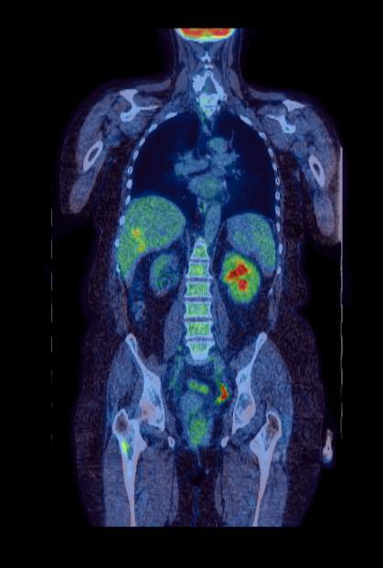
A bone scan may be used to evaluate bone metastasis when clinically indicated. Positron emission tomography (PET) is highly specific for the presence of metastatic TCC but lacks sensitivity. It is best utilised as an imaging modality that assists in diagnostic staging dilemmas (see figure 1).20 Information obtained from pathological and radiological examination is corroborated in order to stratify disease burden on the basis of staging.
The standard staging system is the TNM (tumour, node, and metastasis) system.21 The TNM staging system is applied to urothelial carcinoma and non-urothelial carcinomas. A consensus classification system established by the WHO and International Society of Urological Pathology (ISUP) for urothelial (transitional cell) neoplasms allows further stratification on the basis of grade.
Non-muscle invasive bladder cancer
NMIBC is treated with complete transurethral resection of bladder tumour (TURBT). Those tumours with a high risk of recurrence and progression after TURBT require the use of adjuvant treatments.23-25 This entails instillation of chemotherapeutic agents, usually mitomycin C (MMC), or immunotherapeutic agents such as BCG, either alone or in various combinations.
High-risk tumours could have a second TUR. Cystectomy should be considered in patients with Tis or high grade T1 disease, if BCG has failed.
Muscle invasive bladder cancer
Radical cystectomy with lymphadenectomy (extended) or chemoradiotherapy are the definitive local treatment options. Extended lymphadenectomy may still be curative in patients with limited pelvic lymph node involvement.
Progression-free survival and overall survival have been correlated with the number of lymph nodes removed during surgery.
Urinary diversion options include urostomy (ileal conduit), Mitrofanoff, and Mainz-II. Some highly motivated patients may be suitable for a neo-bladder. Neoadjuvant cisplatin-based chemotherapy has been shown to confer a modest survival benefit, and a recent meta-analysis now suggests a similar benefit for adjuvant chemotherapy in high-risk cases.
Organ preservation using radical radiotherapy is a reasonable alternative to surgery for patients with solitary small volume TCC, with no evidence of CIS elsewhere in the bladder, no hydronephrosis, and good bladder function. They should undergo maximal resection before starting treatment.
It is also considered in patients not fit for surgery, and those who wish to avoid a cystectomy. The addition of radiosensitisers has been shown to improve outcomes.
The BC2001 study demonstrated improved local control with the addition of concomitant chemotherapy to radical radiotherapy.26 The BCON trial demonstrated improved survival with the addition of nicotinamide and carbogen during treatment, the effect being most apparent in tumours demonstrating necrosis.27
Following radical radiotherapy patients need to be willing to undergo regular cystoscopic surveillance, and about 15% will subsequently require a cystectomy because of disease recurrence or development of new bladder tumours.
Advanced and metastatic disease
Cisplatin-containing combination chemotherapy with gemcitabine (GC) or accelerated MVAC (methotrexate, vinblastine, adriamycin and cisplatin) is standard in patients fit enough to tolerate cisplatin. Gemcitabine and cisplatin has been shown to have lower toxicity and is the gold standard first-line treatment in fit patients.
New studies indicate the addition of paclitaxel to GC might prove beneficial.28
Around 50% of patients are unfit for cisplatin-containing chemotherapy because of poor performance status, impaired renal function or comorbidities. Palliation with carboplatin-based regimens or single agent taxane or gemcitabine are reasonable treatment options. Palliative radiotherapy may be used to reduce symptoms such as pain and bleeding.
Follow-up
Follow-up of patients who have received treatment for bladder cancer is imperative either for response evaluation or to detect salvageable recurrence, see table 3.
Response evaluation during chemotherapy is usually scheduled midway through the treatment protocol and radiographic testing is necessary.29
Prognosis
Tumour stage is the most important prognostic factor for progression and overall survival for invasive bladder cancer. In patients with NMIBC, the number of tumours and tumour size also influence prognosis.
Age-standardised relative survival rates for bladder cancer in England during 2005-2009 show that 58.2% of men and 50.2% of women are expected to survive their disease for at least five years. The past decade has seen advances in the diagnosis and treatment of bladder cancer. Mortality rates in Europe appear to have declined in the past decade, by approximately 16% in men and 12% in women. 22 UK statistics for different stages of cancer are unavailable.
Table 4, shows relative survival rates according to tumour stage. (These statistics are from Cancer Research UK and were obtained from different sources including expert opinion.)
Future directions
Bladder cancer is an active research area with ongoing studies into urinary biomarkers that may have a role in diagnosis or detection of recurrence of bladder cancer. Research into intravesical therapies, photodynamic therapy, surgery, radiotherapy and systemic therapy are likely to improve outcomes for patients with bladder cancer.
Summary
Smoking is the main preventable risk factor for bladder cancer. The presence of unexplained haematuria requires rapid evaluation for a possible malignancy.
Cystoscopy is the initial procedure for both the diagnosis and management of urothelial malignancy. A flexible cystoscope is used for diagnostic purposes and rigid cystoscope for tissue biopsy and resection. Surveillance is important either for response evaluation or to detect salvageable recurrence.
A strong patient support network is essential to help patients with bladder cancer. The GP and community palliative care services are important members of the extended multidisciplinary team.
REFERENCES
1 www.cancerresearchuk.org/cancer-info/cancerstats/keyfacts/bladder-cancer
13 Kleinerman RA, Boice JD Jr, Storm HH et al. Second primary cancer after treatment for cervical cancer. An international cancer registries study. Cancer 1995; 76:442-52
14 Boorjian S, Cowan JE, Konety BR et al. Bladder cancer incidence and risk factors in men with prostate cancer: results from Cancer of the Prostate Strategic Urologic Research Endeavor. J Urol 2007; 177:883-87
15 Travis LB, Curtis RE, Glimelius B et al. Bladder and kidney cancer following cyclophosphamide therapy for non-Hodgkin's lymphoma. J Natl Cancer Inst 1995; 87:524-30
16 Talar-Williams C, Hijazi YM, Walther MM et al. Cyclophosphamide-induced cystitis and bladder cancer in patients with Wegener granulomatosis. Ann Intern Med 1996; 124:477-84
17 Groah SL, Weitzenkamp DA, Lammertse DP et al. Excess risk of bladder cancer in spinal cord injury: evidence for an association between indwelling catheter use and bladder cancer. Arch Phys Med Rehabil 2002; 83:346-51
18 Colmers IN, Bowker SL, Majumdar SR, Johnson JA. Use of thiazolidinediones and the risk of bladder cancer among people with type 2 diabetes: a meta-analysis. CMAJ 2012;184:E675-83.doi:10.1503/cmaj. 112102.Epub 2012 Jul 3
19 Stimson CJ, Cookson MS, Barocas DA et al. Preoperative hydronephrosis predicts extravesical and node positive disease in patients undergoing cystectomy for bladder cancer. J Urol 2010;183:1732-37
20 Apolo AB, Riches J, Boyle MG et al. Clinical value of 18F-2-deoxyglucose positron emission tomography (FDG PET) in bladder cancer (abstract). Presented at the 2009 Genitourinary Cancers Symposium.
21 Urinary bladder. In: AJCC Cancer Staging Manual, 7th edn. Springer. New York. 2010
22 Ferlay J, Randi G, Bosetti C et al. Declining mortality from bladder cancer in Europe. BJU Int 2008, 101:11-9. Epub 2007 Oct 30
23 Babjuk M, Burger M, Zigeuner R et al. Guidelines on non-muscle-invasive bladder cancer (TaTl & CIS). European Association of Urology. 2013
24 Pawinski A, Sylvester R, Kurth KH et al. A combined analysis of European Organisation for Cancer Research and Treatment of Cancer and Medical Research Cancer clinical trials for the prophylactic treatment of stage Ta-T1 bladder cancer. J Urol 1996; 156:1934-41
25 Lerner SP, Au JL. Risk-adapted use of intravesical chemotherapy. BJU Int 2008, 102:1247-53
26 Hussain SA, Stocken DD, Peake DR et al. Long-term results of a phase II study of synchronous chemoradiotherapy in advanced muscle invasive bladder cancer. Br J Cancer 2004;90: 2106–2111
27 Hoskin PJ, Rojas AM, Bentzen SM, Saunders MI. Radiotherapy with concurrent carbogen and nicotinamide in bladder carcinoma. J Clin Oncol 2010;28:4912–4918.
28 Bajorin DF, Dodd PM, Mazumdar M et al. Long-term survival in metastatic transitional-cell carcinoma and prognostic factors predicting outcome of therapy. J Clin Oncol 1999;17(10):3173-81
29 Bellmunt J, Orsola A, Wiegel T et al. Bladder cancer: ESMO Clinical Practice Guidelines for diagnosis, treatment and follow-up. Ann Oncol 2011;22 (Suppl 6): vi45-vi49
ARTICLE IN PDF
 Download this article in PDF format
EXTERNAL WEBLINKS
PubMed
NICE
US NCI
Useful links