SEARCH

Practitioner 2010; 254 (1733): 28-32
Be vigilant for secondary periprosthetic joint infection
20 Oct 2010
Article
Deep periprosthetic joint infection (PJI) is one of the most feared complications of hip and knee replacement surgery as it generally requires a combination of surgical intervention(s) and prolonged antibiotic therapy.
In England in 2008, there were 64,722 primary hip and 71,527 primary knee replacements.1 The respective figures for Scotland over a similar period were 6,312 and 6,160.2 The incidence of PJI is 1.5% after knee arthroplasty and 1% after hip arthroplasty.2 The overall cure rate for PJI ranges from 60 to 85%.3 Early postoperative infections are more common and the management of wound problems after joint replacement has previously been discussed.4
PJI is caused by haematogenous spread from a distant primary infection in 70% of all deep infections.5 However, it can potentially be avoided by prompt recognition and treatment of the primary infection focus in susceptible patients. Streptococci are commonly implicated in such secondary infections.
Classification of PJI
PJI can be classified according to the interval between surgery and presentation. This classification is useful, as less radical debridement may be successful in cases where infection has been present for a short period.
Type I: Early infection occurs up to four weeks after surgery and is related to direct contamination of the joint in the perioperative period
Type II: Acute haematogenous infection presents at any time with symptoms present for less than four weeks
Type III: Late infection presents after the immediate postoperative period, but with symptoms present for more than four weeks (although it may have arisen from haematogenous spread originally)
Type IV: A joint may be found to have been infected after a revision operation, for what was initially diagnosed as aseptic failure, when microbiology tests are performed6
Causative organisms
Staphylococcus aureus and Staphylococcus epidermidis tend to cause infection by the primary route through inoculation at the time of surgery. These organisms are responsible for 30-43% of all PJIs.
S. aureus also causes secondary haematogenous infection in cases of bacteraemia, and may affect other sites such as heart valves and bone causing osteomyelitis. Streptococcal infection is responsible for around 10% of PJIs and more commonly causes infection through secondary spread.5
Streptococcus spp. encompass a variety of species of Gram-positive cocci. They are common commensals in the skin and frequently cause skin and soft tissue infections such as cellulitis, erysipelas and impetigo. Rarely, they may lead to necrotising fasciitis where infection affects deeper tissue planes. Other species may cause pneumonia and meningitis. Autoimmune reactions may result in conditions such as glomerulonephritis and rheumatic fever, while haematogenous spread may infect implants such as joint replacements and heart valves.7,8
Streptoccocus spp. are identified in the laboratory by appearance on blood agar such as haemolysis, Gram stain, catalase and determining the presence of Lancefield antigens. These classifications can provide guidance for particular groups of patients at risk of infection and the likely antibiotics required to treat infection.9
Of particular interest are the group A, B, C and G streptococci which can cause invasive, potentially life-threatening infection. We have recently reported our unit's experience with group B streptococcal PJI.10 We undertook a review of all streptococcal PJIs in our hospital (Royal Infirmary of Edinburgh), see box 1, below.
Although historically associated with pregnancy and perinatal infection, group B streptococcal infection is increasingly seen in the general adult population with an incidence rising to 7.3 per 100,000. Risk factors include immunodeficiency, diabetes, and arterial and venous insufficiency. It has a high mortality rate, especially in the elderly.11-16
Risk groups
Host defence mechanisms that prevent infection include physical barriers and immune system mediated protection. These defence mechanisms can be breached through surgical and dental procedures, traumatic wounds or dermatological conditions enabling the development of transient bacteraemia. The immune response can also be blunted by a variety of host factors. Patients with the conditions detailed in table 1 are at greater risk of primary streptococcal infection.8
Presentation
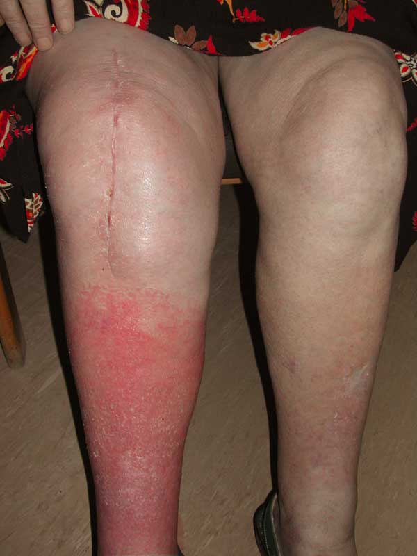
Soft tissue infections generally present with erythema, pain and induration. The absence of abscess formation makes streptococcal cellulitis more likely whereas abscess formation is more commonly seen in staphylococcal disease. The skin may have a raised, indurated border. Any signs such as rapid extension, bullae, pale appearance, severe pain or systemic sepsis should raise the possibility of necrotising fasciitis and indicate the need for prompt referral to hospital.
Other conditions may mimic cellulitis such as thrombophlebitis, olecranon bursitis or other dermatological conditions.
S. pneumoniae may present as pneumonia, or in rare cases, meningitis, while Streptococcus viridans and Streptococcus milleri may present as dental infection.17,18
A swollen hot prosthetic joint without skin changes may be suggestive of primary joint PJI and advice should be sought without delay.
Prevention
Any patient with a joint replacement should be counselled to seek early attention for any soft tissue or dental infection. Soft tissue wounds should be thoroughly cleaned. A course of antibiotics should be considered in any wound in which there has been significant contamination. Animal bites should be covered by a broad spectrum antibiotic such as co-amoxiclav.19
Any subsequent surgical procedure is a risk for bacteraemia. National20 and local guidelines should be followed when using prophylactic antibiotics for subsequent procedures. Prophylactic antibiotics should be administered as a single dose, or for a maximum of 24 hours.20
Historically, antibiotics were administered prophylactically to any patient with a joint replacement (or other prosthetic implant such as a heart valve) when undergoing such procedures. However, recent research has questioned this practice and suggested that prophylaxis is unnecessary for the majority of patients.18 An advisory statement by the American Academy of Orthopaedic Surgeons and the American Dental Association21 suggests that any patient with a joint prosthesis, and especially those who are immunocompromised or have mega-prostheses are at even higher risk and should therefore have prophylactic antibiotics.21
Patients at risk of infection through impaired physical defences such as chronic venous insufficiency should be treated appropriately. Venous ulceration should be treated at an early stage with compression bandaging. Consideration should be given to prophylactic treatment of varicose veins if there are early signs of chronic venous insufficiency e.g. hyperpigmentation in the gaiter area.
Patients with dermatological conditions such as eczema and psoriasis should receive treatment to limit the extent of their disease and risk of secondary infection.
Mild, uncomplicated cellulitis can be treated with oral antibiotics (see figure 1). Attention should be given to local antimicrobial guidelines as these will take into account the most common infectious organisms locally and their susceptibility patterns. In general, simple penicillin agents such as amoxicillin or flucloxacillin should be administered (although low, sub-therapeutic doses should be avoided).
Patients should be referred to hospital for consideration of parenteral antibiotics if they are exhibiting signs of systemic sepsis such as tachycardia, pyrexia or hypotension.8 Early cellulitis and surgical site infection following a knee replacement requires prompt referral and assessment as the joint is very close to the skin and infection can quickly spread through contiguous tissues to the joint (see figure 2).
Initial management
PJI should be suspected if a patient with a joint replacement develops pain in that joint after a soft tissue, respiratory tract or dental infection.
In cases of suspected PJI orthopaedic advice should be sought urgently and antibiotics should be withheld, as the patient requires prompt haematological, biochemical and radiological investigations.
If the patient is systemically well, antibiotics should not be commenced empirically in primary care as this can reduce the sensitivity of subsequent diagnostic tests and jeopardise further management.
Prompt treatment, invariably including surgical intervention, is important for if the PJI persists the bacteria can secrete biofilms. Such biofilms coat implants, and the bacteria become more resistant to antibiotics.
Surgical treatment will include either debridement and prosthesis retention or revision joint replacement. In haematogenous infections that are treated early, implant retention may be more successful than in chronic cases where biofilms have had time to form.3
Conclusion
Patients with previous joint replacement have a lifetime risk of secondary PJI after other common infections. The lifetime risk has been reported to be up to 2% at 20 years after total hip arthroplasty taking into account infection from a subsequent revision procedure as well.22
Streptoccoccus spp. are commonly implicated in these infections. Early recognition and prompt treatment of soft tissue, dental and respiratory infections may also prevent secondary PJI. Acute haematogenous PJI should be suspected if a patient with a joint replacement develops pain in that joint after infection elsewhere. If PJI does occur in this context, optimal outcomes are achieved by early diagnosis, referral and treatment.
REFERENCES
1 National Joint Registry 6th Annual Report (April 2008-March 2009). National Joint Registry. 2009
2 Scottish Arthroplasty Project Annual Report 2009. NHS ISD Scotland. Edinburgh. 2009
3 Esposito S, Leone S. Prosthetic joint infections: microbiology, diagnosis, management and prevention. Int J Antimicrob Agents 2008;32:287-93
4 Dall GF, Huntley JS, Breusch SJ. Managing wound problems following joint replacement. Practitioner 2007;251 (1691):45-50
5 Zimmerli W, Trampuz A, Ochsner PE. Prosthetic-joint infections. N Engl J Med 2004;351:1645-5
6 Tsukayama DT, Estrada R, Gustilo RB. Infection after total hip arthroplasty. A study of the treatment of one hundred and six infections. J Bone Joint Surg Am 1996;78:512-23
7 Weir E, Main C. Invasive group A streptococcal infections. CMAJ 2006;175:32
8 Stevens DL, Eron LL. Cellulitis and soft-tissue infections. Ann Intern Med 2009;150(1):ITC11
9 Lancefield RC. Serological differentiation of human and other groups of hemoytic streptococci. J Exp Med 1933;57:571-95
10 Jenkins PJ, Clement ND, Gaston P et al. Invasive group B streptococcal disease in an orthopaedic unit.
J Hosp Infect 2010; 76:231-33
11 Farley MM, Harvey RC, Stull T et al. A population-based assessment of invasive disease due to group B Streptococcus in nonpregnant adults. N Engl J Med 1993;328:1807-11
12 Farley MM. Group B streptococcal disease in nonpregnant adults. Clin Infect Dis 2001;33:556-61
13 O'Brien KL, Beall B, Barrett NL et al. Epidemiology of invasive group A streptococcus disease in the United States, 1995-1999. Clin Infect Dis 2002;35:268-76
14 Factor SH, Levine OS, Schwartz B et al. Invasive group A streptococcal disease: risk factors for adults. Emerg Infect Dis 2003;9:970-7
15 Phares CR, Lynfield R, Farley MM et al. Epidemiology of invasive group B streptococcal disease in the United States, 1999-2005. JAMA 2008;299:2056-65
16 Skoff TH, Farley MM, Petit S et al. Increasing burden of invasive group B streptococcal disease in nonpregnant adults, 1990-2007. Clin Infect Dis 2009; 49:85-92
17 Deacon JM, Pagliaro AJ, Zelicof SB et al. Prophylactic use of antibiotics for procedures after total joint replacement. J Bone Joint Surg Am 1996;78:1755-7
18 Shanson D. New British and American guidelines for the antibiotic prophylaxis of infective endocarditis: do the changes make sense? A critical review. Curr Opin Infect Dis 2008;21:191-9
19 Medeiros I, Saconato H. Antibiotic prophylaxis for mammalian bites. Cochrane Database Syst Rev 2001-2:CD001738
20 Scottish Intercollegiate Guideline Network. Antibiotic prophylaxis in surgery. Guideline No 104. SIGN. Edinburgh. 2008
21 American Academy of Orthopaedic Surgeons. Information Statement 1033: Antibiotic prophylaxis for bacteremia for patients with joint replacements. American Academy of Orthopaedic Surgeons. 2010
22 Hamilton H, Jamieson J. Deep infection in total hip arthroplasty. Can J Surg 2008;51(2):111-7
USEFUL INFORMATION
British Orthopaedic Association
www.boa.ac.uk
ARTICLE IN PDF
 Download this article in PDF format