SEARCH

Yong ALT. Epidermolysis bullosa requires lifelong monitoring. Practitioner Oct 2019;263(1830):21-25
Epidermolysis bullosa requires lifelong monitoring
24 Oct 2019
AUTHORS
Dr Alicia LT Yong MB ChB MRCP, Clinical Fellow in Dermatology, Department of Dermatology, Royal Victoria Infirmary, Newcastle upon Tyne, UK
Competing interests: None
Acknowledgements: I would like to thank my colleagues Dr Suzy Leech, Consultant Dermatologist, and Dr Stephanie Ball, Post-CCT Fellow in Medical Dermatology, at the Royal Victoria Infirmary, Newcastle upon Tyne for their help with the preparation of the manuscript. I would also like to thank the patient who kindly gave permission for his photographs to be used.
Article
Epidermolysis bullosa (EB) arises from mutations within genes encoding for different proteins which contribute to the structural integrity of the epidermis and basement membrane zone. There are four major EB types: EB simplex (EBS), dystrophic EB (DEB), junctional EB (JEB) and Kindler syndrome. The main cutaneous features of inherited EB are mechanical fragility of the skin and formation of blisters and erosions with minimal trauma. Clinical manifestations can vary widely and this is dependent on the EB subtype. The initial diagnosis of EB is based on the patient’s personal and family history and examination. Skin biopsies are taken from newly induced blisters to identify the level of skin cleavage which helps to determine the subtype. EBS is the most common form of EB. It is almost exclusively autosomal dominant in inheritance. Patients with EBS localised often present with blisters predominantly on the palms and soles following minimal trauma which are exacerbated by warm weather. Onset of symptoms in EBS is usually at birth or shortly after, although in the localised form it may not present until late childhood or even early adulthood. Patients with severe forms of EBS may have widespread cutaneous blistering sometimes leading to death in infancy. JEB is inherited in an autosomal recessive pattern. A hallmark feature of JEB is the presence of enamel hypoplasia. In dominant DEB, generalised cutaneous blistering occurs from birth, with subsequent milia, atrophic scarring and dystrophic nails. Recessive DEB (RDEB) presents with extensive blistering from birth and is associated with anaemia, growth retardation and oesophageal strictures. Patients with RDEB are at risk of developing aggressive cutaneous squamous cell carcinomas.
Epidermolysis bullosa (EB) is a heterogeneous group of genetic disorders characterised by fragility and easy blistering of the skin and mucous membranes.
First described by Koebner in 1886, EB is a rare condition estimated to affect 1 in 17,000 live births with approximately 5,000 people living with EB in the UK.1,2 EB arises from mutations within genes encoding for different proteins which contribute to the structural integrity of the epidermis and basement membrane zone.
There are four major EB types: EB simplex (EBS), dystrophic EB (DEB), junctional EB (JEB) and Kindler syndrome (see table 1). This classification is based on the level of tissue separation within the epidermis and the cutaneous basement membrane zone. In EBS, blistering is confined to the epidermis; in JEB, the lamina lucida (within the basement membrane zone); in DEB, sub-lamina densa (within the uppermost dermis); and in Kindler syndrome, the blistering occurs in multiple levels rather than a specific plane.
EB is clinically diverse with more than 30 subtypes identified. There is a broad spectrum of severity ranging from mild skin fragility localised to extremities, to severe mucocutaneous blistering with extracutaneous complications and mortality in infancy.3
At the international consensus meeting on the diagnosis and classification of EB in 2014 a new classification system was introduced based sequentially on major EB type, phenotypic characteristics, mode of inheritance, targeted protein and gene mutation. Many eponyms for EB subtypes were replaced with descriptive terms.3
The main cutaneous features of inherited EB are mechanical fragility of the skin and formation of blisters and erosions with minimal trauma. However, clinical manifestations can vary widely and this is dependent on the EB subtype and the age of the patient. It is worth noting that there can be significant clinical variability even within the different subtypes.
As a general rule, subtypes with an autosomal recessive inheritance are more severe than autosomal dominant forms with more widespread cutaneous and extracutaneous involvement.
Epidermolysis bullosa simplex
Epidermolysis bullosa simplex (EBS) is the most common form of EB and affects approximately 70% of the EB population.4 It is almost exclusively autosomal dominant. EBS localised (previously Weber-Cockayne) is the mildest and most common form of EB. Patients often present with blisters predominantly on the palms and soles following minimal trauma and symptoms can be exacerbated by warm weather. Onset of symptoms in EBS is usually at birth or shortly after, although in the localised form it may not present until late childhood or even early adulthood.
EBS generalised severe (previously Dowling-Meara) involves widespread cutaneous blistering; a specific feature seen in this condition is herpetiform blistering, where there are clusters of vesicles in an arciform configuration. Palmoplantar hyperkeratosis is seen in all subtypes of EBS and is more confluent in the severe form. Extracutaneous involvement in EBS is uncommon apart from intraoral blistering. Although symptoms tend to improve with age, widespread cutaneous blistering in EBS generalised severe may lead to death in infancy.
Junctional epidermolysis bullosa
Junctional EB (JEB) is inherited in an autosomal recessive pattern. A hallmark of JEB is the presence of enamel hypoplasia, a useful sign once primary teeth have erupted.
JEB generalised severe (previously Herlitz) is the most severe form with extensive, generalised cutaneous blistering present from birth.
The blistering heals with exuberant granulation tissue which is a characteristic feature.
Anaemia and growth retardation are both common features, and involvement of the gastrointestinal, genitourinary, ocular and respiratory systems are seen. JEB generalised intermediate is more common but less severe.
The one-year mortality rate is 40-45% in JEB; common causes include sepsis, failure to thrive and respiratory failure.5
Dystrophic epidermolysis bullosa
In dominant dystrophic EB (DDEB), generalised cutaneous blistering occurs from birth, with subsequent milia (tiny, white bumps under the skin), atrophic scarring and dystrophic nails. Blistering of the oesophagus may also occur which can progress to oesophageal stenosis.
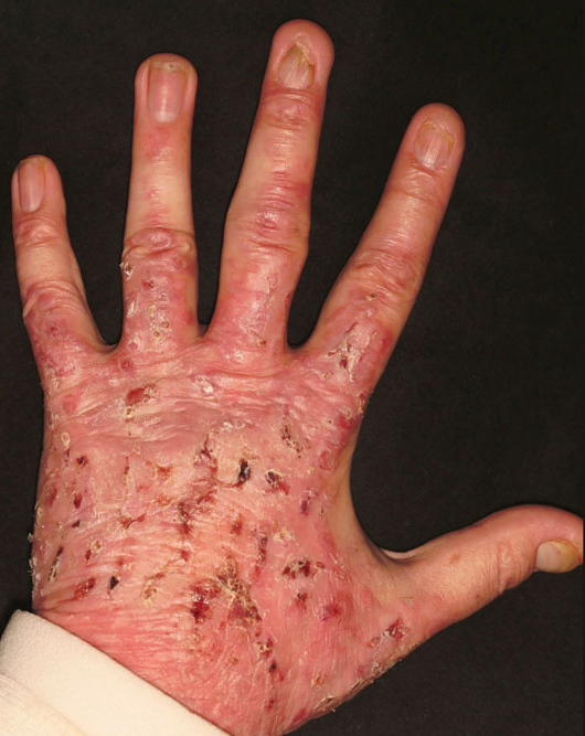
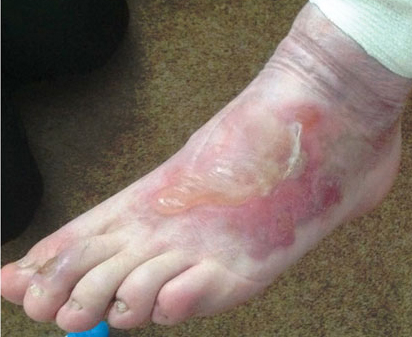
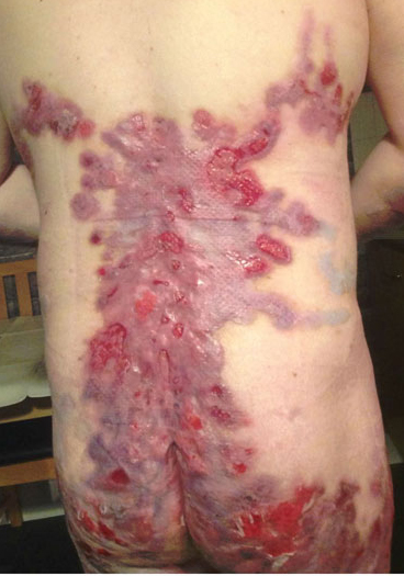
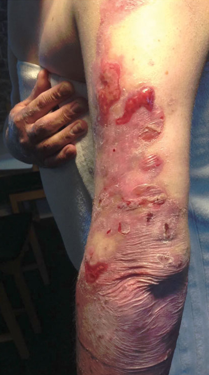
Recessive DEB (RDEB) generalised severe (previously Hallopeau-Siemens) presents with extensive blistering from birth (see figure 1 and figures 2 and 3) and is associated with anaemia, growth retardation and oesophageal strictures.
Pseudosyndactyly (mitten hand deformity) is caused by blistering which heals with scarring. It is a prominent feature with almost 100% of patients developing this deformity by the age of 20.6,7
Other complications include malnutrition, renal failure, cardiomyopathy, delayed puberty and osteoporosis. Patients with RDEB are at risk of developing aggressive cutaneous squamous cell carcinomas (SCC). The risk begins in the mid to late teenage years and increases thereafter, with death commonly occurring from metastatic SCC within five years of diagnosis of the initial primary tumour.3
Kindler syndrome
Kindler syndrome, a rare type of EB, is characterised by generalised blistering and photosensitivity present from birth with gradual development of poikiloderma (mottled hyperpigmented and hypopigmented skin with telangiectasia) and cutaneous atrophy. Blistering, which tends to occur on hands and feet, improves with age. Patients develop extracutaneous features including colitis, oesophagitis, oesophageal strictures and ectropion.
Epidermolysis bullosa acquisita
Epidermolysis bullosa acquisita (EBA) is a rare, acquired immunobullous condition which shares many of the mucocutaneous features of DEB. Both affect type VII collagen within anchoring fibrils of the dermal epidermal junction resulting in a subepidermal split.
This condition usually presents in middle age with blisters and erosions over trauma prone areas of the skin which heal with milia and scarring; mucous membrane involvement may also be present. Bullous pemphigoid is an important differential.8
Diagnosis
The initial diagnosis of EB is based on careful elicitation of the patient’s personal and family history and thorough clinical examination.
Skin biopsies are taken from newly induced blisters for identification of the level of skin cleavage via immunofluorescence antigen mapping or in select cases, transmission electron microscopy. When the level of skin cleavage has been determined, DNA mutational analysis can be performed for further subclassification.
Genetic testing is also performed in the context of prenatal screening and genetic counselling and can aid prognostication.
Routine histology can be done to exclude other differential diagnoses, but it is not usually helpful in making the diagnosis as it may be difficult to differentiate the level at which cleavage occurs using light microscopy alone.9
Management
There is still no cure for EB. There are very few published guidelines or clinical trials. There is general agreement among EB specialists that optimal care requires a multidisciplinary approach. Management is centred on:
• Avoidance of trauma and blistering of the skin
• Good wound care
• Prevention of secondary infection
• Management of extracutaneous complications
Patients with known EB subtypes can be at risk of developing specific extracutaneous complications. Therefore careful surveillance and early intervention should be undertaken to mitigate morbidity and mortality.10
Blisters and erosions can arise in EB from seemingly minimal trauma and can be exacerbated by heat. Steps should therefore be taken to prevent formation of new lesions by the application of dressings to susceptible areas such as the elbows and knees, wearing loose-fitting clothing and keeping cool with air conditioning.
Blisters can be punctured with a sterile needle with the roof left intact to act as a biological dressing. Existing wounds can be dressed with sterile, non-adherent dressings (silicone or hydrocolloid based) to promote healing and to reduce pain and risk of infection. Skin should be cleansed regularly with water and gentle soaps. 10,11
Cutaneous infection is a frequent occurrence in all forms of EB, with Staphylococcus aureus the usual pathogen.6 Wound colonisation is common but infection should be treated to promote healing. Cleansing agents such as normal saline, dilute potassium permanganate, vinegar and diluted bleach have been used. Antibiotics should be prescribed judiciously guided by culture results. Topical antibiotics should only be used for limited periods given the risk of developing resistance. A recent study of 23 children with EB showed that more than half of these patients had mupirocin-resistant S. aureus.12
Pain and itch are burdensome features and can significantly impact on patients’ quality of life. Evidence-based guidelines are available to aid clinicians in managing these symptoms.13 Adequate pain control should be a priority.
Beyond these measures, management should be tailored to the individual patient as complications vary depending on the EB subtype.10
Patients with generalised severe subtypes of EB can develop profound anaemia and growth retardation. Growth is affected by a combination of factors including reduced oral intake, impaired absorption of nutrients through damaged intestinal epithelium and an increased catabolic state. Frequent oral feeding with nutritional supplements can help to increase intake and if still inadequate, enteral feeding
via gastrostomy is recommended. Anaemia, which is multifactorial in origin, is treated with iron supplementation and transfusions.7,10,14
Painful blisters and erosions on the cornea can occur leading to corneal scarring and visual impairment. Corneal dryness should be treated with ophthalmic ointments and artificial tears. Oesophageal strictures can arise from repeated blistering/scarring and dilatation is needed to ensure adequate nutritional intake. Mitten deformity of the hands can be prevented by meticulous wrapping of each digit on a daily basis, however with progression, surgical intervention is often required.7,10,14
EB patients who are prone to contractures and decreased mobility should be referred to occupational therapy for help in maintaining functional independence in activities of daily living.
Osteopenia and osteoporosis should be carefully monitored with serial DEXA scans and treated with calcium and vitamin D supplementation plus bisphosphonates.7,10 The reduction in bone mineral density is multifactorial in origin and results from reduced mobility, decreased nutritional intake, chronic skin infection and high levels of inflammation.
SCC can arise from damaged skin especially in RDEB generalised severe patients; mortality is high with more than 79% of these patients dying from SCC by the age of 55.15 Close monitoring for cutaneous malignancies is of utmost importance and full skin checks by a dermatologist at three-monthly intervals are recommended.
Depression is common among patients who are debilitated by symptoms of EB and psychosocial support should be provided for the entire family unit.
The complications mentioned above are by no means an exhaustive list and patients almost invariably require ongoing multidisciplinary specialist input.
Conclusion
EB rarely presents in primary care as symptoms are usually present at or around birth.
However, GPs can play an integral role in the management of patients with EB. Occasionally, certain forms of EB, for example localised EBS, may present later in life and GPs are in a unique position to consider this rare diagnosis as a possibility, through their knowledge of the patient’s family history and longitudinal approach to medicine.
Local healthcare knowledge surrounding EB can be limited which is often a source of frustration for EB patients. Familiarity with EB enables GPs to assist patients in coordinating care and involvement with specialist centres, local hospital specialties and support agencies such as DEBRA.
Complex EB patients may have common easily treatable conditions which may or may not be related to their primary diagnosis; it is important that these symptoms are not automatically attributed to EB.
Moreover, GPs play a crucial role in providing holistic care including emotional and psychological support for patients and their families.16
Patients are diagnosed and managed in specialist centres which also provide long-term support for affected families. The national EB service is run by four specialist centres in the UK based at: Birmingham Children’s Hospital; Great Ormond Street Hospital for Children; Guys & St Thomas’ Hospital; and Solihull Hospital. For those patients who cannot easily access these centres, there are also outreach clinics several times a year around the country.
The EB multidisciplinary team works closely with DEBRA (see External links), the national charity that funds healthcare and research for patients with EB and provides important resources for patients and healthcare professionals.
EB is a rare and complex genetic condition with multiple subtypes, a phenotypically diverse clinical presentation and differing prognoses. Referral to a specialist centre should be made early if the diagnosis is suspected and management will require a multidisciplinary approach, with patients usually requiring lifelong input and monitoring.
In the past decade, important advances have been made in the development of gene therapy, protein replacement therapy, cell therapies (intradermal injection of allogenic fibroblast and mesenchymal stromal cells), bone marrow stem cell transplantation, gene editing and the use of inducible pluripotent stem cells. Although still in experimental stages, these advances hold promise for potential therapies in the future.17
REFERENCES
1 Mellerio J. Epidermolysis bullosa care in the United Kingdom. Dermatol Clin 2010;28(2):395-96
2 Poster 680. Exploration of gene therapies for dominant dystrophic epidermolysis bullosa. Molecular Therapy 2009;17:S259
3 Fine J, Bruckner-Tuderman L, Eady R. Inherited epidermolysis bullosa: Updated recommendations on diagnosis and classification. J Am Acad Dermatol 2014;70(6):1103-26
4 What is EB? Debra.org.uk. 2019 www.debra.org.uk/what-is-eb/what-is-eb [Last accessed 29 September 2019]
5 Fine J, Johnson L, Weiner M, Suchindran C.
Cause-specific risks of childhood death in inherited epidermolysis bullosa. J Pediatrics 2008;152(2):276-280.e2
6 Fine J, Johnson L, Weiner M. Pseudosyndactyly and musculoskeletal contractures in inherited epidermolysis bullosa: experience of the National Epidermolysis Bullosa Registry, 1986-2002. J Hand Surg 2005;30(1):14-22
7 Fine J, Mellerio J. Extracutaneous manifestations and complications of inherited epidermolysis bullosa: part II. Other organs. J Am Acad Dermatol 2009;61(3):387-402
8 Gupta R, Woodley D, Chen M. Epidermolysis bullosa acquisita. Clin Dermatol 2012;30(1):60-69
9 Fine J. Inherited epidermolysis bullosa. Orphanet J Rare Dis 2010;5:12
10 Ramirez M, Murrell D, Fine J. Management of epidermolysis bullosa. Expert Opinion on Orphan Drugs 2013;1(4):279-93
11 El Hachem M, Zambruno G, Bourdon-Lanoy E. Multicentre consensus recommendations for skin care in inherited epidermolysis bullosa. Orphanet J Rare Dis 2014;9(1):76
12 Singer HM, Levin LE, Garzon MC. Wound culture isolated antibiograms and caregiver-reported skin care practices in children with epidermolysis bullosa. Pediatr Dermatol 2018;35(1):92-96
13 Goldschneider KR, Good J, Harrop E. Pain care for patients with epidermolysis bullosa: best care practice guidelines. BMC Med 2014;12:178. Published online 9 October 2014. doi: 10.1186/s12916-014-0178-2
14 Fine J, Mellerio J. Extracutaneous manifestations and complications of inherited epidermolysis bullosa: part I. Epithelial associated tissues. J Am Acad Dermatol 2009;61(3):367-84
15 Fine J, Johnson L, Weiner M. Epidermolysis bullosa and the risk of life-threatening cancers: The National EB Registry experience, 1986-2006. J Am Acad Dermatol 2009;60(2):203-11
16 Evans W, Rafi I. Rare diseases in general practice: recognising the zebras among the horses. Br J Gen Pract 2016;66(652):550-51
17 Hsu C, Wang S, Lee J, McGrath J. Treatment of hereditary epidermolysis bullosa: Updates and future prospects. Am J Clin Dermatol 2014;15(1):1-6
 Download this article in PDF format