SEARCH

Van-de-Velde V, Tidman M. Diagnosing childhood eczema can be challenging. Practitioner 2017 Sept;261(1807):21-25
Diagnosing childhood eczema can be challenging
25 Sep 2017
AUTHORS
Dr Vanessa Van-de-Velde MRCP, Specialist Registrar in Dermatology
Dr Michael J. Tidman MD FRCP (Ed) FRCP (Lond), Consultant Dermatologist
Department of Dermatology, Royal Infirmary of Edinburgh, Edinburgh, UK
Competing interests: None
Article
Abstract
Atopic eczema is the most common endogenous type of eczema in infants and children and affects around 15-20% of school-age children in the UK. Its prevalence is highest in children under the age of two and subsequently diminishes with age. It has a chronic, relapsing course. Discoid eczema may occur in children with a background of atopic eczema or independently. Allergic contact dermatitis is unusual in infants and children, and the most common example is the localised eczematous eruptions observed around nickel fastenings. However, irritant contact dermatitis is relatively common, a good example being nappy rash (ammoniacal dermatitis) caused by prolonged skin contact with urine and faeces. Those children with a tendency to atopic eczema are more prone to irritant dermatitis. Infantile seborrhoeic dermatitis is common and usually resolves before the child’s first birthday. An emergency referral to a dermatologist or paediatrician should be made via telephone when there is a suspicion of eczema herpeticum or eczema coxsackium. Other indications for referral include diagnostic uncertainty, recurrent secondary infection, when control remains poor despite topical treatments, and for patients with emotional distress or significant sleep disturbance.
The term eczema, which is synonymous with dermatitis, describes a group of inflammatory skin diseases that are often challenging to diagnose accurately and treat effectively, and are frequently encountered by GPs.
Eczema is characterised by erythematous, dry and itchy skin that can also become weepy, crusted, blistered and lichenified.1 Several different types of eczema can present in infants and children.
Atopic eczema
Atopic eczema is the most common endogenous type of eczema in infants and children and affects approximately 15-20% of school-age children in the UK.2 Prevalence is highest in children under the age of two and subsequently diminishes with age. It has a chronic, relapsing course.2
The pattern of atopic eczema may change with age. In babies, cheek and scalp involvement predominate, but other body regions can also be involved. The nappy region is usually spared and widespread xerosis is an important diagnostic clue.
The Dennie-Morgan fold, a prominent crease involving the lower eyelids, is a helpful diagnostic feature.1
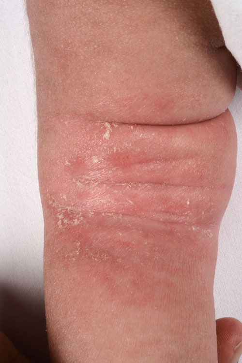
In childhood, flexural involvement predominates with the antecubital and popliteal fossae commonly affected, see figure 1. Lichenification, manifesting as accentuated skin markings, is a common feature.
Post-inflammatory hypopigmentation and hyperpigmentation can develop and may be slow to resolve. After puberty, atopic eczema tends to be less exudative and more chronic, with face, neck and hand involvement common.1
Discoid eczema (nummular dermatitis)
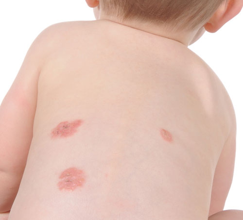
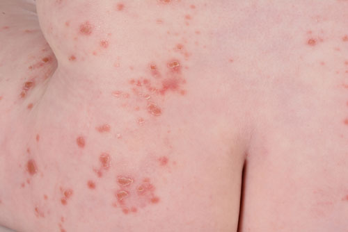
Nummular means coin shaped and describes the morphology of this condition. In infants and children eczematous lesions are often exudative and pruritic with follicular papules that coalesce to form plaques,1 see figure 2. These lesions may occur in children with a background of atopic eczema, or independently.
Contact dermatitis
Compared with adults, allergic contact dermatitis is unusual in infants and children. Nickel allergy is the most common allergen in this age group with localised eczematous eruptions observed around nickel fastenings.1 However, irritant contact dermatitis,
is relatively common in infancy and childhood, nappy rash (ammoniacal dermatitis), caused by prolonged skin contact with urine and faeces, being a good example. Another common irritant in this age group is saliva, because of its alkalinity, and this can cause dribble dermatitis on the chin, anterior neck and upper chest in young children, and lip-licking dermatitis around the mouth in older children. Those children with a tendency to atopic eczema are more prone to irritant dermatitis. It is important to explain to parents that nappy rash is usually not caused by toxins in the nappy, allergy to the nappy, a reaction to washing powders or dermatophyte fungal infections.1
Seborrhoeic dermatitis
Infantile seborrhoeic dermatitis is common, and usually develops in infants around six weeks of age. It may be localised to the scalp (cradle cap) and face, and in rare cases can be very extensive and associated with diarrhoea, internal infections and failure to thrive (Leiner disease). It may be difficult to distinguish from atopic eczema, although affected babies are usually not disturbed by the rash. Furthermore, it is generally characterised by greasy-looking yellow scale, in contrast to the dry scale and excoriations seen in atopic eczema. It usually resolves before the child’s first birthday.1
Assessing disease severity
Although there are a number of validated scoring systems to aid with assessment of severity in atopic eczema such as SCORAD (SCORing Atopic Dermatitis), EASI (Eczema Area and Severity Index) and POEM (Patient Oriented Eczema Measure), these are time consuming and are more useful as research tools than in day-to-day management in primary care.2,3
However, it is important to consider the impact of eczema on quality of life. The effect on everyday activities, schooling, sleep and emotional wellbeing should all be considered. Several validated tools, which are easy to use, can aid with quality of life assessment, such as CDLQI (Children's Dermatology Life Quality Index), IDQoL (Infants' Dermatitis Quality of Life Index) or DFI (Dermatitis Family Impact) questionnaire.2,3
NICE guidelines suggest the most appropriate way to assess severity in primary care is by visual assessment as outlined in table 1.3
Eczema triggers
Superimposed Staphylococcus aureus infection can trigger atopic eczema flares. Weeping, a yellow honeycomb crust, or pustules may indicate staphylococcal infection and can be confirmed with a bacterial swab. Topical corticosteroids can decrease the load of staphylococcal organisms on the skin and co-treatment with oral antibiotics can improve the clinical result.2 MRSA should also be considered and eradication treatment given in line with local guidance if confirmed. Furthermore, treatment of family members who are carriers should be considered.
Parents of children affected with atopic eczema commonly question the role of allergens as triggers. Limited data exist on the benefit of reducing exposure to environmental allergens on improvement or prevention of atopic eczema.4,5 As a result testing for type 1 allergy tends not to be offered routinely in the management of atopic eczema unless there is evidence suggesting an allergic precipitant, such as a convincing history of acute food reactions, or in patients with no improvement despite intensive topical therapy.2,3 For these patients a skin prick test and specific IgE antibody blood tests may be undertaken, following a focused allergy history, by an experienced team.
Patch testing should be undertaken in children when an allergic contact (type 4) allergy is suspected. Patch testing is undertaken in a dermatology department, but children should be referred to the appropriate allergy or paediatric service for skin prick and IgE testing.2,3 Procedures such as elimination diets should only be undertaken with input from paediatric dietitians.2,3
Management
Emollient therapy, topical corticosteroids or calcineurin inhibitors, and antiseptics are the basic components of eczema treatment in most cases.
Emollient therapy
Emollient therapy forms the mainstay of eczema treatment, helping to restore and maintain the skin barrier, in addition to reducing itching and increasing the effectiveness of topical corticosteroids and calcineurin inhibitors.2 Furthermore, emollients can act as a protective barrier in nappy rash and other types of irritant contact dermatitis.
Parents should be advised to use emollients liberally and frequently both during flares and once the skin has settled, aiming for application at least two to four times a day, during and after bathing.2 Choice of emollients should be dictated by patient and parenteral preference with creams, ointments or lotions being offered to aid compliance. Emollients should be smoothed on in the direction of hair growth and emollients or emollient wash products should be used instead of soap.
Topical corticosteroids
A stepped approach can be taken when prescribing topical corticosteroids. The strength of topical corticosteroids should be tailored to the severity of the eczema.2,3 For instance, mild corticosteroids should be used in mild disease and moderate corticosteroids in moderate disease. Parents should be counselled on signs of an eczema flare such as itchy, dry, erythematous skin and increased irritability. They should be advised to commence treatment at the start of a flare and to continue for 10-14 days. Sufficient steroid should be applied to inflamed skin so that the treated area glistens.
A mild corticosteroid should be the first choice for delicate areas such as the face, neck, axillae or groin, the exception is the use of moderate potency corticosteroids during severe flares for 3-5 days.2,3
Treatment should be once daily, tapering down to alternate days, then twice weekly as symptoms improve.
If a child still experiences frequent flares (2-3 per month), a maintenance regimen can be advised: the corticosteroid is applied on two consecutive days of the week.2,3 A mild topical corticosteroid applied once or twice daily for 1-2 weeks to inflamed areas can be useful in nappy rash although a topical antifungal cream such as clotrimazole or ketoconazole applied once or twice daily may be needed if there is a suspicion of Candida albicans, which is often indicated by the presence of satellite lesions.
Topical calcineurin inhibitors
Topical calcineurin inhibitors (tacrolimus or pimecrolimus) can be particularly helpful for patients at risk of side effects from long-term topical corticosteroid use and in those with persistent facial eczema. They are not recommended for children under the age of two and it is important to counsel patients and parents on the stinging sensation that can occur during their initial use.2,3
As with topical steroids, topical calcineurin inhibitors can be used on a maintenance basis to reduce the frequency of flares in atopic eczema.2
Antiseptics
Limited studies have been undertaken looking at the benefit of using antibacterial soaps, bath additives, topical antibiotics or antiseptics in atopic eczema, but nonetheless many clinicians find these adjuvants helpful. Preparations containing benzalkonium chloride can reduce the burden of staphylococcal organisms on the skin and chlorhexidine containing creams or lotions are useful against MRSA.
Bandages and medicated dressings
Dry bandages or localised medicated dressings with emollients or, in the short term, with topical corticosteroids can be very useful in treating chronic lichenified eczema. However, they are not recommended in children with infected atopic eczema.2
Antihistamines
Oral antihistamines should not be used routinely in atopic eczema. Sedating antihistamines can be considered for a 7-14 day course in children over six months old who have sleep disturbance during a flare.2 Non-sedating antihistamines can be offered for a one month trial in children with eczema with severe itch or urticaria and, if beneficial, can be continued but reviewed every three months.2
Infective complications of eczema
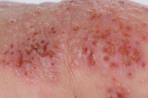
Widespread herpes simplex infection, eczema herpeticum, is a potentially severe complication of atopic eczema. Classically, it appears as numerous punched-out lesions with itchy blisters, see figure 3.
Secondary bacterial infections with staphylococci and streptococci are common and can often be recognised by the hallmark yellow honeycomb crust. Affected children are often unwell and feverish with swollen lymph nodes.
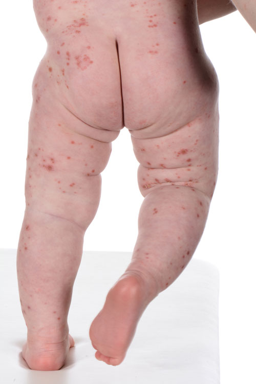
In recent years, an eczema herpeticum-like eruption has been recognised, caused by the enterovirus Coxsackie A6 and A16,6 and named eczema coxsackium. The clinical appearance of eczema coxsackium is similar to eczema herpeticum with erosions and vesicles but lesions are not monomorphous or punched out as seen in eczema herpeticum, see figures 4A and 4B, opposite. Affected children are not as unwell as those with eczema herpeticum. The diagnosis can easily be confirmed with viral swabs.
Referral
The majority of children with eczema can be managed in primary care but in some instances a referral should be sought to consider progression to phototherapy or systemic treatment.2,3
An emergency referral to a dermatologist or paediatrician should be made by telephone when there is a suspicion of eczema herpeticum or eczema coxsackium.2,3
Other indications for referral include an uncertainty about diagnosis, recurrent secondary infection, when control remains poor despite topical treatments, and for patients with emotional distress or significant sleep disturbance.
REFERENCES
1 Schachner L, Hansen R. Pediatric Dermatology, Volume 2. 4th edition. Mosby. New York. 2011
2 Scottish Intercollegiate Guidelines Network. SIGN 125. Management of atopic eczema in primary care. SIGN. Edinburgh. 2011
3 National Institute for Health and Care Excellence. CG57. Atopic eczema in under 12s: diagnosis and management. NICE. London. 2007
4 Boyle RJ, Khan T, Chivinge J et al. Hydrolysed formula and risk of allergic or autoimmune disease: systematic review and meta-analysis. BMJ 2016;352:i974
5 Bath-Hextall F, Delamere FM, Williams HC. Dietary exclusions for established atopic eczema. Cochrane Database Syst Rev 2008; Jan 23(1):CD005203. doi: 10.1002/14651858.CD005203.pub2
6 Mathes EF, Oza V, Frieden IJ et al. “Eczema coxsackium” and unusual cutaneous findings in an enterovirus outbreak. Pediatrics 2013;132(1): e149-e157 doi: 10.1542/peds.2012-3175. Epub 2013 Jun 17
 Download this article in PDF format