SEARCH

Stollery N. Photoguide: Pigmentation disorders. Practitioner 2015; 259(1786):30-31
Photoguide: Pigmentation disorders
21 Oct 2015
AUTHORS
Dr Nigel Stollery MB BS DPD, GPwSI Dermatology, Kibworth, Leicestershire, UK
Article
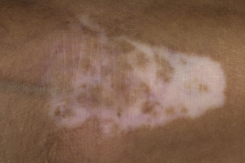
Vitiligo
Vitiligo is a common acquired disorder of skin and mucous membrane pigmentation affecting up to 2% of the population. The most common age of onset is 20. Affected areas have well demarcated white or hypopigmented macules which can range in size from a few millimetres to vitiligo affecting the whole body. The condition is progressive with lesions enlarging centrifugally at unpredictable rates. Vitiligo can be classified into three main types: localised, generalised and universal (where all the skin is affected). The diagnosis is usually made on clinical examination without a biopsy. Treatment can be divided into non-surgical (phototherapy, laser, steroid therapy, topical therapy and depigmentation therapy) and surgical (grafting, melanocyte cultures and noncultured suspensions).
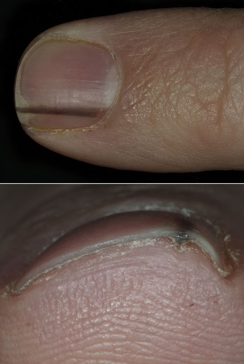
Melanonychia
This is a common condition in which brown or black discoloration of the nail occurs usually as a linear streak, so-called longitudinal melanonychia. In most cases it arises from a benign naevus but it can be related to an underlying melanoma in the nail matrix which most commonly occurs in the thumb, index finger or first toe. Melanonychia occurs more frequently in those with darker skin and is more common with increasing age. Other causes of nail pigmentation include exposure to tobacco, silver nitrate and potassium permanganate, subungual haemorrhage and fungal and Gram-negative bacterial infections. Dermoscopy can be helpful but if there is diagnostic doubt a biopsy should be undertaken from the nail matrix rather than just the nail.
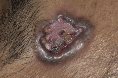
Pigmented basal cell carcinoma
Basal cell carcinomas (BCC) as a family are the most common type of cancer. They pose little threat to life and are more common in fair-skinned individuals with skin types l and ll who have had a lot of sun exposure. BCC may be nodular, superficial, morphoeic or pigmented. Pigmented BCC are a brown, blue or grey colour and may be mistaken for melanomas. Histologically they resemble the more common nodular or superficial types.
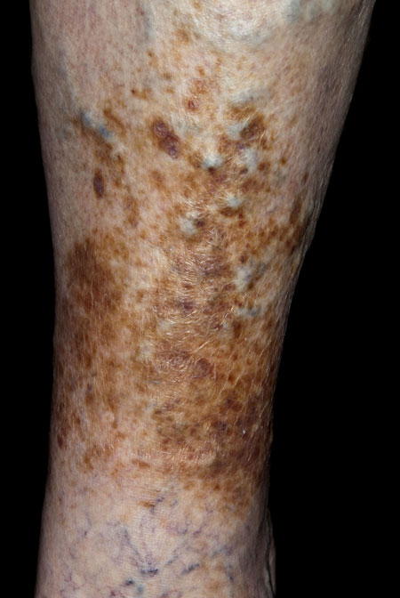
Haemosiderin deposition
Haemosiderin is an iron storage complex containing ferritin, denatured ferritin and other materials. It is found in blood cells, but not circulating blood, most commonly in macrophages and is especially abundant following haemorrhage. When blood leaves ruptured vessels the red blood cells die leaving haemoglobin which is then broken down by macrophages leaving haemosiderin deposited in tissue. Haemosiderin hyperpigmentation is caused by the deposition of haemosiderin and occurs in haemochromatosis, haemorrhagic disease, purpura and, in this case, stasis dermatitis.
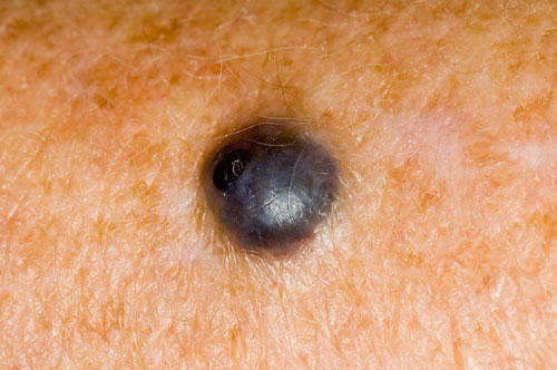
Blue naevus
Blue naevi appear blue in colour because the melanocytes are situated deeper within the tissue than normal brown naevi. They can be found anywhere on the body, but are most common on the scalp, face, hands and feet. Blue naevi are more common in children and teenagers although they can occur at any age. The lesions are usually round and less than 2-3 mm in diameter. Occasionally they are elevated but are usually flat. If there is any doubt regarding the diagnosis, excision and histological examination is advisable, but in most cases excision is only undertaken for cosmetic reasons.
Lead pellet
This man presented with a pigmented lesion of the knee. He had suddenly noticed the lesion a few months before and had recently lost weight. The overlying skin was normal and no pigment network was visible when the lesion was examined under the dermatoscope. A provisional diagnosis of a blue naevus was made. An excision was undertaken and a foreign body revealed. On further questioning, the patient recalled being shot in the area with a lead pellet as a child some 70 years ago. Histology confirmed the presence of a lead pellet.
ARTICLE IN PDF
 Download this article in PDF format